Skin
Question 1. What is skin? List its functions.
Answer.
The skin (integument) is the outer covering of the body with a total area of about 20 square feet. It is the largest organ (area wise) of the body constituting about 16% of the body weight.
Functions
- Protection of the body from heat, cold, ultraviolet rays, etc.
- Prevention of loss of the body fluids and absorption of water within the body.
- Regulation of the body temperature.
- Acts as a sensory organ.
- Synthesis of vitamin D with the help of ultraviolet rays.
- Absorption of lipid-soluble materials, e.g. vitamins like A, D, E and K; solvents like acetone; and heavy metals like arsenic, lead and mercury.
Question 2. What are the layers of the skin?
Answer.
The skin consists of two layers:
- Epidermis: It is the superficial layer. It consists of stratified squamous epithelium and is derived from ectoderm. It is avascular.
- Dermis: It is the deep layer. It is made up of connective tissue and is derived from mesoderm. It is highly vascular and contains glands, nerve endings and hair follicles.
Question 3. Enumerate the layers of epidermis.
Answer.
The thick skin presents the following five layers, from deep to superficial:
- Stratum basale/stratum germinatum/basal layer
- Stratum spinosum/spiny layer
- Stratum granulosum/granular layer
- Stratum lucidum/clear layer
- Stratum corneum/cornified layer

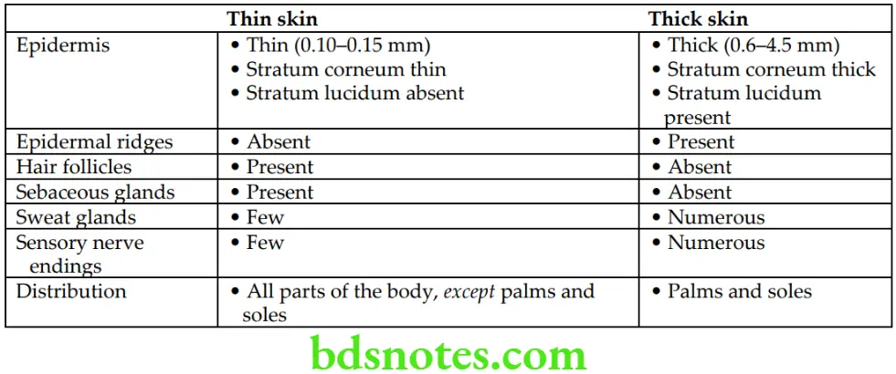
Question 4. What are the differences between thin and thick skin?
Answer.
The differences between thin and thick skin.
Differences Between Thin and Thick Skin
Question 5. What are the layers of dermis?
Answer.
The dermis consists of two layers:
- Papillary layer: It is superficial and forms one-fifth of the total thickness of dermis. It sends finger-like projections (called dermal papillae) towards epidermis.
- Reticular layer: It is a deep layer and contains course bundles of collagen fibres. It also contains blood vessels and nerves.
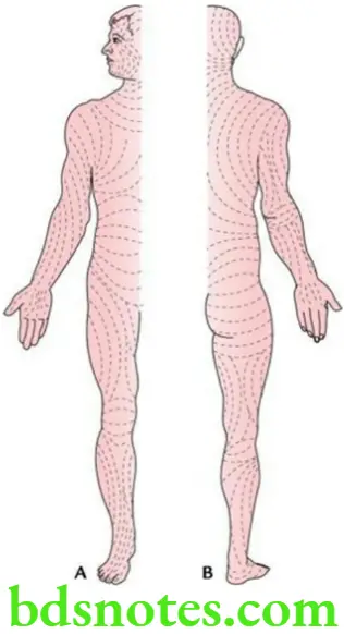
Question 6. What are cleavage lines/Langer’s lines? Mention their clinical significance.
Answer.
These are lines on the surface of the skin. They are produced by the pull of collagen fibres present within the dermis, and radiate in definite directions. They correspond to the natural orientation of collagen fibres in dermis. In general, the Langer’s lines tend to run longitudinally in the limbs and circumferentially in the neck and the trunk.
Clinical significance
The knowledge of orientation of these lines is of special interest to surgeons as:
- Incisions made parallel to these lines heal rapidly and produce hair-line scar (due to formation of less scar tissue).
- Incisions made across these lines heal poorly and produce wide scar (due to formation of more scar tissue).
Question 7. What are the appendages of the skin?
Answer.
Appendages of the skin:
- Hair
- Sweat glands
- Sebaceous glands
- Nails
Superficial fascia
Question 1. What is superficial fascia?
Answer.
- It is a layer of loose connective tissue located deep to skin. It connects the skin to the underlying deep fascia. The superficial fascia contains subcutaneous fat, nerves and vessels. It is mostly heavily infiltrated with fat in females and children, which is the main factor responsible for smooth external contours of the body in females and children. AN4.3
- It allows mobility of the skin on the underlying structures.
- It acts as a distributary layer in which blood vessels, lymphatics and nerves can travel before entering the dermis.
Read And Learn More: Selective Anatomy Notes And Question And Answers
- It forms a kind of insulating layer over the body surface and accounts for the increased resistance of the females to cold in comparison with the males.
- It is extremely thin and devoid of fat in the eyelids, the external ear, penis and scrotum.
- In palms, soles, back of neck and scalp, it is made up of dense connective tissue, which firmly bind it to the underlying structures.
Question 2. Enumerate the sites of subcutaneous injections.
Answer.
The sites of subcutaneous injectionare:
- Posterior aspect of arm
- Anterior aspect of forearm
- Anterior abdominal wall
- Anterior aspect of thigh
Question 3. What is panniculus carnosus? Enumerate the muscles that represent panniculus carnosus in humans.
Answer.
The panniculus carnosus is a thin sheet of striated muscle present in the superficial fascia of lower animals. Its fibres are inserted into the skin.
The following muscles in the human body represent the panniculus carnosus:
- Muscles of the scalp
- Muscles of the facial expression
- Platysma (in neck)
- Subareolar muscle (of breast)
- Palmaris brevis (in hand)
- Dartos muscle (of scrotum)
- Corrugator cutis ani (around the anal orifice)
Deep fascia
Question 1. What is deep fascia? Mention its clinical significance.
Answer.
- It is a dense, inelastic fibrous membrane that separates the superficial fascia from the underlying structures. It is made up of regularly arranged collagen fibres.
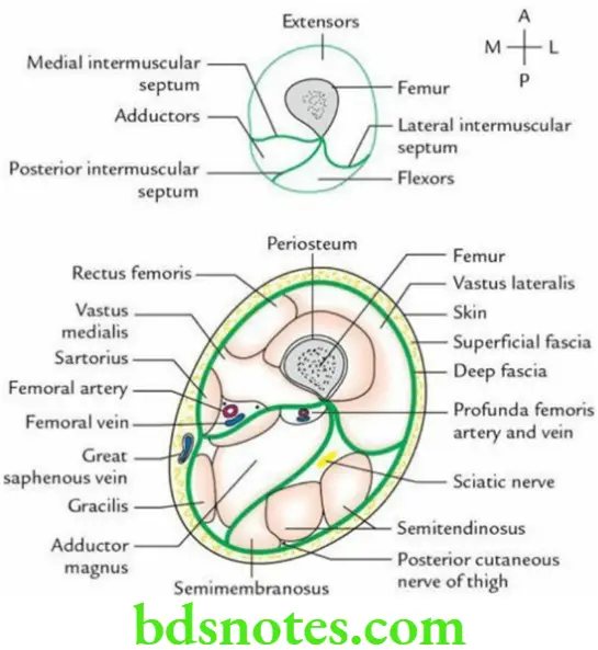
- It sends septa between muscles from its deep aspect forming intermuscular septa.
- It ensheaths the muscles, vessels and nerves. The sheath around the muscles forms tunnels within which muscles can slide independent of the adjacent muscles.
- It forms thickened bands – the retinacula – at certain sites, such as wrist and ankle, which hold the tendons in place and prevent their bow stringing during the movements of the hand and feet at these sites.
Clinical significance
The deep fascia forms fascial planes that are of special interest to surgeons because:
- They can operate along the fascial planes easily with minimal injury to adjoining structures.
- Deep fascia provide better understanding of the location and the routes of spread of pus as pus tracks along the fascial planes (i.e. paths of least resistance).
Question 2. Enumerate the modifications of deep fascia.
Answer.
- Intermuscular septa, in limbs to form fascial compartments
- Retinacula, i.e. extensor and flexor retinacula around wrist and ankle
- Fibrous flexor sheaths in digits of hand and feet
- Aponeurosis, i.e. palmar aponeurosis in palm and plantar aponeurosis in sole
- Ligaments to connect the bones at joints
- Fascial sheath around certain muscles
- Interosseous membranes in forearm and leg

Leave a Reply