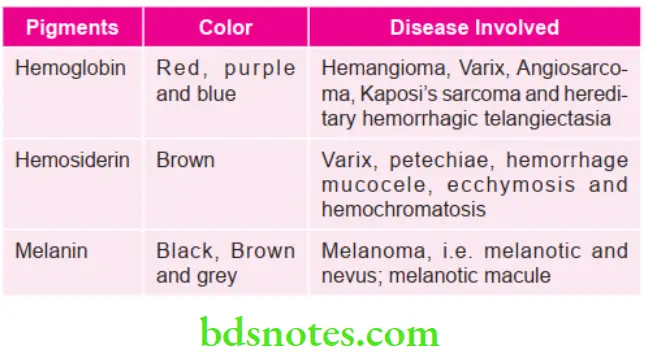
Oral Pigmentation Question And Answers
Question 1. Classify and enumerate causes of pigmentation of oral mucosa.
Answer.
Classification of Pigmentation of Oral Mucosa Occupational
Endogeneous Pigmentation
- Blue/purple vascular lesion
- Hemangioma
- Angiosarcoma
- Kaposi sarcoma
- Brown melanotic lesion
- Melanoma
- Melanoplakia
- Addison disease
- HIV oral melanosis
- Drugs in ductal melanosis
- Brown heme associated lesions
- Jaundice
- Hematoma
- Hemochromatosis
- Ecchymosis and petechiae
.Read And Learn More: Oral Medicine Question And Answers
- Exogenous Pigmentation
- Accidental pigmentation (Foreign substance embedded)
- Articles of road surface embedded in gingiva
- Charcoal containing toothpowder
- Graphite tattoos
- Iatrogenic pigmentation
- Pigmentation due to drugs and metals
- Bismuthism
- Plumbism
- Mercurialism
- Argyria
- Arsenism
- Auric stomatitis
- Copper, chromium, zinc and cadmium pigmentation
- Localized pigmentation
- Chlorhexidine stains (yellow brown to brown colour)
- Hairy tongue (Green to brown to black)
- Tobacco stains (Dark brown or black stain coal tar)
- Accidental pigmentation (Foreign substance embedded)
Causes of Pigmentation of Oral Mucosa
- Extrinsic Causes
- Habits—tobacco, catechu
- Oral drugs
- Iatrogenic
- Chlorhexidine mouthwash
- Medicaments such as silver nitrate, iodine, iron
- Tattoo made by patient
- Poor oral hygiene
- Intrinsic Causes
- Neonatal jaundice
- Tetracycline therapy during formation of teeth
- Osteogenesis imperfecta
- Dentinogenesis imperfecta
- Amelogenesis imperfecta
- Fluorosis
- Nonvital teeth or internal resorption
- Turner’s teeth
- Congenital porphyria
- Erythroblastosis fetalis
- Cystic fibrosis
- Enamel opacities
- Congenital heart disease
- Acute exanthematous disease.
Question 2. Enumerate the various causes of oral pigmentation. Describe in detail various endogenous pigmentation.
Or
Write short note on endogenous pigmentation.
Answer. For causes refer to Ans l of the same chapter.
Endogenous Pigmentation

Lesions Consisting of Hemoglobin Pigments
Hemangioma Clinical Features
- It is a most common tumor in children.
- It has biphasic growth showing initial rapid growth with gradual involution over 5 to 7 years.
- It is more common in girls.
- It is commonly seen in skin, subcutaneous tissue but can occur anywhere in the body like liver, brain, lungs and other organs.
- It grows rapidly in first year and 70% involutes in 7 years.
- Early proliferative lesion is bright red, irregular; deep lesion is bluish colored. Involution causes color fading, softness, shrinkage leaving crepe paper like area.
- Commonly, it is central; common in head and neck region.
- Often large haemangiomas may be associated with visceral anomalies. Head and neck hemangioma is associated with ocular and intracranial anomalies; sacral with spinal dysraphism.
- Multiple cutaneous hemangiomas may be associated with hemangioma of liver causing hepatomegaly, cardiac failure (CCF), anemia.
- Ulceration, bleeding, airway block and visual disturbances are common complications.
- A definitive even though rare, but important lifethreatening complication is platelet trapping and severe thrombocytopenia presenting as ecchymosis, petechiae, intracranial hemorrhage massive gastrointestinal bleed.
Varices
It is defined as the pathological dilatation of the vein.
Varices Clinical Features
- This is reddish to purple in color and is nodular.
- It is seen over tongue, lip or cheek and is more common in old age, i.e. in sixth and seventh decades of life.
- Borders are sharply delineated and are smooth.
Thrombus
- Thrombus is a blood clot.
- In terms of injury, thrombus is physiologic while in terms of pathology, it is thrombus.
Thrombus Clinical Features
- Seen commonly on lower lip and buccal mucosa.
- If thrombus is present in varix, then it appears as a bluish purple nodule.
Kaposi’s Sarcoma
- Clinically Kaposi’s sarcoma is of four types, i.e.
- Classic (Chronic)
- Endemic (Lymphadenopathic; African)
- Immunosuppression associated (Transplant)
- AIDS-related
- Classic Type
- Development of cutaneous multifocal blue red nodules on the lower extremities.
- Lesion slowly increases in size and number with some of the lesions extinguishing and new ones forming on adjacent or distinct skin.
- Orally, soft bluish nodules occur on palatal mucosa or gingiva.
- Lymphadenopathic
- Present in young African children.
- There is generalized or localized enlargement of lymph node chain which includes cervical lymph nodes.
- Disease follow fulminant course with visceral involvement and minimum skin or mucous membrane involvement.
- Immunosuppression Associated
- It is usually seen in renal transplant patients.
- It occurs 1 to 2 years after transplantation
- Progression of disease is directly proportional to loss of cellular immunity of host.
- AIDS Related
- Homosexual AIDS patients have maximum chances of developing Kaposi’s sarcoma.
- Lesions occur on cutaneous lesions, i.e. along lines of cleavage and tip of nose.
- Oral lesions can occur anywhere in oral cavity, but predilection is for palatal mucosa and gingiva.
- Early oral mucosal lesions are flat and slight blue, red or purple plaques, either focal or diffuse and may be completely asymptomatic.
- Later on these lesions may be more deeply discoloured and there is development of surface papules and nodules which may become exophytic and ulcerated. These lesions can also bleed.
- Cervical lymphadenopathy and salivary gland enlargement is seen.
- Angiosarcoma
- It is a malignant vascular tumor.
Angiosarcoma Clinical Features
- It is seen over cheek, lip, palate, tongue and gingiva.
- Mandible is mostly affected.
- It is an aggressive lesion which ulcerates.
- Margins are ill defined.
- On manipulation, lesion bleeds spontaneously and its consistency is firm.
Hemosiderin Pigments Lesions Consisting
Ecchymoses and Petechiae
Both the terminologies have minor difference, i.e. ecchymosis is greater than 2 cm in size while petechiae are small pinpoint hemorrhages.
Ecchymoses and Petechiae Clinical Features
- These are seen on face as well as lips.
- They are red in color and are macular. As hemoglobin get converted to hemosiderin lesion appear brown in color.
- On applying pressure, lesion does not blanch.
- It is fluctuant.
Hemochromatosis
- It is also known as bronze diabetes.
- Hemochromatosis is the combination of four diseases, i.e. liver cirrhosis, diabetes mellitus, heart failure and bronze skin.
- In this, iron gets deposited inside the body which leads to sclerosis.
- In hemochromatosis, tanning is due to increase in melanin production.
Hemochromatosis Clinical Features
- Men predilection is commonly seen.
- Pancreas, adrenal gland, liver and skin are the organs which get affected.
- Skin becomes bluish grey over face, arms and genitals.
- At the junction of hard and soft palate there is presence of black-brown pigmentation which is diffuse.
Hematoma
It defined as the local collection of blood in an organ or tissue, which is clotted.
Hematoma Clinical Features
- Hematomas in mucosa occur due to trauma.
- Initially, hematoma is bluish in color and later on i.e. after 24 hours as clotting get completed it becomes brownish or hard black in color.
- While applying pressure hematomas does not show blanching.
- Hematoma just occurring after trauma is fluctant, rubbery in consistency and its outline is discrete.
- Mucosa which is overlying hematoma is movable.
- Hematoma is painless, but if get infected it become painful.
- Hematoma occurring on tongue may lead to dysphagia or interferes with speech.
Brown Melanotic Lesions
Melanotic Macule
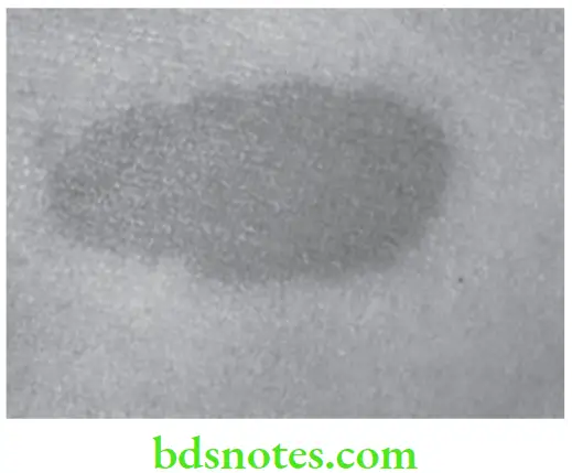
It is a benign pigmented lesion which consists of increased melanin pigmentation at the basal cell layer of epithelium as well as lamina propria.
Melanotic Macule Clinical Features
- Most commonly occurs during middle age.
- Female predilection is present.
- Most commonly seen over the vermilion border of lower lip. At times, it may also be seen over gingiva, buccal mucosa and palate.
- It can be blue, brown, grey or black in color.
- Mostly the lesion is less than 1 cm in size but at times, it may be larger in size.
- It can either be oval or irregular in shape.
Melanoacanthoma
Oral melanoacanthoma is a benign and is a rare occurring entity.
Melanoacanthoma Clinical Features
- Blacks are most commonly affected and females are most commonly affected as compared to males.
- It is commonly seen on buccal mucosa and mostly occurs in middle-aged individuals. Besides buccal mucosa other sites involved are lip, palate and gingiva.
- Lesion is dark brown to black in color and is smooth.
- Lesion is asymptomatic but at times, it increases in size in few months.
Melanoplakia
Melanoplakia is a flat, localized lesion which is black or brown in color.
Melanoplakia Clinical Features
- It occurs most commonly in black people.
- Its color ranges from light brown to blue.
Melanoma
It is also known as melanocarcinoma and is a malignant neoplasm.
Clinical Types of Malignant Melanoma
- Superficial spreading melanoma: Exists in a radial growth phase. Lesion present as tan, brown or black admixed lesion on sun-exposed skin. Radial growth phase may last for several months to years.
- Nodular melanoma: It exists in a vertical growth phase. It presents sharply delineated nodule with varying degrees of pigmentation. They may be pink or black.
- Lentigo maligna melanoma: Exists in a radial-growth phase. The lesion occurs as macular lesion on malar skin of Caucasians.
- Acral lentiginous melanoma: Melanoma developing on the palms and soles as well as toe and fingers. It is characterized by macular lentiginous pigmented area around nodule.
- Mucosal lentiginous melanoma: Develops from mucosal epithelium that lines respiratory, gastrointestinal and genitourinary systems. It is more aggressive.
- Amelanotic melanoma: It is an erythematous or pink sometimes eroded nodule.
Melanoma Clinical Features
- Oral melanomas initiate as macular pigmented focal lesions.
- Most of the lesions are pigmented excepting few nonpigmented lesions which referred to as “amelanotic melanomas”, which appear as “slightly” inflamed looking areas.
- Pigmented lesions are often dark-brown, bluish-black or simply black in appearance.
- The initial macular lesions grow very rapidly and often result in a large, painful, diffuse mass.
- Surface ulceration is very common and beside; this, hemorrhage, paresthesia and superficial fungal infections are often present.
- As the tumor continues to grow, small satellite lesions can develop at the margin of the primary tumor.
- Like other epithelial malignant tumors, melanomas exhibit little or no induration at the periphery.
- Oral melanomas often cause rapid invasion and extensive destruction of bone. This often results in loosening and exfoliation of the regional teeth in the jaw.
- Widespread dissemination of the tumor cells occurs frequently in the lymph nodes as well as in the distant sites, e.g. the lung, liver, bone and brain, etc.
Addison’s Disease
It is also known as chronic adrenal insufficiency.
Addison’s Disease Clinical Features
- In Addison’s disease pigmentation occurs early. Melanin gets deposit over the skin and mucous membrane at pressure points. Cheek is the commonly affected area.
- Pigmentation is blue black to pale brown in color.
Question 3. Write short note on bismuth pigmentation.
Answer. It is an exogeneous pigmentation.
Bismuth Pigmentation Causes
- Medicinal use of bismuth containing preparation.
- Many proprietary drugs contain bismuth salt and bismuth containing pastes may result in bismuth pigmentation.
Mechanism of Action
There is bacterial degeneration of the organic material of food which produced bisthmuth compound and hydrogen sulfide and pigmentation is because of bismuth sulfide granules which produce blue-black color.
Bismuth Pigmentation Clinical Features
- Vague gastrointestinal tract disturbances, nausea, bloody diarrhea, bismuth grippe and jaundice.
- Bismuth line: Sometime in long bone, white bands of increased density appear in the end of diaphysis immediately adjacent to epiphyseal lines. This is called bismuth lines.
Oral Manifestations
- Patient complains of metallic taste with burning sensation in oral cavity and annoying gingivostomatitis with symptoms similar to ANUG.
- Large extremely painful shallow ulcerations are seen at times on cheek mucosa in molar region, regional
- lymphadenopathy may be present.
- Tongue is enlarged and sore.
- Blue-black bismuth line appears to be well-demarcated to eye on gingival papilla.
Management
- Establishing and maintenance of oral hygiene.
- Stoppage of use of bismuth.
- Management of painful ulcerative lesions should be done by topical application of lignocaine hydrochloride gel and other treatment procedure followed in ANUG.
Question 4. Write short note on differential diagnosis of endogenous pigmentation.
Answer. Following is the differential diagnosis of endogenous pigmentation
Lesions Consisting of Hemoglobin Pigments
Hemangioma
- Varicosity: In varicosity superficial vein become prominent while in hemangioma, there is presence of dome-shaped mass.
- Mucocele, superficial cyst and ranula: Hemangioma, i.e. cavernous hemangioma is non-fluctuant and gets emptied on applying pressure while the mucocele, superficial cyst and ranula are fluctuant and does not get empty on applying pressure.
Varices
- Hemangioma: Hemangioma is congenital while varices occur in older age. Hemangioma regresses spontaneously while varices do not. Varix has limited growth while hemangioma has finite growth potential.
- Ranula and superficial cyst: Ranula as well as superficial cyst does not become empty on applying digital pressure while varices get empty.
- Nevus: Nevi does not blanch on applying the pressure while varices blanch while applying pressure.
Kaposi’s Sarcoma
- Hemangioma: Hemangioma get blanched on applying pressure while Kaposi’s sarcoma does not.
- Nevi: Aggressiveness of Kaposi’s sarcoma is more as compared to nevi.
- Purpura: In purpura, numerous papules are seen over the soft palate while in Kaposi’s sarcoma soft bluish nodules occur on palate.
- Melanoma: Melanoma does not show multicentric growth while Kaposi’s sarcoma shows multicentric growth.
Lesions Consisting of Hemosiderin Pigments
Ecchymoses and Petechiae
- Junctional nevus, Amalgam tattoo, Melanotic macule, melanoma: In all these lesions, color of lesion changes from bluish brown to green and then to yellow colour while in ecchymosis or petechiae color changes from bright red to brown.
- Trauma due to coughing and vomiting: If trauma is due to coughing and vomiting, there is presence of bruising which is broad and is red or bluish in color.
- Trauma due to fellation: Initially in fellation trauma, the color is blue, then it get changed to green and later on yellow.
- Infectious mononucleosis: In infectious mononucleosis, Paul Bunnell test is positive while it is negative in ecchymosis and petechiae.
Hematoma
- Amalgam tattoo, and melanoma: In these lesions, color of lesion changes from bluish brown to green and then to yellow color while hematoma is bluish in color and later on, i.e. after 24 hours as clotting get completed it becomes brownish or hard black in color.
- Hemangioma: Hemangioma is deep red or bluish red in color and is dome-shaped.
Brown Melanotic Lesions
Melanotic Macule
- Amalgam tattoo: Associated with amalgam filling while melanotic macule is genetic or racial or due to environmental factors.
- Melanoplakia: Size of melanoplakia is large and most commonly occur in black people.
- Ecchymosis: It is brown in color and regress spontaneously.
- Superficial spreading melanoma: This occurs in old age and has circumferential growth. Palate is most commonly affected in superficial spreading melanoma while melanotic melanoma occur in middle age and vermilion border of lower lip is most commonly affected.
Melanoacanthoma
Melanoma: Melanoma is malignant while melanoacanthoma is benign. Melanoma occurs commonly in men, while melanoacanthoma occurs commonly in black females.
Melanoplakia
- Amalgam tatto: In this amalgam, filling is seen in tooth.
- Melanoma: Melanoma is malignant while melanoplakia is benign in nature.
- Junctional nevus: It rarely occurs in mouth.
Melanoma
- Melanotic neuroectodermal tumor of infancy: As its name suggests, it most commonly occurs in infants while melanoma occurs in middle age.
- Ecchymosis: It regresses spontaneously while melanoma does not.
- Oral melanotic macule: Melanoma shows asymmetry while oral melanotic macule does not; borders of melanoma are irregular while oral melanotic macule has regular borders; color of melanoma ranges from shades of brown to black and at times white, red and blue too while color of oral melanotic macule is brown, black, blue or grey; diameter of melanoma is greater than 1 cm while size of oral melanotic macule is less than 1 cm.
- Melanoplakia: Consistency of melanoplakia is firm.
Addison’s Disease
- Hyperpituitarism: Urine test is done for assessment of levels of 17-ketosteroids. In hyperpitutarism the levels are high while in Addison’s disease, levels are low.
- Peutz-Jeghers syndrome: In this, bluish black macule is present over the skin while in Addison’s disease generalized darkening of skin is present.
- Von Recklinghausen’s disease: Brown spots are present on skin which are known as café-au-lait spots.
Question 5. Write short note on Peutz-Jeghers syndrome.
Answer. It is also known as hereditary intestinal polyposis syndrome.
- Peutz-Jeghers syndrome is an autosomal dominant disorder.
- It is a precancerous syndrome.
- Syndrome consists of presence of pigmented macules on skin and lips and presence of gastrointestinal polyps.
Peutz-Jeghers Syndrome Features
- Occurs during the childhood age.
- Intestinal polyps are the major signs to be detected and are seen all over gastrointestinal tract. Intestinal polyps are present in large numbers.
- As polyps become symptomatic from 1st to 3rd decade of life they lead to ulcers which bleed frequently.
- Pigmented macules are seen in perioral region. Gingiva, buccal mucosa and labial mucosa are also affected. Orally color of macules is brown while on skin macules are bluish black in color.
- Pigmented areas are asymptomatic
- Area of pigmentation ranges from 2 to 10 mm.
- Tuberous sclerosis is seen in Peutz-Jeghers syndrome.
Question 6. Enumerate causes of endogenous melanin pigmentation.
Answer. Following are the causes of endogenous melanin pigmentation:
- Physiologic, i.e. melanotic macule
- Developmental, i.e. pigmented nevus
- Idiopathic
- Neoplastic, i.e. melanoma
- Reactive, i.e. melanoacanthoma
- Drug induced
- Hormones, i.e. hypofunction of pituitary gland, in pregnancy
- Genetic, i.e. melanotic macule, melanoplakia, Neurofibromatosis
- Autoimmune, i.e. Addison’s disease
- Infectious, i.e. HIV oral melanosis
Question 7. Write short note on differential diagnosis of intrinsic discoloration of teeth.
Answer. Following is the differential diagnosis of intrinsic discoloration of teeth:
- Aging: Discoloration is yellowish brown in color and it has less translucency.
- Death of pulp: It is associated with trauma to the tooth. Tooth become grayish black in color and has less translucency.
- Fluorosis: Tooth either appears white or yellow brown or brown or mottled.
- Tetracycline staining: Teeth appear yellow brown in color and also show yellow fluorescence.
- Internal resorption: Associated tooth appear pink in color.
- Calcific metamorphosis: Teeth appears yellow in color.
- Dentinogenesis Imperfecta: Teeth appear bluish grey in color and are translucent.
- Amelogenesis imperfecta: Teeth appear yellow brown in color.
- Congenital erythropoietic porphyria: Teeth appear either yellow or brown red. Teeth also show red fluorescence.
- Erythroblastosis fetalis: Teeth appear yellow or greenish in color.
Question 8. Write short note on café-au-lait pigmentation.
Or
Write short note on café-au-lait spots.
Answer. Meaning of café-au-lait is “coffee with milk”.
- Solitary idiopathic café-au-lait spots are occasionally observed in the general population, but multiple café-au-lait spots are often indicative of an underlying genetic disorder.
- Café-au-lait pigmentation may be identified in a number of different genetic diseases, including neurofibromatosis type l, McCune-Albright syndrome and Noonan’s syndrome.
- Café-au-lait spots typically present as tan or brown colored, irregularly shaped macules of variable size. They may occur anywhere on the skin, although unusual, examples of similar appearing oral macular pigmentation have been described in some patients.
- Neurofibromatosis type l is an autosomal dominant disease caused by a mutation or a deletion of the NF 1 gene l. The size, number, and age at onset of the cutaneous cafe-au-lait spots are of diagnostic importance for this disease.
- In McCune-Albright syndrome the café-au-lait spots appear distinct from those associated with neurofibromatosis. The borders of the pigmented macules are irregularly outlined, whereas in neurofibromatosis, the borders are typically smooth.
- Noonan’s syndrome is an autosomal dominant disorder, the classic-appearing cafe-au-lait spots are more characteristically seen in patients with the Noonan’s phenotype.
- Microscopically, when compared with adjacent uninvolved skin, genetic café-au-lait spots exhibit basilar melanosis without a concomitant increase in the number of melanocytes. In contrast, when compared with similar appearing lesions in otherwise normal patients, genetic café-au-lait spots do exhibit increased numbers of melanocytes.
- Café-au-lait spots do not require any treatment and are not associated with any risk for malignant transformation.

Question 9. Write short note on nevi.
Answer. Nevi is defined as a congenital, developmental, tumor like malformation of either skin or mucous membrane.
Nevi is a true mesodermal structure which is composed of dermal melanocytes which rarely undergo malignant transformation.
Nevi is the congenital or acquired benign tumor of nevus cells which occur on skin. Oral nevi occur rarely
Nevi are of four types, i.e. intramucosal, junctional, compound and blue nevus.
Nevi Clinical Features
- Cutaneous nevi occur more commonly.
- Occurrence of nevi is predilected towards males as compared to females.
- Oral melanocytic nevi are rare but present as solitary lesions and are common in females.
- Pigmented nevi do not blanch on pressure.
- Nevi are asymptomatic and present as small, solitary, brown or blue well-circumscribed nodule or macule.
- As the nevi reaches at its size, its growth ceases, and it may remain static indefinitely.
- Oral nevi can develop at any age but most common age of occurrence is over age of 30 years.
- Hard palate is the most common site for nevus which is followed by buccal and labial mucosa and gingiva.
Nevi Diagnosis
- Biopsy is mandatory for the confirmation of nevi as clinical diagnosis includes variety of focal pigmented lesions.
Nevi Differential Diagnosis
- Amalgam tattoo: Amalgam restoration is seen adjacent to pigmentation.
- Vascular lesions: Hematoma, varix and hemangioma are the vascular lesions. History rules out hematoma while varix and hemangioma are ruled by diascopy.
- Melanoma: Rare in oral cavity
Nevi Treatment
- Complete but conservative surgical excision is done for oral lesions.
- Laser therapy and intense pulse light therapies are used successfully in treatment of cutaneous nevi.

Leave a Reply