Describe the oculomotor nerve under the following headings:
- Oculomotor Nerve Functional components,
- Oculomotor Nerve Origin, course distribution and
- Oculomotor Nerve Applied anatomy.
Answer. The oculomotor nerve is purely motor and responsible for the movements (largely) and accommodation of the eye.
“Understanding the anatomy and function of the oculomotor nerve through FAQs: Q&A explained”
Oculomotor Nerve Functional Components
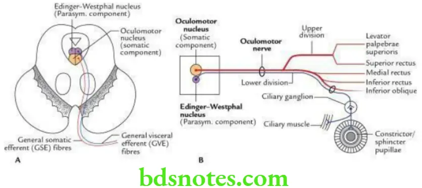
- General somatic of event (GSE) fibres, supply the extrinsic muscles of the eyeball. They arise from the large somatic component of the oculomotor nucleus.
- General visceral efferent (GVE) fibres supply the intrinsic muscles of the eyeball (ciliaris and sphincter pupillae). They arise from the small parasympathetic component of the oculomotor nucleus (Edinger–Westphal nucleus).
- General somatic af agent (GSA) fibres carry proprioceptive sensations from the muscles (vide supra).
Read And Learn More: Selective Anatomy Notes And Question And Answers
“Importance of studying the oculomotor nerve function for medical students: Questions explained”
“Common challenges in understanding oculomotor nerve function effectively: FAQs provided”
Oculomotor Nerve Origin, Course And Distribution The oculomotor nerve arises from the oculomotor nucleus, located in the upper part of the midbrain. The nerve emerges from the midbrain in the interpeduncular fossa, then runs between the posterior cerebral and superior cerebellar arteries, and passes on to the lateral side of the posterior communicating artery.
Now it runs forwards and upwards piercing the dura mater near the posterior clinoid process and travels forwards in the lateral wall of the cavernous sinus. After emerging from the cavernous sinus, it divides into upper and lower divisions which enter the orbit through a superior orbital fissure where:
- The upper division supplies the superior rectus of the eyeball and levator palpebrae superioris.
- The lower division supplies the medial rectus, inferior rectus, and inferior oblique of the eyeball. The nerve to the inferior oblique gives a motor root to the ciliary ganglion.
“Factors influencing success with oculomotor nerve knowledge: Q&A”
Oculomotor Nerve Applied Anatomy
The lesion of the oculomotor nerve (intranuclear lesion) leads to:
- Ptosis, due to paralysis of levator palpebrae superioris
- Loss of accommodation, due to involvement of ciliary muscle
- Lateral squint, due to paralysis of the medial rectus muscle and unopposed action of healthy lateral rectus
- Diplopia, due to paralysis of the medial rectus muscle
- Dilatation of pupil, loss of pupillary reflex, due to paralysis of sphincter pupillae
Note: In paralysis of the oculomotor nerve, the person cannot look upwards, downwards and medially.
Leave a Reply