Fungal Infections Question And Answers
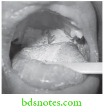
Question 1. Write short note on Thrush.
Answer.
- Thrush is the prototype of oral infection caused by yeast like fungus. It is the superficial infection of upper layer of oral mucus membrane and result in formation of patchy white plaque on mucosal surface.
- It is also known as pseudomembranous candidiasis.
Thrush Clinical Features
In Infants
- In neonates, oral lesions starts between 6th and 10th day after birth.
- The lesions in infants are described as soft white or bluish white, adherent patches on oral mucosa areas.
- They are painless and are removed with little difficulty.
In Adults
- The common sites are roof of mouth, retromolar area and mucobuccal fold. It is more common in female.
- Prodromal symptoms are rapid onset of bad taste. Spicy food will cause discomfort.
- Patient may complain of burning sensation, and there is history of dryness of mouth.
- Inflammation, erythema and painful eroded areas are present.
- Multiple, curdy, loosely adherent patches on any part of oral mucosa.
- Mucosa adjacent to lesion appears red and moderately swollen.
- White patches are easily wiped out with wet gauge which leaves either normal or erythematous areas.
- Deeper invasion by organism leaves ulcerative lesion on removal of patch.
Read And Learn More: Oral Medicine Question And Answers
Thrush Differential Diagnosis
- Lichen planus (Plaque form): By gauze lesion of acute pseudomembranous candidiasis is wiped away.
- Leukoplakia: Cannot be wiped off by gauze.
- Genodermatoses: Cytological smear helps in confirmation of diagnosis.
- Gangrenous stomatitis: Pseudomembrane formed in gangrenous stomatitis has dirty color. The pseudomembrane is not seen raising above the surface.
- Chemical burn: Burn in oral mucosa is thin as compared to pseudomembrane of candidiasis.
Thrush Treatment
Oral candidiasis can be treated either topically or systemically. Treatment should be maintained for 7 days.
- Removal of cause: In patients with denture sore mouth and angular cheilitis, replacement of denture or relining of denture or addition of mycostatin suspension is done. Cleaning of denture is carried out thoroughly and regularly, it should be left out of the mouth in night , in the hypochlorite solution
- Topical treatment: They are given to decrease the systemic absorption of drug, but their effects depend on the compliance of patient. Following are the topical agents used:
- Clotrimazole: 10mg of tablet should be dissolved in mouth 5 times a day. It is used less frequently, one vaginal troche can be dissolved in mouth daily.
- 1% gentian violet can be used, but it leads to staining.
- Nystatin preparations: It consists of suspension, vaginal tablet and oral pastille. Nystatin vaginal tablet 1 lakh units should be dissolved in mouth three times a day.
- Nystatin oral pastilles i.e. one or two pastilles should be dissolved slowly in mouth 5 times a day. One teaspoon of Nystatin oral suspension is mixed with one-fourth cup of water is used as oral rinse.
- Amphotericin B: 5 to 10 mL of solution is used as oral rinse and then expectorated 3 to 4 times a day.
- Systemic treatment: Following are the systemic agents used:
- Nystatin should be given as 250 mg thrice a day for 2 weeks which is followed by one troche daily for third week.
- Ketoconazole: 200 mg of tablet should be taken with food once daily.
- Fluconazole: 100 mg of tablet should be taken once daily for period of two weeks
- Itraconazole: It should be given 200 mg daily orally for two weeks.
Question 2. Write short note on Candidiasis.
Or
Write short note on oral candidiasis.
Or
Write short answer on treatment of oral candidiasis
Or
Write short answer on candidiasis.
Or
What is candidiasis? Discuss predisposing factors, clinical features and management of candidiasis.
Answer. Candidiasis is a superficial infection of upper layer of oral mucus membrane and result in formation of patchy white plaque or flecks on mucosal surface.
Classification of Oral Candidiasis
Primary Oral Candidiasis
- Acute form
- Pseudomembranous candidiasis
- Erythematous candidiasis
- Chronic form:
- Hyperplastic candidiasis
- Erythematous candidiasis
- Pseudomembranous candidiasis
- Candida associated lesion:
- Denture stomatitis
- Angular stomatitis
- Median rhomboid glossitis
- Keratinized primary lesion super-infected with candida.
- Leukoplakia
- Lichen planus
- Lupus erythematosus
Secondary Candidiasis
Candidal endocrinopathy syndrome.
Etiology and Predisposing Factors
- Changes in oral microbial flora: Marked changes owing to administration of antibiotics, excessive use of antibacterial mouth rinses.
- Local irritant, i.e. denture, orthodontic appliance and heavy smoke.
- Drug therapy: Administration of corticosteroids, cytotoxic drugs and immunosuppressants.
- Acute to chronic disease: Acute and chronic disease such as leukemia, lymphoma, diabetes and TB.
- Malnutrition states: Malnutrition states such as low serum vitamin A, pyridoxine and iron levels.
- Age: Infancy, pregnancy and old age.
- Endocrinopathy: Endocrinopathies such as hyperparathyroidism, hypoparathyroidism and Addition’s disease.
- Immunodeficiency states: Primary and acquired immunodeficiency state.
- Others: Tight and close-fitting garments encourage growth of candida.
Candidiasis Clinical Features
In Infants
- It start between 6th and 10th day after the birth.
- Infection is contracted from maternal vaginal canal where Candida albicans flourish during pregnancy.
- Lesions are soft white or bluish white, adherent patches on oral mucosa.
- Lesion is painless and can be removed with little difficulty.
In Adults
- Common sites are roof of mouth, retromolar area and mucobuccal fold. It is common in women.
- Rapid onset of bad taste, spicy food causes discomfort.
- Patient may complain of burning sensation and there may be dryness of mouth.
- There is presence of pearly white or bluish white plaques on oral mucosa. All of these resemble as cottage cheese or curdled milk. Patches are loosely attached to mucosa.
- Mucosa adjacent to it appears red and moderately swollen. Area can be painful
- White patches are easily wiped out with wet gauge which leaves normal or erythematous area.
- Deeper invasions by organism leave an ulcerative lesion on removal of patch.

Candidiasis Investigations
- On staining with periodic acid Schiff (PAS) method, candidal hyphae are readily identified. Organisms are identified by bright magenta color. The candidal hyphae are 2μm in diameter, vary in length and may show branching.
- About 10–20% KOH is also used to identify organisms readily.
- Cultures can be obtained readily on Sabouraud’s medium and on ordinary bacteriological culture media. Colonies are creamy white, smooth with a yeasty color.
- On corn-meal agar medium C. albicans form chlamydospores.
Candidiasis Differential Diagnosis
- Leukoplakia: History of recent administration of antibiotics favor diagnosis of candidiasis.
- Gangrenous stomatitis: Pseudomembrane dirty in color and not raised above surface.
- Chemical burns: Superficial white material burns of oral mucosa appears thin and delicate as compared to pseudomembranous candidiasis.
- Lichen planus (Plaque form): Candidal lesion can be wiped with gauzes
Candidiasis Treatment/Management
Oral candidiasis can be treated either topically or systemically. Treatment should be maintained for 7 days.
- Removal of cause: In patients with denture sore mouth and angular cheilitis, replacement of denture or relining of denture or addition of mycostatin suspension is done. Cleaning of denture is carried out thoroughly and regularly, it should be left out of the mouth in night , in the hypochlorite solution
- Topical treatment: They are given to decrease the systemic absorption of drug, but their effects depend on the compliance of patient. Following are the topical agents used:
- Clotrimazole: 10 mg of tablet should be dissolved in mouth 5 times a day. It is used less frequently, one vaginal troche can be dissolved in mouth daily.
- 1% gentian violet can be used, but it leads to staining.
- Nystatin preparations: It consists of suspension, vaginal tablet and oral pastille. Nystatin vaginal tablet 1 lakh units should be dissolved in mouth three times a day. Nystatin oral pastilles i.e. one or two pastilles should be dissolved slowly in mouth 5 times a day. One teaspoon of Nystatin oral suspension is mixed with one-fourth cup of water is used as oral rinse.
- Amphotericin B: 5 to 10 mL of solution is used as oral rinse and then expectorated 3 to 4 times a day.
- Systemic treatment: Following are the systemic agents used:
- Nystatin should be given as 250mg thrice a day for 2 weeks which is followed by one troche daily for third week.
- Ketoconazole: 200mg of tablet should be taken with food once daily.
- Fluconazole: 100mg of tablet should be taken once daily for period of two weeks
- Itraconazole: It should be given 200mg daily orally for two weeks.
Question 3. Enumerate fungal lesions of orofacial region. Describe etiology, predisposing factors, classification, clinical features, investigation and management of oral candidiasis.
Answer.
Fungal Lesions of Orofacial Region
- Candidiasis
- Histoplasmosis
- North American blastomycosis
- South American blastomycosis
- Paracoccidioidomycosis
- Coccidioidomycosis
- Cryptococcosis
- Zygomycosis
- Aspergillosis
- Toxoplasmosis
- Sporotrichosis
- Rhinosporidiosis
For etiology, predisposing factor, classification, clinical features, investigations and management of oral candidiasis refer to Ans 2 of same chapter.
Question 4. Describe in detail about etiopathogenesis, clinical features, differential diagnosis and treatment of acute pseudomembranous candidiasis.
Answer.
Etiopathogenesis of Acute Pseudomembranous Candidiasis
- People-to-people acquired infections mostly happen in hospital settings where immunocompromised patients acquire the yeast from healthcare workers
- C. albicans containing various known virulence factors which helps in the spreading of infections in human beings and favors its pathogenicity.
- The virulence factors of the C. albicans have the great role in the pseudohyphae formation by attached with epithelial cells, endothelial cells, hyphal switching, surface recognition molecules, phenotypic switching and extracellular hydrolytic enzyme, i.e. proteinase and phospholipase production have been suggested to be virulence attributes for Candida.
- Extracellular hydrolytic enzymes seem to play an important role in candidal overgrowth, as these enzymes facilitate adherence and tissue penetration and hence invasion of the host, among the most important hydrolytic enzymes produced by Candida are phospholipases and secreted aspartyl proteinases (mainly by C. albicans and C. tropicalis).
- The hemolysin is another most common virulence factor which contributes to candidal pathogenesis. The Candida including all species secretes the aspartyl proteinase 5 and 9 (SAP5 and SAP9). The maximum amount is secreted by C. albicans followed by C. tropicalis, C. kefyr andC. krusei.
- Due to their virulence factors such as adhesions property the colonization of the Candida is take place in superficial tissue (local site) or it invade the deeper into the host tissue in yeast form but they transformed into the hyphal form during active infection.
For clinical features and differential diagnosis and treatment, refer to Ans 2 of same chapter

Leave a Reply