Question. Describe the Trigeminal Nerve under the following headings:
- Trigeminal Nerve functional components,
- Trigeminal Nerve Origin and course,
- Trigeminal Nerve Distribution and
- Trigeminal Nerve Applied anatomy.
Answer. The trigeminal nerve is a mixed nerve but mainly sensory.
Trigeminal Nerve Functional Components
Read And Learn More: Selective Anatomy Notes And Question And Answers
“Understanding the anatomy and function of the trigeminal nerve through FAQs: Q&A explained”
- Special visceral efferent (SVE) fibres to muscles of mastication.
- General somatic afferent (GSA) fibres to carry:
- Pain and temperature sensations from the head and face
- Proprioceptive sensations from the muscles of mastication
Trigeminal Nerve Origin And Course It arises from two roots:
- The large sensory root arises from the sensory nuclei of the trigeminal nerve located in the brainstem and upper part of the spinal cord.
- The small motor root arises from the motor nucleus of the trigeminal nerve located in the pons.
“Importance of studying the trigeminal nerve for medical and anatomy students: Questions explained”
The sensory and motor roots emerge from the anterior surface of the pons, the motor root lying medial to the sensory root. After emerging from the brainstem, the nerve passes upwards, forward and laterally in the posterior cranial fossa.
On reaching the depression on the apex of the petrous temporal bone in the middle cranial fossa, it expands to form the trigeminal ganglion. (Remember motor root passes deep to the ganglion, whereas the sensory root forms the ganglion.)
The anterior border of the trigeminal ganglion gives rise to three divisions of the trigeminal nerve:
- Ophthalmic division: It is purely sensory and enters the orbit through the superior orbital fissure.
- Maxillary division: It is purely sensory and enters the pterygopalatine fossa through the foramen rotundum.
- Mandibular division: It enters the infratemporal fossa through foramen ovale. It is joined by the motor root (which also passes through the foramen ovale) just below this foramen. Hence, it is both sensory and motor.
“Common challenges in understanding trigeminal nerve anatomy effectively: FAQs provided”
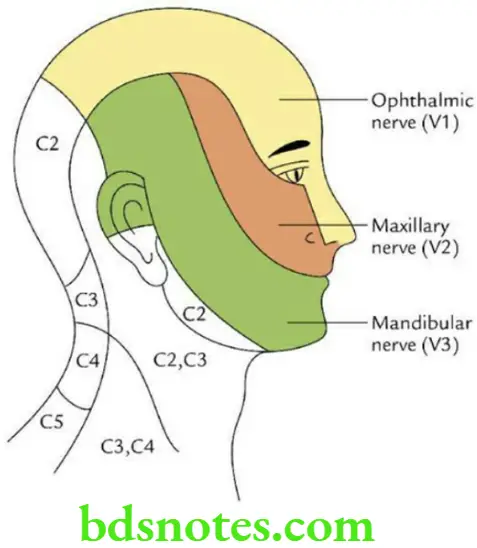
Trigeminal Nerve Distribution
Sensory distribution:
- Most of the skin of the head and face
- The mucous membrane of nasal cavities, oral cavity and paranasal air sinuses
- The teeth of both jaws
- Cornea and conjunctiva of the eye
Motor distribution: Muscles of the mastication
Trigeminal Nerve Applied anatomy
Trigeminal neuralgia (tic douloureux): A clinical condition characterized by intermittent attacks of severe lancinating pain in the region of sensory distribution of one or more divisions of trigeminal nerve in the face, usually the 2nd and 3rd divisions.
“Factors influencing success with trigeminal nerve knowledge: Q&A”
“Steps to explain the anatomy of the trigeminal nerve: Divisions vs branches vs nuclei: Q&A guide”
Question: Write a short note on the nuclei of the trigeminal nerve.
Answer. The trigeminal nerve has the following four nuclei:
- The principal sensory nucleus lies in the dorsolateral region of the tegmentum of the upper part of the pons lateral to the motor nucleus of the trigeminal nerve. It receives all sensory fibres of the trigeminal nerve.
- The mesencephalic nucleus lies in the midbrain above the main sensory nucleus. It receives proprioceptive impulses from muscles of mastication, TMJ and teeth.
- The spinal nucleus lies in the spinal cord below the main sensory nucleus. It receives pain and temperature sensations from the face.
- The motor nucleus lies in the upper part of the bones. It gives motor fibres to muscles of mastication.
“Role of the ophthalmic division in sensory innervation of the forehead: Questions answered”
Mandibular Nerve Origin and course
The mandibular nerve is the largest division of trigeminal nerve. It arises from the trigeminal ganglion and enters the infratemporal fossa through foramen ovale. In the foramen ovale, it is joined by the small motor root of the trigeminal nerve, and thus emerges from the skull as a mixed nerve. After emerging from foramen ovale, it divides almost immediately into the anterior and posterior divisions.
“Early warning signs of gaps in understanding trigeminal nerve basics: Common questions”
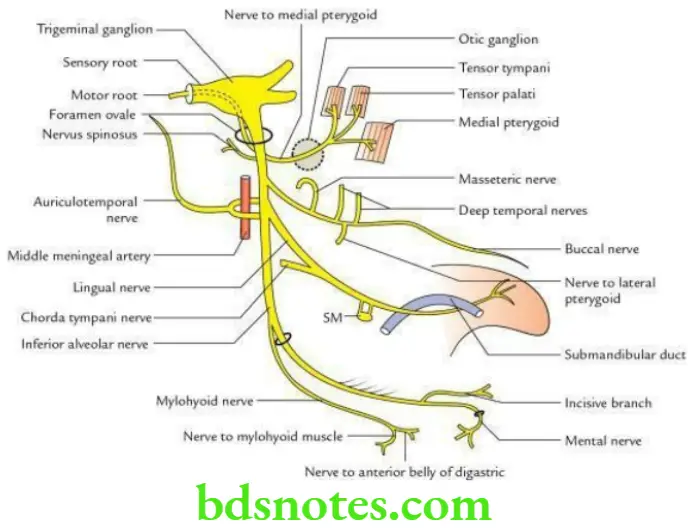
Mandibular Nerve Branches and distribution
From the trunk
- Meningeal branch (nervus spinosus), which enters the skull through foramen spinosum and supplies the dura mater.
- Nerve to medial pterygoid, it passes through otic ganglion and supplies the medial pterygoid muscle. In addition to it, it also supplies twigs to the tensor palati and tensor tympani muscles.
From the anterior division
- Muscular branches to the temporalis (deep temporal nerves), masseter (masseteric nerve) and nerve to the lateral pterygoid.
- Buccal nerve (sensory to the skin and mucosa of the cheek).
From the posterior division
- Auriculotemporal nerve (sensory to the auricle and temple).
- Lingual nerve (sensory to the anterior two-third of tongue).
- Inferior alveolar nerve: Gives the mylohyoid nerve and then enters into the mandibular canal to supply sensory fibres to the lower teeth and gums. It gives mental nerve, which supplies the skin of the chin, and skin and mucosa of the lower lip.
Note: The nerve to mylohyoid supplies mylohyoid muscle and anterior belly of digastric.
“Asymptomatic vs symptomatic effects of ignoring trigeminal nerve principles: Q&A”
Trigeminal Nerve Anatomy
Functional components of trigeminal nerve:
1. Sensory components
- Sensations of pain, temperature, touch & pressure travel along axons
- Their cell bodies lie in the trigeminal ganglion
- Peripheral processes forms
- Ophthalmic nerve
- Maxillary nerve
- Mandibular nerve
- Central processes form sensory root
“Can targeted interventions improve outcomes using trigeminal nerve knowledge? FAQs provided”
2. Motor component
- The motor nucleus receives impulses from
- Right & left cerebral hemispheres
- Red nucleus
- Mesencephalic nucleus
- It supplies
- Muscles of mastication
- Tensor veli palatini
- Tensor tympani
- Mylohyoid
- Anterior belly of digastric
- It supplies
“Differential applications of sensory vs motor functions of the trigeminal nerve: Questions answered”
Question 2. Mention boundaries & contents of Infratemporal Fossa
Answer:
Infratemporal Fossa Boundaries:

“Steps to apply trigeminal nerve knowledge in clinical practice: Diagnosis vs treatment: Q&A guide”
- The anterior & medial walls are separated in their upper parts by the pterygomaxillary fissure
- The upper end of this fissure is continuous with the anterior part of the inferior orbital fissure

Leave a Reply