X-Ray Films And Accessories
Question 1. Write short note on intensifying screen.
or
Write short note on composition and function of intensifying screen.
Answer.
- Intensifying screen is a device that transfers X-ray energy into visible light; the visible light, in turn exposes the screen film.
- These screens intensify the effect of X-rays on the film, so less radiation is required to expose a screen film, and hence the patient is exposed to less radiation.
- In extraoral radiography, a screen film is sandwiched between the two intensifying screens of matching size and secured in a cassette.
- An intensifying screen is a smooth plastic sheet coated with minute fluorescent crystals called as phosphors. When exposed to X-rays, the phosphors fluoresce and emit visible light in the blue or green spectrum, the emitted light then exposes the film.
- Combination of X-ray film with an intensifying screen results in an image receptor system which is 10–60 times more sensitive to X-rays than the film alone. Thus, the duration of exposure is reduced, the contrast is improved and radiation back scatter minimized. Intensifying screen also reduces the patient dose by 85 to 90%.
Read And Learn More: Oral Radiology Question And Answers
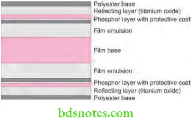
Intensifying Screen Composition
- Base: It is made of either a stiff sheet of cardboard or polyester plastic material. It is about 0.25 mm thick. The base is the supporting component of the screen.
- Reflecting layer: This is a thin layer of white material, i.e. magnesium oxide or titanium dioxide between the base and the luminescent layer. It serves to redirect to the film a large fraction of the emitted visible light which is moving away from the film and which would therefore otherwise be lost. So, it increases the sensitivity but some degree of unsharpness is created because of divergence of light reflected back to the film.
- Phosphor layer: This layer consists of light sensitive phosphor crystals suspended in a plastic material. When these crystals are struck by photons they fluoresce, i.e. they emit visible light photons which expose the X-ray film.
- Coat: This layer protects the phosphor layer from mechanical insult such as abrasion, scratching, etc.

Intensifying Screen Functions
- Intensifying screens are used in all extraoral radiography. They are used inside a cassette on both sides.
- Intensifying screens are not used in intraoral radiography. Screens are used in intraoral radiography in endodontics.
- Rare earth screens are four times much better than calcium screen.
- Intensifying screens reduces duration of exposure, improves contrast and minimize radiation back scatter.
- Intensifying screen also reduces the patient dose by 85 to 90%.
- Intensifying screen reduces tube and generator loading.
- Intensifying screen also reduces patient motion artifacts.
Question 2. Write short note on grid.
Answer.
Grid
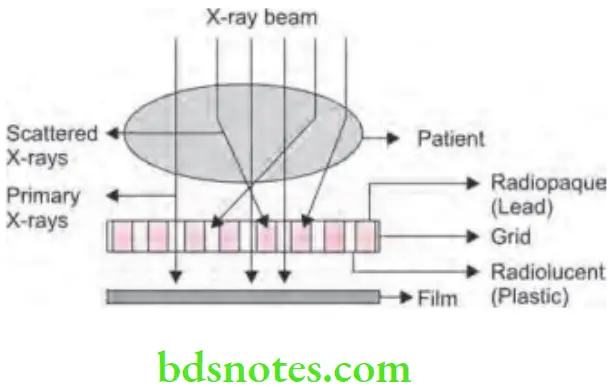
- These are devices which reduces the amount of scattered radiation reaching the film and still allowing the primary beam to reach the film.
- Thus, grid prevent scattered radiation which reduces fog and increases contrast.
Grid Composition
A grid is composed of alternative strips of radiopaque and radiolucent material
- Radiopaque material is lead.
- Radiolucent material is plastic.
- The radiopaque material absorbs the scattered radiations while the radiolucent material allows passage of primary radiation.

Types of Grid
Following are the grids used, i.e.
- Stationary grids and moving grids
- Focused grids and non-focused grids
Stationary Grids
Stationary grids do not move.
- Linear grid
-
- In this, strips of lead are used which are placed strictly parallel to each other.
- Here the primary radiations travel radially from X-ray tube focal spot to the film will encounter the grid obliquely. So grid lines appear wider.
- While using the linear grid, cut-off of the beam may occur as some primary radiation can be absorbed by lead which is situated in peripheral region.
- For all practical applications, central beam in the plane is parallel with grid lines.
- Focused grid
- Here strips of lead are angled progressively from center to the edge so the inter spaces are directed at the focal spot.
- Focused grid helps in prevention of primary cut-off.
- Focused grid minimizes the disadvantages of linear grid.
- Pseudo-focused grid
- Here the height of strips is reduced progressively from centre which leads to reduction in grid ratio from center to edge.
- Crossed grid
- Linear grids are placed mutually at the right angles so even the small amount of radiation is blocked.
Moving Grids
- These grids move sideways across the film during the exposure.
- These grids reduce the white lead lines in the radiographic image.

Grid Advantages
Grids remove the scattered radiation efficiently which leads to decrease in film fog and increases radiographic contrast.
Grid Disadvantages
- The radiopaque material (lead) may absorb some primary radiation and produces overall pattern of thin parallel white lines on the film called grid lines.
- The exposure time must be increased.
Question 3. Write short note on Potter-Bucky diaphragm.
Answer.
Potter-Bucky diaphragm
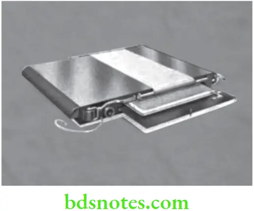
Potter-Bucky diaphragm is also known as moving grid.
- It was discovered by Hollis E Potter in 1920.
- Potter-Bucky diaphragm is a moving grid which prevents scattered radiation from reaching to the film, and secure better contrast and definition.
- In this the grid is moved sideways across the film during exposure. This leads to blurring out of the shadows of grid strips, thus they are not visible on the film.
- Image of the radiopaque grid lines on the film can be deleted by mechanically moving the grid in a direction of 90° to the grid lines (but not the object) during exposure. This results in evening (blurring) out the radiolucent lines and resulting in a more uniform exposure.
- Potter-Bucky diaphragm eliminates most of the scatter while allowing most of primary radiation through.
- Lead sheets cast a shadow on the image and this is removed by moving the grid at the time of exposure.
- In older days, the shape of diaphragm is curved but nowadays shape of diaphragm is flat.

Potter-Bucky diaphragm Advantages
- Removes the scattered radiation and decreases fog as well as increases contrast.
- It eliminates white lead lines efficiently.
Potter-Bucky diaphragm Disadvantages
- It increases exposure time due to slow movement.
- It increases patient dosage.
- It can cause failure.
- It is costly.
Question 4. Discuss composition of dental film packet in detail.
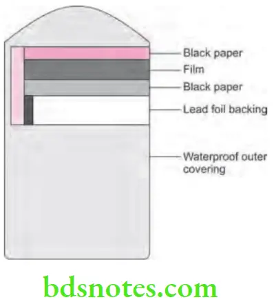
Answer. Dental film and its surrounding packaging is known as a film packet. Intraoral X-ray film packets consist of four basic components, i.e.
- X-ray film
- Paper film wrapper
- Lead foil sheet
- Outer film wrapping.
X-ray Film
- An intraoral X-ray film is a double emulsion film.
- Single film packet can consist of one film, i.e. one-film packet or two films, i.e. two-film packet
- Two-film packet gives two identical radiographs with the same amount of exposure which is necessary to produce a single radiograph. This is indicated when there is need of duplicate record of a radiograph.
- A small raised dot which is known as identification dot is located at one corner of the intraoral X-ray film. The raised dot distinguishes between the left and right side of the patient after the film gets processed. So the identification dot helps in film orientation, mounting and interpretation.
Paper Film Wrapper
- It is a protective sheet of black paper which covers the film inside the film packet.
- It provide shield to the film from light leak.
Lead Foil sheet
- It is a single thin piece of lead foil in the film packet which is located behind the film wrapped inside the black protective paper.
- The sheet is positioned behind the film.
- Lead foil sheet absorbs most of the X-rays that passes through the film and prevents them from reaching the tongue and other oral tissues.
- It also absorbs back scattered or secondary radiation and prevents film fog.
- A secondary function of lead foil sheet is to provide sufficient strength to the whole film packet.
- If the film packet is placed reverse in the mouth, the shadow of the foil is seen on radiograph as ‘tyre-track’ marks or ‘Herring bone’appearance which is the embossed pattern placed on the lead foil by the manufacturer.
Outer Plastic Wrapping
- Outer package wrapping is a soft-vinyl or a paper wrapper that hermetically seals the film packet, protective black paper and lead foil sheet.
- This outer wrapper serves to protect the film from exposure to light and saliva.
- The outer wrapper of the film packet has two sides, i.e. tube side and label side.
- Tube side: The tube side of the plastic cover is solid white and bears and raised dot, this raised dot or identification dot should be placed towards the X-ray tube. When inserted in the mouth, the edge carrying the dot should be placed at the incisive or occlusal margin of the tooth.
- Label side: This side of the film packet has a flap which is used to open the film packet to remove the film prior to processing. Label side is color-coded to identify films outside of the plastic packaging container. Color codes distinguish between one film and two film packets and between film speeds. When placed in the mouth, the color coded side of the film packet must face the tongue.
- Following information is printed on the label side of the film packet:
- A circle or dot that corresponds with the raised identification dot on the film.
- The statement “opposite side toward tube”
- The manufacturer’s name200 Mastering the BDS IVth Year-I (Last 25 Years Solved Questions)
- The film speed
- The number of films enclosed

Question 5. Write short note on filter and grid.
Answer. For grid refer to Ans 2 of same chapter.
Filter
- Filter in X-ray machine removes the unwanted radiation leaving wanted radiation undiminished.
- Aluminum filters are the most commonly used filters in X-ray machine.
Ideal Requirements of Filter Material
- It should discriminate against the lower energy photos.
- Material should not consist of absorption edge at energy close to the energies of photons which are desired to be use.
- Thickness of material should not be very small because in such cases due to irregularity in thickness the beam produced should be non uniform. Pin holes occur easily without producing tiny completely unfiltered and unacceptable beams.
Filter Materials
Following are the materials used:
- Aluminum till 30 KV to 120 KV
- Copper till 100 KV to 250 KV
- Tin till 200 KV to 600 KV
- Lead till 600 KV to 2 mV.
Filter Types
Profile Wedge Filter
- Reducing the radiographic density of soft tissue profile can be accomplished by absorbing and removing some of the X-rays out of the part of X-ray beam reaching profile soft tissue.
- Absorbing device is profile wedge filter which has a vertical bar which is wedge shaped.
Wedge Filters
Used with megavoltage radiations as well as in radiotherapy when it is desired to treat one side of patient only.
Question 6. Write short note on composition of intraoral X-ray film.
or
Write short note on composition of X-ray film.
Answer. X-ray film or intra-oral X-ray film used in dentistry consists of four basic components:
- Film base
- Adhesive layer
- Film emulsion
- Protective layer
Film Base
- It is a transparent supporting material upto which the emulsion is coated.
- Film base is a flexible piece of polyester plastic, i.e. polyethylene terephthalate which is 0.2 mm in thickness and is constructed to withstand heat, moisture and chemical exposure.
- It exhibits a slight blue tint which is used to emphasize contrast and enhance image quality. It also provides optimal viewing conditions.
- Main objective of the film base is to provide stable support for delicate emulsion.
- Film base also provides the strength.
Adhesive Layer
- Adhesive layer is a subcoating which consists of a thin layer of adhesive material which covers both sides of the film base.
- Adhesive layer is added to the film base before the emulsion is applied and it ensures good adhesion between the sensitive emulsion and the film base.
Film Emulsion
- Film emulsion is coated on both sides of the film base to provide the film greater sensitivity towards the X-ray radiation.
- Emulsion is a homogeneous mixture having two principal components, i.e. silver halide crystals and Gelatin matrix
- Silver halide crystals: Halide is a chemical compound which is sensitive to radiation or light. Halides used in Intra – oral X-ray film are made up of element silver and a halogen (bromine or iodine).
- Silver bromide and silver iodide are two types of silver halide crystals present in the film emulsion. Typical emulsion is 80 to 99% silver bromide and l to 10% silver iodide. Presence of silver iodide aids to the sensitivity to the film emulsion and decreases radiation dose which is required to produce an adequate diagnostic image.
- Silver halide crystals absorb radiation at the time of X-ray exposure and store energy from the radiation.
- Gelatin matrix: Gelatin is used to support silver halide crystals which are suspended in gelatin framework over the film base.At the time of film processing, gelatin absorbs the processing solutions and permit the chemicals to react with the silver halide crystals.
Protective Layer
- It is a thin, non-abrasive, transparent supercoat placed over the emulsion.
- It protects the emulsion surface from manipulation as well as mechanical and processing damage by forming additional layer of gelatin.
Question 7. Write short note on film holders.
Answer. These are also known as film-holding devices.
Types of Film holders
- Blade type
- Throat stick
- Acrylic blade with slot
- Snap-A-ray.
- Bite Block
- Styrofoam block
- Snap-A-ray.
- Artery forceps with bite block.
- Positioning indicating device
- Rinn XCP (extension cone paralleling) with normal or rectangular collimator.
- Precision X-ray holder.
- Snap–A–Ray Film Holder
- It is a simple plastic film holder which can be used in both anterior and posterior region of oral cavity.
- It does not consists of film backing with it so the film can band and cause image distortion.
- It is very useful as it can be used in the mouth of patients who cannot tolerate film backing.
- Fitzgerald Hemostat
- This hemostat comes complete with a rubber bite block and suitable metal film backings.
- It is especially useful in patients who have a limited degree of mouth opening.
- Patient retains the forceps within the mouth by biting against the rubber bite block.
- After the patient bites on the rubber bite block, the film and the film backing are rotated until it is parallel to the long axis of the teeth being radiographed.
- Film should be placed against the palate, and usually parallel to the midline.
- Bite Blocks
- They are used for the anterior teeth and mandibular premolars.
- In the anterior projection, the film is inserted into the slot in the small end of the wooden bite block and the patient bites on the large end of the film block while in case of the mandibular anterior and premolar, the film is inserted in the slot in the large end of the block and the patient bites on the small end of the block.
- Rinn XCP Instruments
- This is a set of two instruments—an anterior and a posterior instrument.
- Each instrument consists of three parts, i.e.
- Anterior and posterior bite blocks: These are designed to retain the film packet by means of tension created by a semiflexible plastic backing.
- Indicator rod: These are made of stainless steel and are used to align the X-ray cone with the film. There is an anterior rod offset and a posterior right-angled rod designed to insert into the receptacle holes of the respective bite blocks.
- Locator ring: They are made for sliding into the rod to establish alignment of the cone and rectangularshaped extension cone with the film. This also prevents “cone-cutting”.
- Rinn XCP instruments have been modified to include a rectangular, shaped locator ring which can be rotated on its own axis.
- This decreases exposure to the patient and also simplifies cone and film alignment.
Advantages of Film holders
- These offer protection to the patient, because their use often reduces frequency of retakes, as the film can be positioned more accurately in the patient’s mouth.
- They also provide an external guide to indicate the film position.
- The possibility of misaligning the X-ray tube and partially missing the film (cone-cut), is also reduced.
- Some of the holders also collimate the beam to the size of the film being used, which further reduces patient exposure.
- The exposure to the patient fingers is also reduced, as the patient does not have to hold the film.
Disadvantages of Film holders
- Due to the presence of the bite block resting upon the teeth, the film may not extend far enough beyond the apical region to allow any latitude for examination of the apical tissues and structures.
- The mouth closing over the block prevents the operator from checking the position of film in the mouth.
- It is difficult to angulate the tube to meet abnormal conditions. In many cases, exposures with the use of film holders result in distortion of the teeth.
Question 8. Classify intraoral radiography films. Write in detail about composition of radiographic film.
or
Write short note on X-ray film composition and classification of X-ray film.
Answer.
Classification of Intraoral Radiography Films
On the Basis of Use
Intra-oral films: Plated intraorally for imaging
- Periapical films
- No 0 for children
- No 1 for anterior adult projection
- No 2 for standard adult projection
- Occlusal films
- Bitewing films
On Basis of coating of Emulsion
- Single coated: Produce better and sharper images but exposure to patient is more.
- Double coated: Film consist of emulsion on both the sides. Exposure to patient is less.
On the Basis of speed of Film
- Slow speed: Consists of very small grain of silver bromide and emulsion is over one side. Exposure required is more.
- Fast speed: Consists of larger grain size and emulsion lies over both the sides.
- Hyper speed G: It is a 800-speed film which half the patient exposure without blurring quality of image.
On the Basis of Packaging
- Single film packet
- Double film packet
Barrier Envelops
- With barrier envelopes: It ensures that there is not gross contamination in darkroom.
- Without barrier envelopes.
Composition of Radiographic Film
X-ray film or intraoral X-ray film used in dentistry consists of four basic components:
- Film base
- Adhesive layer
- Film emulsion
- Protective layer
For details refer to Ans 6 of same chapter.
Question 9. Write short note on intraoral X-ray films.
Answer. An intraoral X-ray film serves as a recording medium or image receptor. A latent image is recorded in the X-ray film when it is exposed to information carrying X-ray photons.
- Intraoral films are used in the oral cavity.
- These films are small in size and are coated over both the sides which cause few radiations to make an image.
- Commonly single film packets and sometimes double film packets are used.
- Only D–speed and E–speed films are used for intra- oral radiography. E or Ekta speed films should be used in the clinics as they allow good radiographic visualization with the minimum radiation exposure.
- Intraoral films are available in plastic film packet.
Classification of an X-ray Film
For details refer to Ans 9 of same chapter
composition of an X-ray Film
For details refer to Ans 6 of same chapter.
Types of intraoral Films
Mainly the intraoral films are divided on the basis of their clinical use, i.e.
Periapical Films
- They are usually used to record crowns, roots and periapical areas related to tooth.
- Periapical films are given various numbers, i.e.
- No 0 for children (22 × 35 mm)
- No 1 for anterior adult projection (24 × 40 mm)
- No 2 for posterior adult projection (31 × 41 mm)
Bitewing Films
- They are used to record the crown of maxillary and mandibular teeth in one film.
- These films consists of a paper tab which project from middle of the film on which the patient bite to support the film.
- This films helps in detection of interproximal caries, visualizing alveolar crest and in assessment of periodontal disease.
- Various sizes of the film are:
- Size 0 for child (posterior) (22 × 35 mm)
- Size 1 for child(anterior) (24 × 40 mm)
- Size 2 for adult (posterior) (31 × 41 mm)
- Size 3 for adult (anterior) (27 × 54 mm)
Occlusal Films
- Size of this film is four times the routine periapical films, i.e. 60 × 75 mm.
- It shows larger areas of maxilla and mandible.
- It is held in position by letting the patient bites lightly on the film to support it between occlusal surface of each jaw.
Question 10. Write short note on X-ray film.
Answer. An X-ray film serves as a recording medium or image receptor. A latent image is recorded in the X-ray film when it is exposed to information carrying X-ray photons.
Classification of an X-ray Film
For details refer to Ans 9 of same chapter
Composition of an X-ray Film
For details refer to Ans 6 of same chapter.
Storage of an X-ray Film
- X-ray film gets adversely affected by the heat, humidity and radiation. So following points are considered for storing the X-ray films:
- X-ray film must be kept in a cool and dry place to prevent from film fog.
- Optimum temperature to store X-ray film ranges from 50 to 700 F.
- Optimum humidity level to store X-ray film ranges from 30 to 50%.
- X- ray film should be stored in the areas which are shielded from source of radiation.
- Lead lined or radiation-resistant storage boxes are used to prevent film fog.
- X-ray films must be used before expiry date.
- Oldest film in the stock must be used before using any new film.
Question 11. Write short note on IOPA.
Answer. Full form of IOPA is intra oral periapical X-ray.
Size of IOPA Films
- Size 0: (22 × 33 mm): Used in children for both periapical and bitewing films, used for small mouths.
- Size 1: (24 × 40 mm): Used for the adult anterior periapical.
- Size 2: (31 × 41 mm): Used for the adult posterior periapicals and can also be used for occlusal films in children.
- Size 3: (57 × 76 mm): Used for occlusal films (mainly in adults).
Contents of ioPa Film Packet
- One corner of IOPA film has a small raised dot which is used for orientation film. Indentation dot is present on film cover and film.
- As film is placed in mouth of the patient, side of the film with raised dot is positioned facing X-ray tube and towards the occlusion.
- IOPA film packets consist of one or two sheets of film. Film is encased in the protective black paper wrapper and then in plastic wrapping which is resistant to moisture.
- Outer wrapping indicates location of raised dot and identifies which side of film should be directed towards X-ray tube.
- Between wrapper there is thin lead foil backing which is embossed in the pattern. Foil is positioned in film packet behind the film away from tube.
Structures seen in IOPA
- Tooth
- Periapical structures
- Lamina dura
- Alveolar bone surrounding the tooth
- Inferior dental canal
- Maxillary antrum outline in relation to upper molars
- Outline of nasal cavity
Indications of IOPA
- Detection of apical infection/inflammation
- For assessing the periodontal status
- After trauma to assess the teeth and alveolar bone
- Assessment of presence and position of unerupted tooth
- Assessment of root morphology before extraction
- During endodontic therapy
- For preoperative assessment and postoperative appraisal of periapical surgery
- For detailed evaluation of apical cysts and other lesions within alveolar bone.
- For assessment of position and prognosis of implants.
Question 12. Write short note on film speed.
Answer. Film speed refers to the amount of radiation required to produce a radiograph of standard density.
- Film speed is indicated over the label side of intra-oral film packet and also on the outside of film box or container.
- Factors determining the film speed are:
- Size of silver halide crystals
- Thickness of emulsion
- Presence of special radiosensitive dyes.
- Film speed should determine that how much radiation and the exposure time are necessary to produce an image on the film.
- For example, a fast film needs less radiation exposure because film responds more quickly; response of fast film is quick because silver halide crystals in the emulsions are larger. Larger are the crystals, faster is the film speed.
- For identifying the speed of film an Alphabetical classification system is used. X-ray films are given speed ratings which range from A speed i.e. slowest to F speed i.e. fastest. Only D and F speed films are used for intraoral radiography. E-speed films are discontinued by Kodak.
- American Dental Association and American Academy of Oral and Maxillofacial Radiology currently recommend the use of F-speed film.
- F-speed film need 60% of exposure time of D-speed film and has comparable image contrast and resolution.
- Use of F-speed film leads to less radiation exposure for the patient.
- F-speed film is faster as compared to D-speed due to the larger crystals and increased amount of silver bromide inside the emulsion.
- Recently used F-speed films not only reduce the radiation dose to the patient but also provide stable contrast characteristics under various processing conditions.
- Ekta speed films (E – speed films) are the only E speed films which are used in clinics, as they allow good radiographic visualization with minimum radiation exposure.

Leave a Reply