Joints Of The Upper Limb
Question 1. Enumerate the components of shoulder complex.
Answer.
The shoulder complex consists of the following five components:
- Glenohumeral joint (shoulder joint proper)
- Acromioclavicular joint
- Sternoclavicular joint
- Subacromial joint (between coracoacromial arch and subacromial bursa)
- Scapulothoracic joint (linkage between scapula and thoracic wall)
The last two components are functional joints.
Read And Learn More: Selective Anatomy Notes And Question And Answers
Question 2. Describe the Shoulder Joint (Glenohumeral Joint) under the following headings: (a) classification, (b) articular surfaces, (c) ligaments, (d) relations, (e) nerve supply, (f) movements and (g) applied anatomy.
Answer.
Shoulder Joint (Glenohumeral Joint) Classification
Synovial joint of ball and socket type.
Shoulder Joint (Glenohumeral Joint) Articular surfaces
They are formed by the large hemispherical head of humerus and shallow glenoid cavity of scapula.
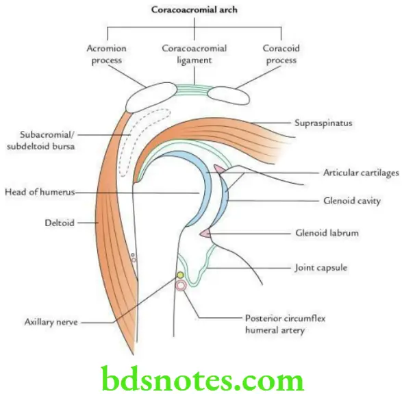
The glenoid articular surface is deepened by the glenoid labrum.
Glenoid labrum
- It is a rim of fibrocartilage attached to the peripheral margin of the glenoid cavity.
- It is triangular in cross-section and deepens the shallow glenoid cavity.
Ligaments
Capsular ligament
- Attachments
- Medially: To peripheral margin of glenoid cavity outside the glenoid labrum. The supraglenoid tubercle is intracapsular.
- Laterally: To anatomical neck of humerus except on medial side where it descends about 2–3 cm on the shaft, up to the surgical neck of humerus.
- Muscles strengthening the capsule: In general, the capsule is loose and lax, but it is strengthened by the musculotendinous (rotator) cuff formed by the following muscles:
- Openings in the capsule: The capsule presents two openings/deficiencies:
- One in front, for communicating with subscapular bursa
- One at the intertubercular sulcus to provide the passage for the tendon of long head of biceps brachii
Transverse humeral ligament: This ligament bridges across the bicipital groove.
Glenohumeral ligaments: These are thickenings in the anterior part of the capsule and are seen when the capsule is exposed from the behind. They are three in number and named superior, middle and inferior glenohumeral ligaments according to their location.
Coracohumeral ligament: It is a wide, strong fibrous band on the superior surface of the joint, extending from the base of coracoid process to the anterior aspect of greater tubercle of humerus.
Coracoacromial ligament: It extends between the lateral side of coracoid process and the medial border of acromion.
Shoulder Joint (Glenohumeral Joint) Relations
Superiorly
- Coracoacromial arch
- Subacromial bursa
- Supraspinatus
- Tendon of long head of biceps brachii (intracapsular)
- Deltoid
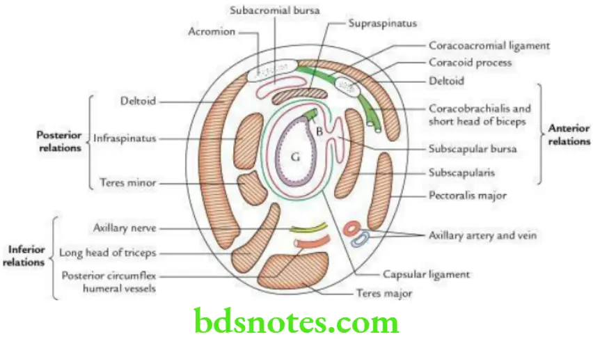
Anteriorly
- Subscapularis
- Coracobrachialis
- Short head of biceps
- Deltoid
Posteriorly
- Infraspinatus
- Teres minor
- Deltoid
Inferiorly
- Long head of triceps
- Axillary nerve
- Posterior circumflex humeral vessels
Nerve supply
The nerves supplying the joint are
- Suprascapular nerve
- Axillary nerve
- Musculocutaneous nerve
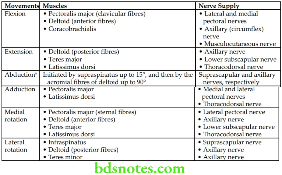
Movements
The movements and muscles producing them, with their nerve supply.
Movements of Shoulder Joint and Muscles Producing Them with Their Nerve Supply
Shoulder Joint (Glenohumeral Joint) Applied anatomy
Dislocation of shoulder joint
- The shoulder joint is the most commonly dislocated joint in the body due to (i) disproportionate size of articular surfaces – head of humerus and glenoid cavity of the scapula (the head of humerus is much larger to fit properly into smaller glenoid cavity of scapula [4:1 ratio]) and (ii) laxity of joint capsule.
- Dislocation most commonly occurs inferiorly because the joint is least supported below.
Frozen shoulder (adhesive capsulitis)
It is a clinical condition characterized by painful and uniform restriction of all movements of shoulder joint. It occurs due to shrinkage of joint capsule leading to adhesion between rotator cuff and head of humerus.
Question 3. Enumerate the factors providing stability to shoulder joint.
Answer.
- Musculotendinous (rotator) cuff
- Glenoid labrum
- Long head of biceps brachii muscle
- Coracoacromial arch
Question 4. Enumerate the synovial bursae related to the shoulder joint.
Answer.
- Subscapular bursa: Lies deep to subscapularis and communicates with joint cavity through a gap between superior and middle glenohumeral ligaments.
- Subacromial bursa: Lies deep to acromion and upper part of deltoid. It is the largest synovial bursa in the body.
- Between tendons of infraspinatus and teres major.
- Between coracoid process and joint capsule.
- Between teres major and long head of triceps brachii.
Question 5. Write a short note on Coracoacromial Arch.
Answer.
It is bony-fibrous arch lying above the shoulder joint formed by (a) coracoid process, (b) coracoacromial ligament and (c) acromial process.
The coracoacromial ligament is a flat triangular ligament which extends from the medial border of acromion (narrow end) in front of acromioclavicular articulation to the lateral border of the coracoid process (broad end).
Just below this arch, the supraspinatus muscle passes above the head of humerus on its way to greater tubercle of humerus for insertion. The coracoacromial arch is separated from this muscle by subacromial bursa to allow free movements of shoulder during contraction of supraspinatus.
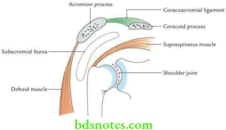
Coracoacromial Arch Applied Anatomy: The coracoacromial arch acts as a secondary socket of shoulder joint and prevents its superior dislocation.
Question 6. What are various types of sternoclavicular joint?
Answer.
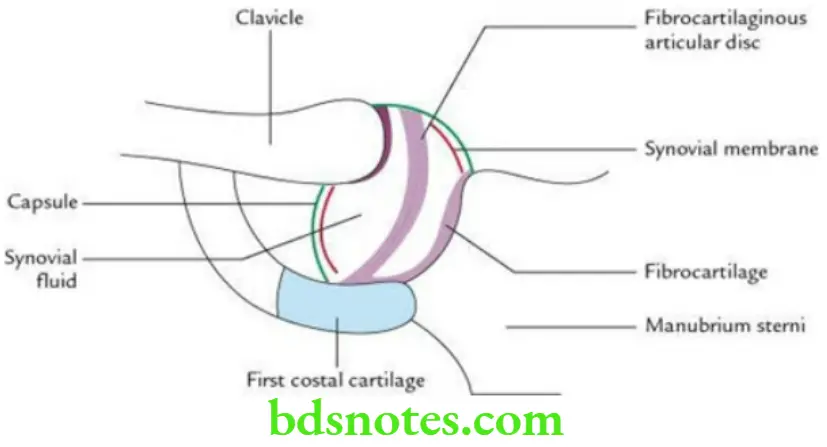
The sternoclavicular joint is of following types: synovial, saddle, compound and complex. The details are as under:
- Synovial, because it has synovial cavity filled with synovial fluid
- Saddle, because articulating surfaces are concavo-convex in shape
- Compound, because more than two bones are articulating, i.e. sternal end of clavicle, clavicular notch of manubrium sterni and upper surface of 1st costal cartilage (last two form a continuous concavo-convex surface)
- Complex, because its articular surfaces are covered by fibrocartilage and its cavity is divided into two parts by an intra-articular fibrocartilaginous disc
Question 7. Write a short note on abduction at shoulder joint.
Answer.
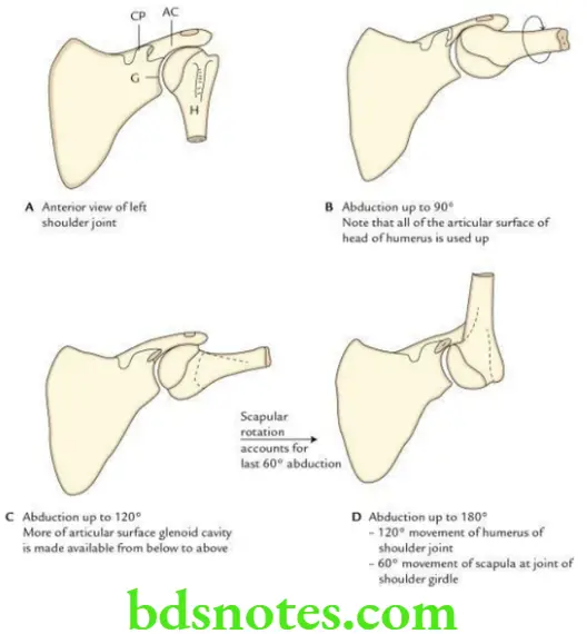
- The abduction at shoulder joint is a complex movement.
- It is performed by conjoint action of both prime movers and synergists.
- Prime movers are
- Middle fibres of deltoid
- Supraspinatus
- Synergist muscles are
- Subscapularis
- Infraspinatus
- Teres minor
The sequence of events occurring during abduction at shoulder is as follows:
- Natural range of abduction at shoulder joint is only 60°.
- When the humerus is rotated medially, range of movement of abduction is increased up to 90°.
- When humerus moves in the plane of body of scapula, range of movement is increased up to 120°.
- When humerus is rotated laterally by contraction of infraspinatus and teres minor, range of movement is increased up to 180°.
Question 8. Describe the elbow joint under the following headings: (a) classification, (b) articular surfaces, (c) ligaments, (d) relations, (e) nerve supply, (f) movements and (g) applied anatomy.
Answer.
Classification
Synovial joint of hinge variety.
Articular surfaces
It is a compound joint consisting of two articulations.
Humeroradial: Between capitulum of humerus and head of radius.
Humeroulnar: Between trochlea of humerus and trochlear notch of ulna.
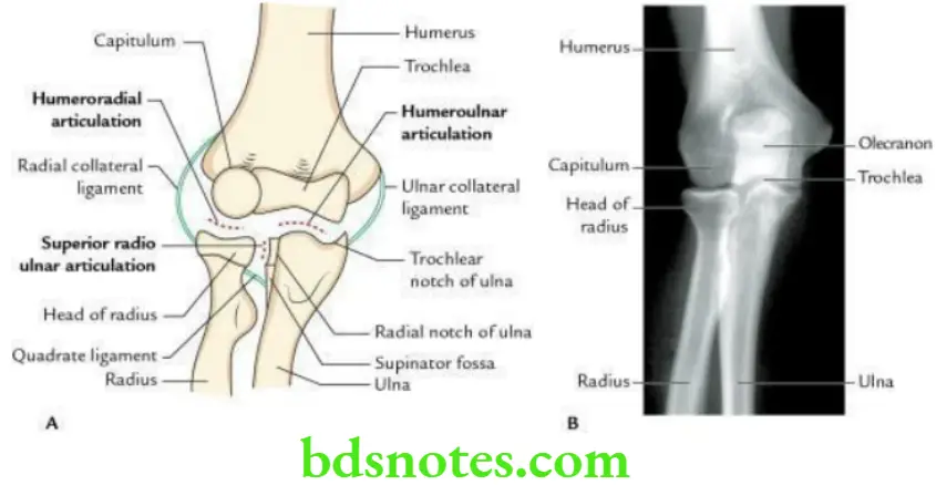
The elbow joint communicates with superior radioulnar joint.
Ligaments
Capsular ligament
- Attachments
- In front
- Superiorly, it is attached to the humerus above the coronoid and radial fossae.
- Inferiorly, it is attached to the coronoid process of ulna and annular ligament of superior radioulnar joint.
- Behind
- Superiorly, it is attached to the margins of the olecranon fossa.
- Interiorly, it is attached to the upper margins of olecranon process and annular ligament of superior radioulnar joint.
- In front
- On either side: The capsule becomes continuous with medial and lateral collateral ligaments of the elbow joint.
Medial (ulnar) collateral ligament:
It is triangular in shape and consists of the following three parts:
- Anterior part, which extends from front of medial epicondyle of humerus to the medial margin of coronoid process of ulna
- Posterior part, which extends from back of medial epicondyle of humerus to the medial margin of the olecranon process of ulna
- Inferior part, which extends between lower ends of anterior and posterior parts and stretches between the olecranon and coronoid processes of ulna
Lateral (radial) collateral ligament:
It extends from lateral epicondyle of humerus above to the annular ligament below.
Relations
Anterior
- Brachialis
- Tendon of biceps brachii
- Median nerve
- Brachial artery
Posterior
- Tendon of triceps brachii
- Anconeus
Medial
- Flexor carpi ulnaris
- Ulnar nerve
- Common flexor origin
Lateral
- Supinator
- Common extensor origin
Nerve supply
- Radial nerve
- Musculocutaneous nerve
- Median nerve
- Ulnar nerve
Movements
The movements and muscles producing them with their nerve supply.
Movements of the Elbow Joint and Muscles Producing Them with Their Nerve Supply

Applied anatomy
Dislocation:
The dislocation of elbow joint usually occurs posteriorly and is often associated with fracture of coronoid process.
Effusion:
The effusion of elbow joint causes distension on the posterior aspect of elbow because the joint capsule is weak posteriorly.
Tennis elbow (lateral epicondylitis)
It presents with pain and tenderness over lateral epicondyle of humerus. It occurs due to:
- Sprain of lateral collateral ligament of the elbow joint
- Tearing of fibres of extensor carpi radialis brevis (ECRB)
- Inflammation of bursa underneath the ECRB
- Tear of common extensor origin
Question 9. What is carrying angle? Give its clinical significance.
Answer.
- It is an angle formed between long axes of arm and forearm when the elbow is fully extended.
- This angle occurs because the medial flange of trochlea lies 6 mm lower than that of lateral flange of trochlea.
- It varies from 10° to 15° in males and 15° to 30° in females.
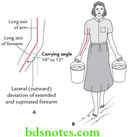
Functional significance
- It helps in holding and carrying the objects.
- It prevents rubbing of forearm with pelvis in females.
- It helps to put food in mouth during eating.
Applied anatomy
An increase in carrying angle causes cubitus valgus deformity of the elbow.
Question 10. Give a brief account of radioulnar joints and discuss the movements occurring at these joints.
Answer.
There are three radioulnar joints, i.e. superior, middle and inferior.
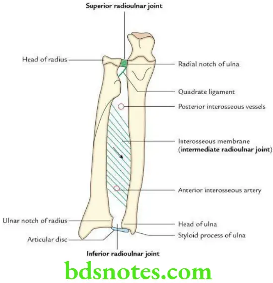
- Superior Radioulnar Joint: It is a pivot type of synovial joint between head of radius and radial notch of ulna. The annular ligament surrounds the head of radius and keeps it in contact with the radial notch of ulna. A fibrous band – the quadrate ligament extending from inferior border of radial notch of ulna to the neck of radius – closes the joint from below.
- Intermediate Radioulnar Joint: It is a fibrous joint of syndesmosis type between the shafts of radius and ulna.
Here radius and ulna are joined by an interosseous membrane whose fibres are directed downwards and medially.
It binds the radius and ulna but allows some movement. - Inferior Radioulnar Joint: It is also a pivot type of synovial joint between head of ulna and ulnar notch of radius.
A triangular articular disc of fibrocartilage extends from depression near the styloid process of ulna to the articular margins of ulnar notch of radius. It separates this joint from wrist joint.
The synovial membrane of inferior radioulnar joint projects upwards to form a pouch in front of interosseous membrane – the recessus sacciformis.
Movements
Movements occurring at radioulnar joints are
- Supination
- Pronation
Question 11. Write a short note on interosseous membrane.
Answer.
- It is a fibrous membrane which extends between interosseous borders of radius and ulna.
- The direction of fibres in this membrane is downwards and medially towards ulna.
- It forms intermediate (middle) radioulnar joint of syndesmosis type.
- Superiorly the gap between oblique cord and upper border of membrane, provides passage to posterior interosseous artery to go into the posterior compartment.
- Inferiorly interosseous membrane blends with capsule of inferior radioulnar joint.
- An opening in the inferior part of membrane provides passage to anterior interosseous artery to enter the posterior compartment.
- Membrane is related anteriorly to flexor pollicis longus (FPL), flexor digitorum profundus (FDP) and pronator quadratus (PQ).
- Membrane is related posteriorly to supinator, abductor pollicis longus (APL), extensor pollicis longus (EPL), extensor pollicis brevis (EPB), extensor indicis and posterior interosseous nerve and vessels.
Functional significance
Interosseous membrane: (a) helps in transmission of force from the radius (received from wrist joint) to the ulna for onward transmission to the humerus and (b) helps in supination and pronation.
Question 12. Give a brief account of movements of supination and pronation and discuss their functional significance.
Answer.
Movements
- Supination: It is the movement of forearm in which the palm of hand is turned forwards/upwards.
- Pronation: It is the movement of forearm in which the palm of hand is turned backwards/downwards.
The details of movements of supination and pronation.
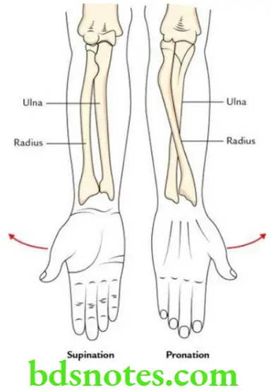
Movements of Supination and Pronation

Functional significance
- They help in picking up food and putting it into the mouth.
- They are used in mechanical jobs, e.g. opening and tightening the screws with screw driver.
Question 13. Describe the wrist joint under the following headings: (a) classification, (b) articular surfaces, (c) ligaments, (d) relations, (e) nerve supply, (f) movements and (g) applied anatomy.
Answer.
Classification
Structural:
Synovial joint of ellipsoid variety.
Functional:
Diarthrosis.
Articular surfaces
Proximally
- Inferior articular surface of the lower end of the radius
- Inferior surface of the articular disc of the inferior radioulnar joint
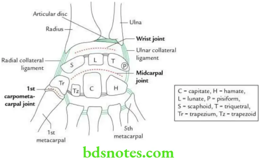
Distally
- Scaphoid
- Lunate
- Triquetral
Ligaments
Capsule:
It is attached above to the peripheral margins of the proximal and distal articular surfaces including the articular disc. Distally, it blends with the palmar and dorsal radiocarpal ligaments.
Radial collateral ligament:
It extends from the styloid process of radius to the lateral aspects of scaphoid and trapezium.
Ulnar collateral ligament:
It extends from the styloid process of ulna to the medial aspects of triquetral and pisiform bones.
Palmar and dorsal radiocarpal ligaments:
These are the thickenings on the palmar and dorsal aspects of the fibrous capsule.
Relations
Anterior
- Superficial: Tendons of flexor carpi radialis and palmaris longus
- Intermediate: Radial artery median nerve and flexor digitorum superficialis
- Deep: Tendons flexor pollicis longus and flexor digitorum profundus
Posterior:
Extensor tendons of the wrist and fingers with their synovial sheaths.
Lateral:
Radial artery.
Nerve supply
- Anterior interosseous nerve
- Posterior interosseous nerve
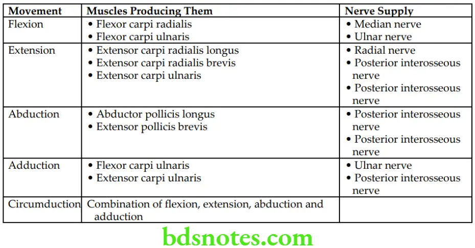
Movements
Movements and muscles producing them with their nerve supply.
Movements of the Wrist Joint
Applied anatomy
Colles fracture:
It is fracture of distal end of radius due to fall on outstretched hand with distal fragment being displaced upwards and backwards.
Smith fracture:
It is reverse of Colles fracture due to fall on the back of the hand with distal fragment being displaced upwards and forwards (i.e. palm is flexed).
Question 14. Enumerate the range of movement at the wrist joint.
Answer.
- Flexion = 60°–85°
- Extension = 50°–60°
- Abduction = 15°
- Adduction = 30°–45°
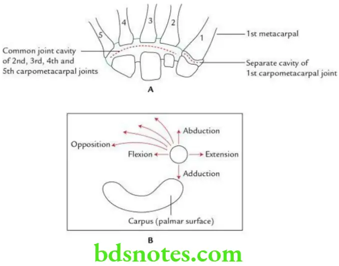
Question 15. Write a brief note on the 1st carpometacarpal joint.
Answer.
Classification
Structural: Synovial joint of saddle variety
Functional: Diarthrosis
Articular surfaces
Proximally: Distal surface of trapezium
Distally: Proximal surface of the base of 1st metacarpal (MC) bone
Ligaments
- Capsular ligament: It is a loose fibrous sac, which encloses the joint cavity. It is thickest dorsally and laterally.
- Lateral ligament: Broad fibrous band extending from the lateral surface of trapezium to the lateral surface of 1st MC.
- Anterior ligament: Extends obliquely from the palmar surface of trapezium to the ulnar side of base of 1st MC.
- Posterior ligament: Extends obliquely from the dorsal surface of trapezium to the ulnar side of 1st MC.
Relations
- Anterior: Muscles of thenar eminence
- Posterior: Long and short extensors of thumb
- Medial: First dorsal interosseous muscle
- Lateral: Abductor pollicis longus
- Posteromedial: Radial artery
Nerve supply
Median nerve
Movements
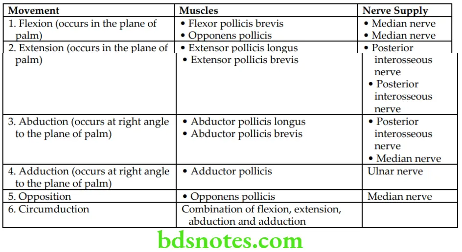
The movements and muscles producing them with their nerve supply.
Movements of 1st Carpometacarpal Joint
Question 16. Give the classification of metacarpophalangeal (MP), proximal intraphalangeal (PIP) and distal interphalangeal (DIP) joints, and discuss the movements occurring at these joints.
Answer.
The classification and movements of MP, PIP and DIP joints.
Classification and Movement of MP, PIP and DIP joints

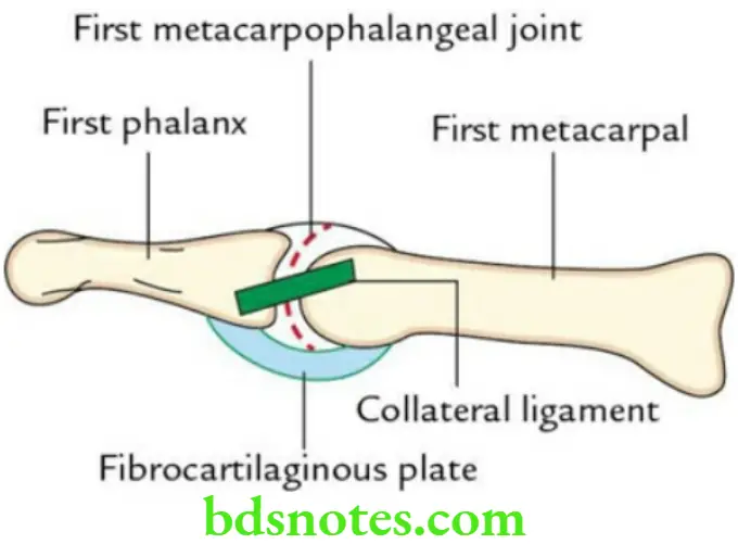
Question 17. Write a short note on 1st metacarpophalangeal joint.
Answer.
- Type: Ellipsoid type of synovial joint
- Articular surfaces: Convex articular head of 1st metacarpal and curved articular base of proximal phalanx
- Ligaments: Capsular ligaments, palmar ligaments, and medial and lateral collateral ligaments
- Capsular ligament: Thick in front and thin behind
- Palmar ligament: Fibrocartilaginous plate which replaces deep transverse metacarpal ligaments
- Medial and lateral collateral ligament: Oblique bands extending downwards and forwards from head of metacarpal to the base of proximal phalanx
Movements and muscle producing them
- Flexion: Flexor pollicis brevis
- Extension: Extensors of thumb
- Abduction: Abductor pollicis brevis
- Adduction: Adductor pollicis
Question 18. Enumerate the chief flexors of MP, PIP and DIP joints.
Answer.
- MP joints: Lumbricals and interossei
- PIP joints: Flexor digitorum superficialis
- DIP joints: Flexor digitorum profundus
Question 19. Which movements of thumb are tested to confirm the integrity of radial, ulnar and median nerves?
Answer.
- To test integrity of radial nerve: Test extension of thumb, because this movement of thumb is lost if radial nerve is damaged.
- To test integrity of ulnar nerve: Test adduction of thumb, because this movement of thumb is lost if ulnar nerve is damaged.
- To test integrity of median nerve: Test abduction and opposition of thumb, because both these movements of thumb are lost if median nerve is damaged.

Leave a Reply