Question 1. Describe the origin, course, relations & branches of maxillary artery (or) Give an account of lateral pterygoid muscle
Answer:
Maxillary Artery Origin:
- It is the largest terminal branch of external carotid artery arising behind the neck of the mandible within the substance of the parotid gland
Maxillary Artery Course and relations:
Within the substance of the parotid gland the artery is divided into three parts
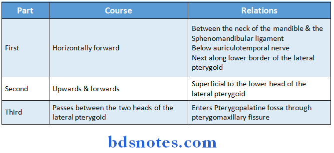
Read And Learn More: BDS Previous Examination Question And Answers
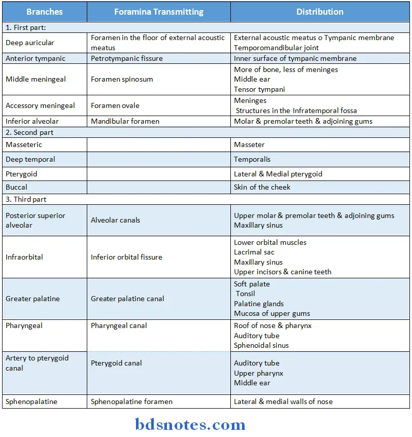
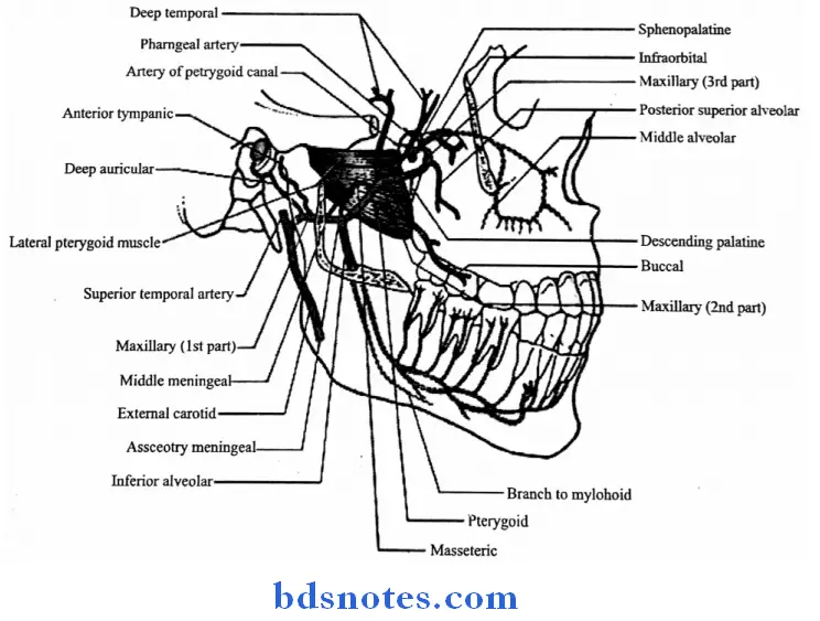
Question 2. Describe the muscles of mastication under following headings. Name the muscle attachments, Nerve supply, Action (or) Give an account of lateral pterygoid muscle (or) Muscles of mastication (or) Attachments, relations, nerve supply & action of lateral pterygoid muscle (or) Muscles of mastication
Answer:
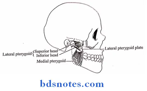
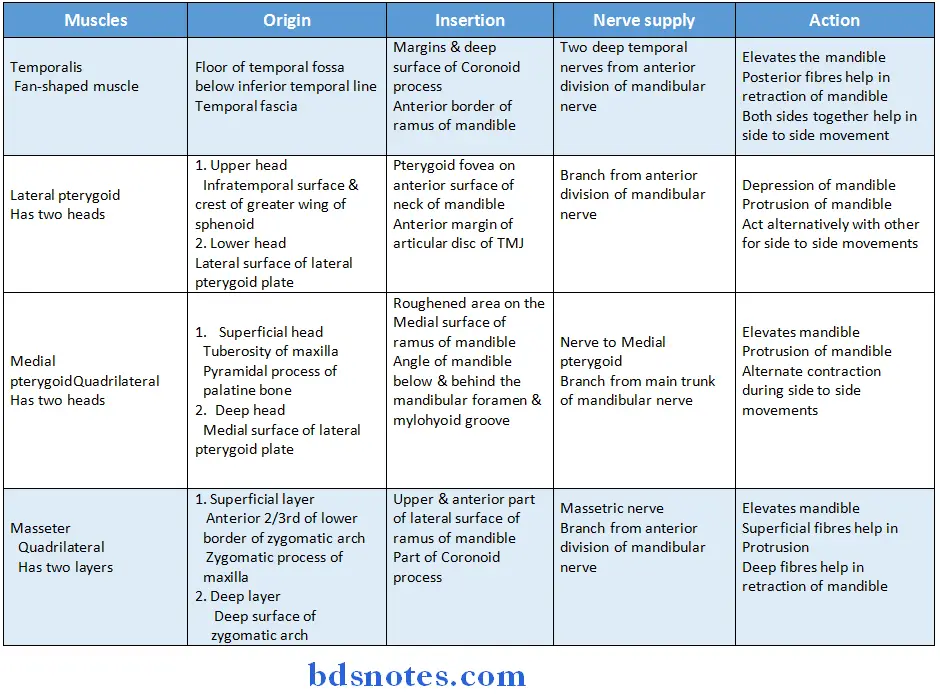
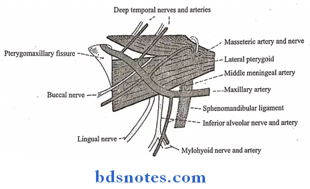
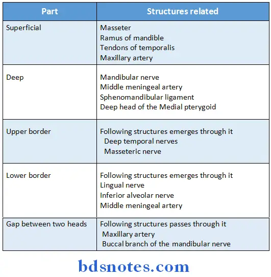
Pterygoid Muscle Relations:
Question 3. Describe the mandibular nerve under following headings, Origin, Root fibres, Branches, Termination, Relations (or) Inferior alveolar nerve
Answer:
Mandibular Nerve Origin:
- Mandibular nerve originates in the middle cranial fossa through its roots
Mandibular Nerve Root fibres:
- It has
- Sensory root
- It is large root
- Arises from the lateral part of the trigeminal ganglion
- Motor root
- Smaller root
- Lies deep to the trigeminal ganglion
- Sensory root
Mandibular Nerve Branches:
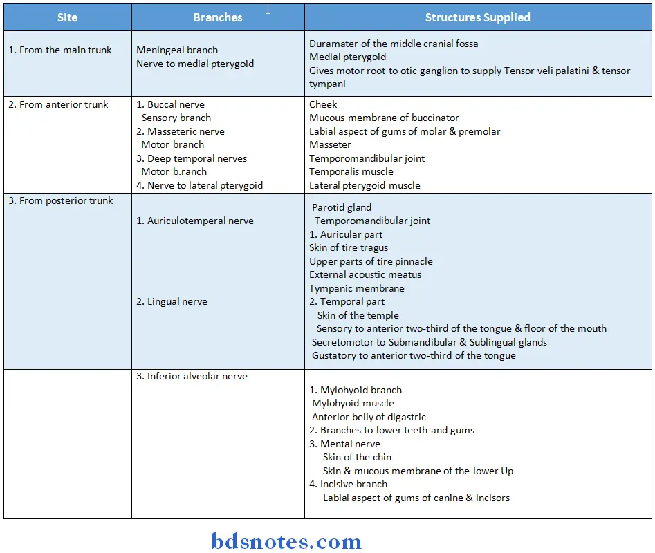
Mandibular Nerve Termination:
- It teminates by dividing into small anterior trunk and large posterior trunk
Mandibular Nerve Relations:
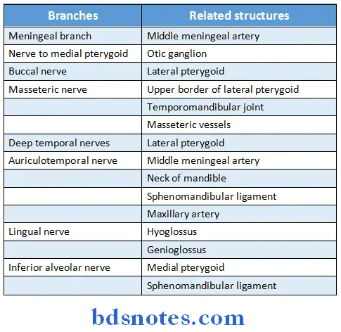
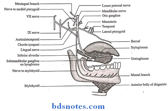
Question 4. Describe the formation, course, branches and distribution of mandibular division of trigeminal nerve
Answer:
Trigeminal Nerve Formation:
- It is nerve of first branchial arch,
- It is formed by sensory & motor root
Trigeminal Nerve Course:
- It begins on the middle cranial fossa through
- Sensory root
- It is large root
- Arises from the lateral part of the trigeminal ganglion
- Leaves the cranial cavity through the foramen ovale
- Motor root
- Smaller root
- Lies deep to the trigeminal ganglion
- Passes through foramen ovale
- Joins sensory root
- Forms main trunk
- After a short course, main trunk divides into
- Small anterior trunk
- Large posterior trunk
- Sensory root
Question 5. Describe origin, course, relation, distribution & applied anatomy of lingual nerve
Answer:
Lingual Nerve Origin:
- It is one of the terminal branch of the posterior division of trigeminal nerve
Lingual Nerve Course:
- It begins one cm below the skull
- About 2 cm below skull, it is joined to the Chorda tympani nerve at an acute angle
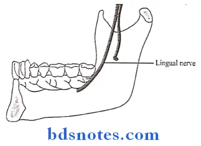
- Then lies in contact with mandible
- Medial to 3rd molar tooth
Lingual Nerve Relations:
- It is related to
-
- Lower 3rd molar tooth
- Hyoglossus
- Genioglossus
Lingual Nerve Distribution:
- Sensory to anterior twothird of the tongue & floor of the mouth
- Secretomotor to Submandibular & Sublingual glands
- Gustatory to anterior twothird of the tongue
Lingual Nerve Applied anatomy:
- During improper extraction of the lower third molar or fracture of angle of mandible, the lingual nerve may get damaged
- This results in loss of all sensations from anterior twothird of the tongue
Question 6. Describe functional components of Trigeminal Nerve. Write in detail about mandibular nerve
Answer:
Functional components of trigeminal nerve:
1. Sensory components
- Sensations of pain, temperature, touch & pressure travel along axons
- Their cell bodies lie in the trigeminal ganglion
- Peripheral processes forms
- Ophthalmic nerve
- Maxillary nerve
- Mandibular nerve
- Central processes form sensory root
2. Motor component
- The motor nucleus receives impulses from
-
- Right & left cerebral hemispheres
- Red nucleus
- Mesencephalic nucleus
- It supplies
- Muscles of mastication
- Tensor veli palatini
- Tensor tympani
- Mylohyoid
- Anterior belly of digastric
- It supplies
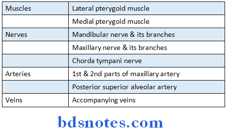
Question 7. Mention boundaries & contents of Infratemporal Fossa
Answer:
Infratemporal Fossa Boundaries:
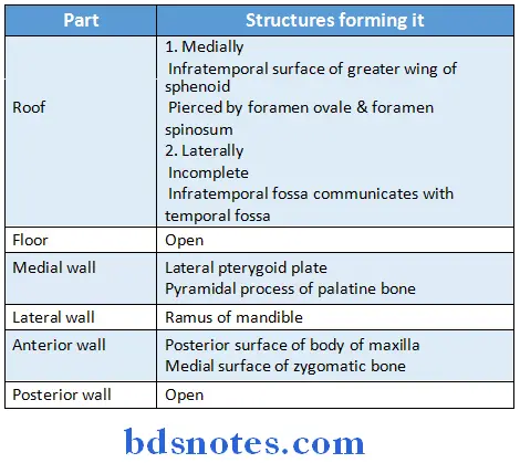
- The anterior & medial walls are separated in their upper parts by the pterygomaxillary fissure
- The upper end of this fissure is continuous with the anterior part of the inferior orbital fissure
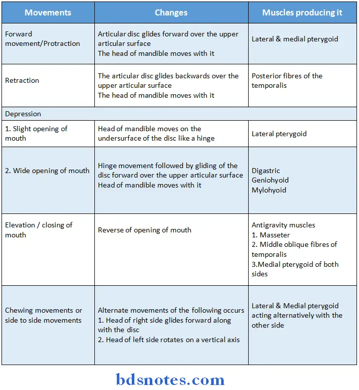
Question 8. Describe the movements of temporomandibular joint & mention the muscles producing these movements. What are the factors responsible for its stability (or) Articular disc of TMJ
Answer:
Movements & muscles producing TMJ
TMJ Factors effecting stability:
- Stress
- Position of teeth when they are engaged
- Loss of posterior teeth reduces the support of the jaw & joint activity
- While the remaining produces greater strain on the joint
Question 9. Describe temporomandibular joint under following headings: type of joint, articular surfaces, ligaments, nerve supply, movements & muscles producing movements
Answer:
Type of Temporomandibular Joint :
- It is a type of bicondylar joint, synovial type
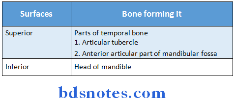
Temporomandibular Joint Ligaments:
- The ligaments of TMJ are as follows:
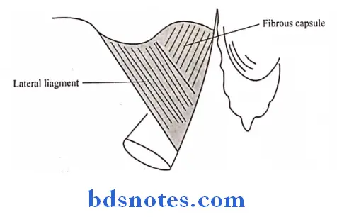
1. Fibrous capsule:
Temporomandibular Joint Attachments:
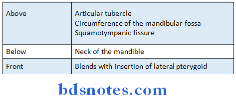
- The synovial membrane lines the fibrous capsule
2. Lateral or temporomandibular ligament:
- It blends with the lateral part of fibrous capsule
- It reinforces & strengthens the lateral part of fibrous capsule
- It extends from the tubercle of root of zygoma to lateral surface of neck of mandible
3. Sphenomandibular ligament:
- It is accessory ligament of TMJ
- It is remnant of dorsal part of Meckel’s cartilage
Temporomandibular Joint Location:
- It lies on a deep plane away from the fibrous capsule of TMJ
Temporomandibular Joint Attachments:
- Superiorly: To the spine of sphenoid
- Inferiorly: To the lingula of mandibular foramen
Temporomandibular Joint Relations: It is related
- Laterally to
- Lateral pterygoid muscle
- Auriculotemporal nerve
- Maxillary artery
- Medially to
- Chorda tympani nerve
- Wall of pharynx
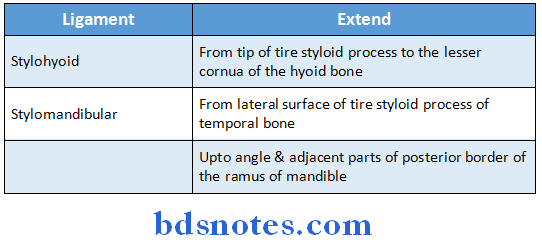
4. Stylomandibular ligament:
- It is accessory ligament of temporomandibular joint
- It is derived from investing layer of deep cervical fascia
Temporomandibular Joint Extend:
- From lateral surface of the styloid process of temporal bone
- Upto angle & adjacent parts of posterior border of the ramus of mandible
Temporomandibular Joint Nerve Supply:
- It is supplied by
-
- Auriculotemporal nerve
- Masseteric nerve
Question 10. Describe temporomandibular joint under following headings: articulating bones & surfaces, capsule, intraarticular disc, ligaments, movements with muscles producing it & applied anatomy
Answer:
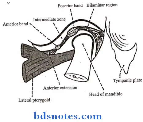
Temporomandibular Joint Intraarticular disc:
- It is an oval plate of fibrocartilage & divides the joint into two compartments
- It represents the primitive insertion of lateral pterygoid muscle
- Posteriorly the disc splits into upper & lower lamellae by venous plexus
-
- Upper lamellac
- Attached to Squamotympanic fissure
- Lower lamellae
- Attached to posterior surface of neck of the mandible
- Parts
- Structurally the disc contains five parts
- Anterior extension
- Anterior thick band
- Intermediate zone
- Posterior thick band
- Bilamellar region
- Structurally the disc contains five parts
- Upper lamellac
Temporomandibular Joint Applied anatomy:
- Dislocation of mandible
- Occurs during excessive opening of the mouth
- Head of mandible may slip into Infratemporal fossa
- Reduced by exerting downward traction of ramus of mandible & elevation of chin
- Derangement of the articular disc
- May occur due to overclosure or malocclusion
- Results in clicking & pain during movements of the jaw
- Operations of TMJ
- May damage VII nerve & Auriculotemporal nerve
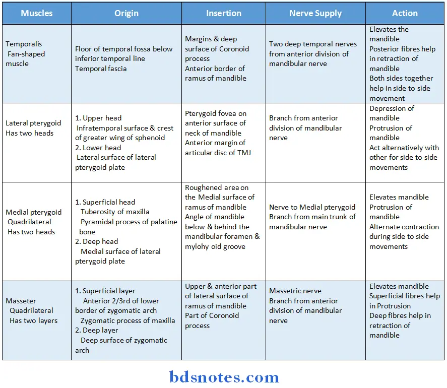
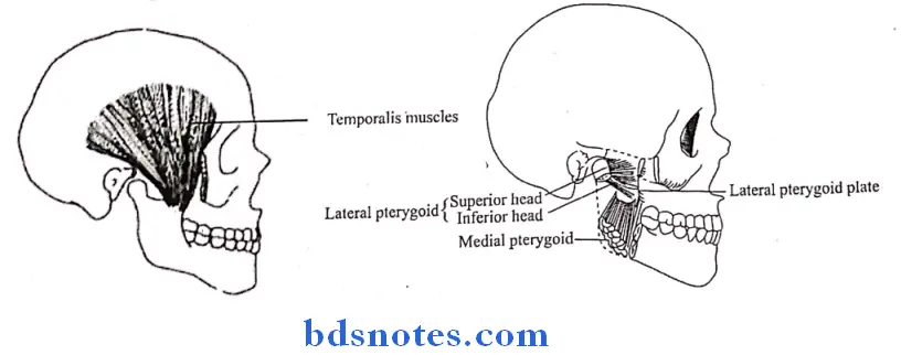
Question 11. Describe muscles of mastication under headings:
Origin Insertion Nerve supply Action (e) Applied aspect
Answer:
muscles of mastication Applied Aspects:
- Masseter muscle
- It can be palpated both intraorally and extraorally
- Common muscle involved in myositis ossificans
- It shows dual action in complete denture
- It commonly undergoes hypertrophy in bruxism
- Temporalis muscle
- Sudden contraction of it results in coronoid fracture
- Lateral pterygoid
- Commonly involved muscle in MPDS
- Only muscle of mastication which has attachment to the TMJ
- It forms roof of pterygomandibular space
- Medial pterygoid
- It can be palpated only intraorally
- Commonly involved in MPDS
- Trismus following inferior alveolar nerve block is mostly due to involvement of medial pterygoid muscle
Question 12. Describe mandibular nerve under following headings:
Origin Division and branches Course and relations Applied aspects
OR
Describe origin, course, relations, branches and applied anatomy of mandibular nerve.
Answer:
Mandibular Nerve:
- It is nerve of first branchial arch
Mandibular Nerve Origin:
- Mandibular nerve originates in the middle cranial fossa through its roots
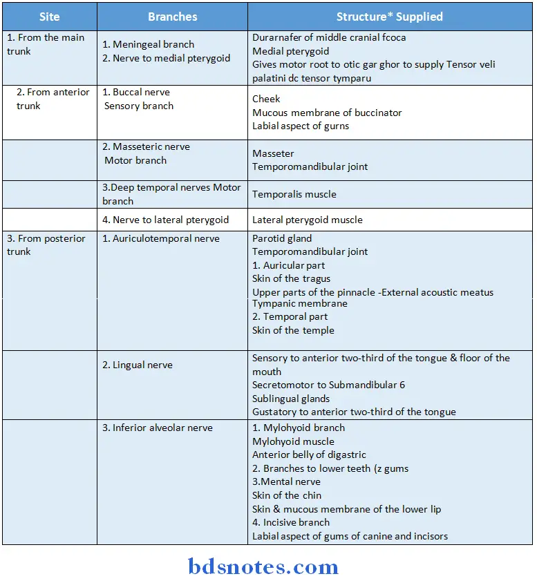
Mandibular Nerve Branches:
- Sensory root
- It is large root
- Arises from the lateral part of the trigeminal ganglion
- Leaves the cranial cavity through the foramen ovale
- Motor root
- Smaller root
- Lies deep to the trigeminal ganglion
- Passes through foramen ovale
- Joins sensory root
- Forms main trunk
- After a short course, main trunk divides into
- Small anterior trunk
- Large posterior trunk
Mandibular Nerve Termination:
- It terminates by dividing into small anterior trunk & large posterior trunk
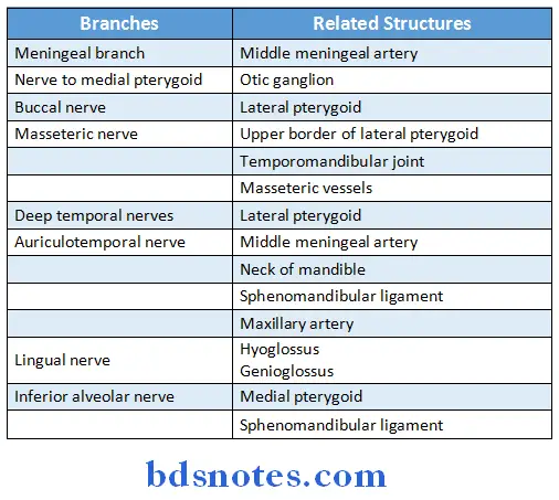
Mandibular Nerve Relations:
Mandibular Nerve Applied Aspects:
- Motor root of mandibular nerve is tested by asking the patient of clench her/his teeth
- In carcinoma of tongue pain is radiated to ear and temporal fossa
- To treat trigeminal neuralgia, sensory root of nerve is divided behind the ganglion
- Mandibular nerve supplies efferent and afferent loops of jawjerk reflex
- During extraction of mandibular teeth inferior alveolar nerve block is given
Question 13. Articular disc
Answer:
- It is an oval plate of fibrocartilage & divides the joint into two compartments
- Upper compartment
- Permits gliding movements
- Lower compartment
- Permits gliding & rotatory movements
- It represents the primitive insertion of lateral pterygoid muscle
- Posteriorly the disc splits into upper & lower lamellae by venous plexus
- Upper lamellae
- Attached to Squamotympanic fissure
- Lower lamellae
- Attached to posterior surface of neck of the mandible
- Upper lamellae
- Upper compartment
Articular disc Parts:
- Structurally the disc contains five parts
- Anterior extension
- Anterior thick band
- Intermediate zone
- Posterior thick band
- Bilamellar region
Articular disc Surfaces:

Question 14. Inferior Alveolar Nerve
Answer:
- It is a branch of posterior division of mandibular nerve
Inferior Alveolar Nerve Course:
- Emerges under lower border of the lateral pterygoid
- Passes downward & forward between the ramus of mandible & Sphenomandibular ligament Enters the mandibular foramen
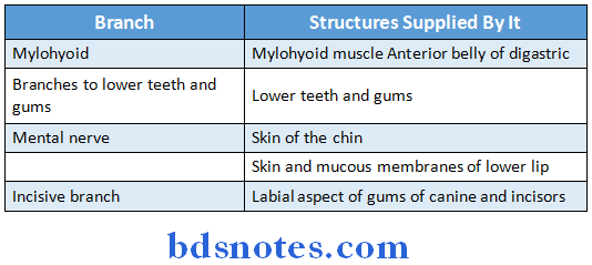
- Runs in a bony canal below the teeth & finally divides into incisive & mental nerves
Question 4. Styloid Process Of Temporal Bone
Answer:
- It is long, slender & pointed bony process projecting downwards, forwards & slightly medially from the temporal bone
- It descends between the external & internal carotid arteries to reach the side of the pharynx.
- It is interposed between the parotid gland laterally & internal jugular vein medially
- The styloid process with its attached structures is called styloid apparatus
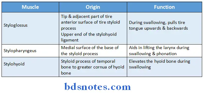
Structures attached to Styloid Process Of Temporal Bone :
1. Muscles:
2. Styloid Process Of Temporal Bone Ligaments:
Question 15. Pterygoid plexus of veins (or) Pterygoid plexus of veins
Answer:
Pterygoid Plexus of Veins Location:
- Lies around & within the lateral pterygoid muscle

Pterygoid Plexus of Veins Communications:
Question 16. First part of maxillary artery
Answer:
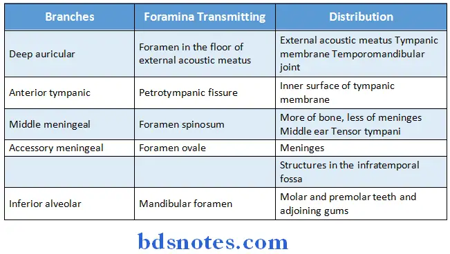
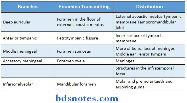
- Maxillary artery is the largest terminal branch of external carotid artery arising behind the neck of the mandible within the substance of parotid gland
- During its course it is divided into three parts
First part of maxillary artery:
Question 17. Third part of maxillary artery
Answer:
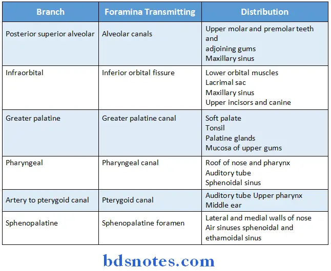
- Maxillary artery is the largest terminal branch of external carotid artery arising behind the neck of the mandible within the substance of parotid gland
Question 18. Mandibular nerve
Answer:
Mandibular nerve Origin:
- Originates in the middle cranial fossa through its roots
Mandibular nerve Root fibres:
- It has two roots
-
- Sensory rootlarge root
- Motor rootsmall root
Mandibular nerve Branches:
- From the main trunk
- Meningeal branch
- Nerve to medial pterygoid
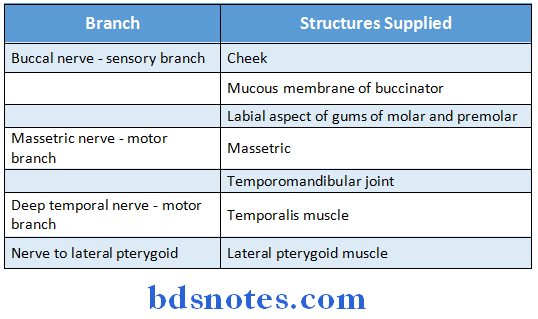
- From anterior trunk
- Buccal nerve
- Masseteric nerve
- Deep temporal nerve
- Nerve to lateral pterygoid
- From posterior trunk
- Auriculotemporal nerve
- Lingual nerve
- Inferior alveolar nerve
Mandibular nerve Termination:
- It terminates by dividing into small anterior trunk and large posterior trunk
Mandibular nerve Applied aspects:
- Motor root of mandibular nerve is tested by asking the patient of clench her/his teeth
- In carcinoma of tongue pain is radiated to ear and temporal fossa
- To treat trigeminal neuralgia, sensory root of nerve is divided behind the ganglion
- Mandibular nerve supplies efferent and afferent loops of jawjerk reflex
- During extraction of mandibular teeth inferior alveolar nerve block is given
Question 19. Masseter muscle
Answer:
- It is quadrilateral in shape
- It is muscle of mastication
- It is two layers superficial and deep
Masseter muscle Origin:
- Superficial layer
- Anterior 2/3rd of lower border of zygomatic arch
- Zygomatic process of maxilla
- Deep layer
- Deep surface of zygomatic arch
Masseter muscle Insertion:
- Upper and anterior part of lateral surface of ramus of mandible
- Part of coronoid process
Masseter muscle Nerve supply:
- Massetric nerve
- Branch from anterior division of mandibular nerve
Masseter muscle Actions:
- Elevates the mandible
- Superficial fibres help in protrusion
- Deep fibres help in retraction of mandible.
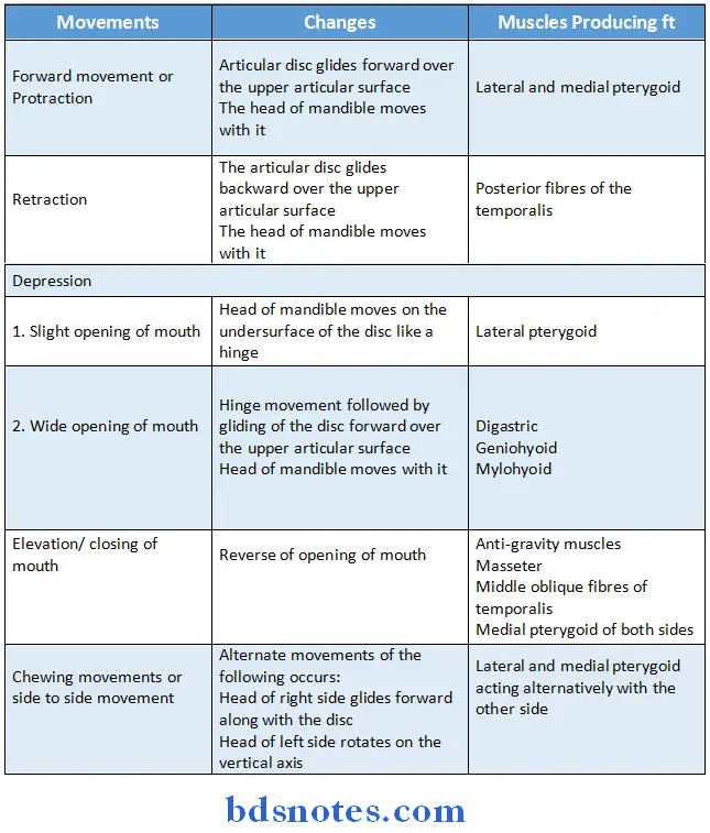
Question 20. Movements of temporomandibular joint
Answer:
Question 21. Lingual nerve
Answer:
Lingual nerve Origin:
- It is one of the terminal branch of the posterior division of trigeminal nerve
Lingual nerve Distribution:
- Sensory to anterior twothird of the tongue & floor of the mouth
- Secretomotor to Submandibular & Sublingual glands
- Gustatory to anterior twothird of the tongue
Question 22. Auriculotemporal nerve
Answer:
Auriculotemporal nerve Course:
- It arises by two roots
- Runs backwards
- It is encircled by middle meningeal artery
- Unites to form a single trunk
- Runs between the neck of mandible & sphenomandibular ligament, above maxillary artery
- Next turns upwards & ascends on temple
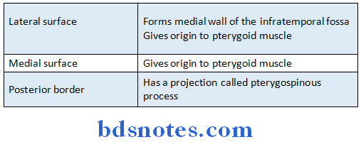
Question 23. Lateral pterygoid plate
Answer:
- The lateral pterygoid plate is directed backwards & laterally
- It has medial & lateral surfaces & a free posterior border
Question 24. Name any four branches of first part of maxillary artery
Answer:
- Deep auricularsupplies:
- External acoustic meatus
- Tympanic membrane
- Temporomandibular joint
- Anterior tympanicsupplies:
- Inner surface of tympanic membrane
- Middle meningealsupplies:
- More of bone, less of meninges
- Middle ear
- Tensor tympani
- Accessory meningealsupplies:
- Meninges
- Structures in the Infratemporal fossa
- Inferior alveolarsupplies:
- Molar & premolar teeth & adjoining gums
Question 25. What are the branches of third part of maxillary artery?
Answer:
- Posterior superior alveolarsupplies
- Upper molar & premolar teeth & adjoining gums
- Maxillary sinus
- Infraorbitalsupplies
- Lower orbital muscles
- Lacrimal sac
- Maxillary sinus
- Upper incisors & canine teeth
- Greater palatinesupplies
- Soft palate
- Tonsil
- Palatine glands
- Mucosa of upper gums
- Pharyngealsupplies
- Roof of nose & pharynx
- Auditory tube
- Sphenoidal sinus
- Artery to pterygoid canalsupplies
- Auditory tube
- Upper pharynx
- Middle ear
- Sphenopalatinesupplies
- Lateral & medial walls of nose
- Air sinusessphenoidal & ethamoidal sinus
Question 26. Name the contents of the temporal fossa
Answer:
- Temporalis muscle
- Middle temporal artery
- Zygomaticotemporal nerve
- Zygomaticotemporal artery
- Deep temporal nerve
- Deep temporal artery
Question 27. Temporalis muscle
Answer:
- It is fan shaped muscle
Temporalis muscle Origin:
- Floor of temporal fossa below inferior temporal line
- Temporal fascia
Temporalis muscle Insertion:
- Margins and deep surface of coronoid process
- Anterior border of ramus of mandible
Temporalis muscle Nerve supply:
- Two deep temporal nerves from anterior division of mandibular nerve
Temporalis muscle Action:
- Elevates the mandible
- Posterior fibres helps in retraction of mandible
- Both sides together help in side to side movements
Question 28. Three features of synovial joint
Answer:
- Synovial joint joins bones with a fibrous joint capsule that is continuous with the periosteum of the joined bones
- It is filled with synovial fluid
- They are most common and most movable type of joint
- Synovial Joint contains:
- Synovial cavity
- Joint capsule
- Articular cartilage
- Other structures present are:
- Articular discs
- Articular fat pads
- Accessory ligaments
- Bursae
Synovial Joint Types:
- Gliding/Plane joints
- Hinge joint
- Pivot joint
- Saddle joint
- Ellipsoidal / Condyloid joint
- Ball and socket joint
- Compound joint
Movements produced by them:
- Abduction
- Adduction
- Extension
- Flexion
- Rotation
Question 29. Name the branches from the anterior trunk of mandibular nerve.
Answer:
Question 30. Terminal branches of inferior alveolar nerve
Answer:

Leave a Reply