Instrument
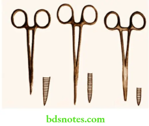
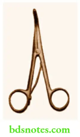
Artery Forceps (Haemostat)
- It is also called Spencer Well’s artery forceps. It has a ratchet and two blades with uniform serrations.
- It is used to control bleeding, not only from arteries but from the veins and capillaries. Once the bleeding points are caught, they are coagulated or ligature is applied. Curved artery forceps are commonly used.
Read And Learn More: Clinical Medicine And Surgery Notes
- The smaller version of this is called Mosquito forceps. This is extremely useful in the repair of harelip, cleft palate, or other plastic surgery operations.
- It is also available as a straight artery forcep to hold stay sutures.
Allis Tissue Holding Forceps
- It has a ratchet and triangular expansion at the tip, where the serrations are present.
- It can be used to hold tough structures like fascia, aponeurosis, etc.
- Even though it can cause trauma, because of its better grip, it can be used to hold the duodenum for duodenal closure during gastrectomy.
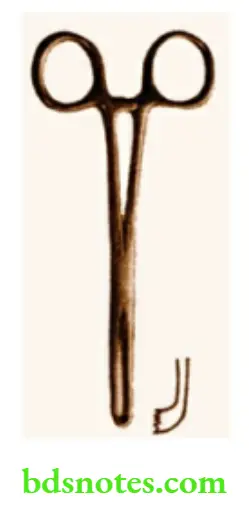
Kocher’s Forceps
- This is similar to an artery forceps with serrations. It is available as curved and straight.
- There is a sharp tooth at the tip of the instrument. Hence, it has a better grip.
- Kocher’s forceps can be used to hold tough structures like aponeurosis, fascia, etc.
- During thyroidectomy, it can be used to hold the strap muscles for dividing them.
- Theodor Kocher, the German surgeon, got the Nobel Prize for his contribution to thyroid surgery.

Remember
- Kocher’s forceps
- Kocher’s test
- Kocher’s thyroid dissector
- Kocher’s vein
- Kocher’s subcostal incision
- Kocher’s gland holding forceps
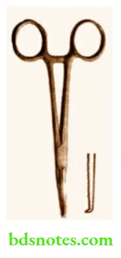
Sinus Forceps
- This is like an artery forceps that has no ratchet.
- Serrations are confined to the tip so as to hold the wall of an abscess cavity for biopsy.
- In Hilton’s method of drainage of an abscess, once the incision is made, the sinus forceps are thrust into the abscess cavity, and by opening the blades in all directions, the loculi are broken.
- To facilitate the free opening of the blades, sinus forceps have no ratchet.
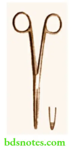
Swab (Sponge) Holding Forceps
- This has a ratchet and two long blades.
- The operating end is rounded with serrations.
- It is used to hold the swab (gauze pieces) to prepare the parts with antiseptic agents at the time of surgery.
- This instrument can also be used as a blunt ‘dissector’ with the swab while dissecting at a depth, e.g. lumbar sympathectomy, vagotomy, etc.
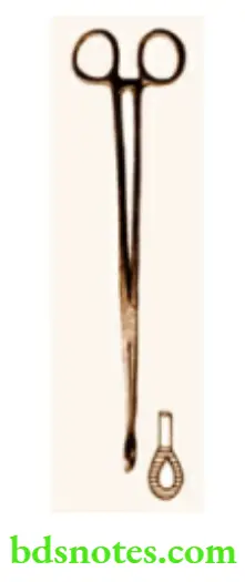
Babcock’s Forceps
- An instrument with a ratchet and a triangular expansion with fenestrations at the operating end. It does not have any teeth. Thus, it is used to hold intestines during anastomosis or resection.
- This instrument can also be used to hold many other structures such as the thyroid gland, mesoappendix, uterine tubes, etc.
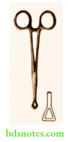
Lane’s Forceps
- This is similar to Babcock’s forceps, but the tip is broader and expanded with a bigger opening.
- It is used to hold the appendix.
- However, it does not seem to have any additional advantage when compared to Babcock’s forceps.
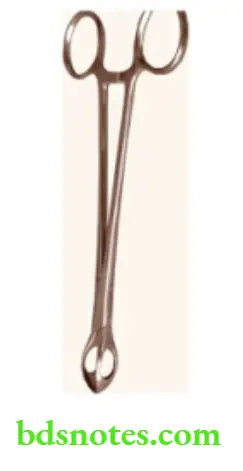
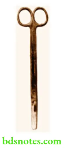
Dissecting Scissors
- This is also called Mayo’s scissors.
- This instrument does not have a ratchet and the operating end is sharp.
- This is used to dissect tissue planes during surgical operations and to cut or divide important structures.
- It is popularly called tissue scissors.
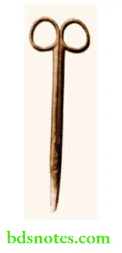
Straight Scissors
- It is used to cut sutures or knots. Hence, called suture-cutting scissors.
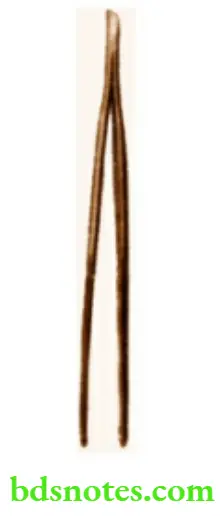
Dissecting Forceps
- This is a toothed forceps. It is also available as non-toothed forceps.
- Dissecting forceps with dissecting scissors makes a good ‘tool’ for a surgeon to develop a tissue plane in the majority of surgeries.
- The forceps are very useful to ‘pick’ individual layers such as serosa, seromuscular layer, mucosa, etc. during anastomosis.
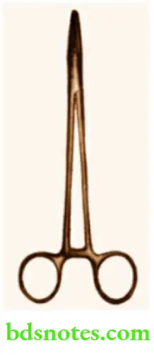
Needle Holder
- This is a long instrument with a ratchet at the non-operating end.
- The operating end has two small blades with serrations.
- It is used to hold curved needles while suturing.
- A firm grip is essential to apply proper sutures.
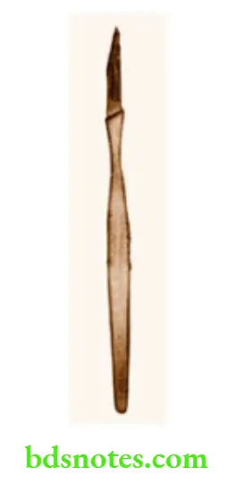
Scalpel With Blade
- Popularly called the ‘surgeon’s knife’.
- This is used to incise the skin and subcutaneous tissue.
- Due to its sharp nature, it can be used to divide a major vascular pedicle once ligatures are applied.
Needles
- The curved needles can have a round body or may be of cutting type.
- Round body needles are used to suture subcutaneous tissue, intestines, etc.
- Cutting needles are used to suture skin or tough fibrous structures.
- A straight needle is used for skin sutures.
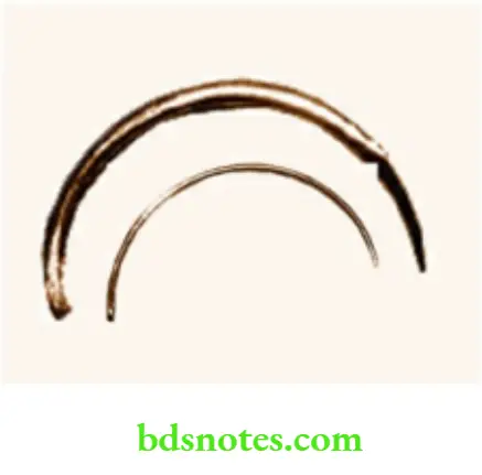
Czerny Retractor
- This is a double-hooked retractor on one side and a single blade on the other side.
- This is a superficial retractor, that can be used to retract layers of the abdominal wall, muscles, etc.
- Thus, during appendicectomy, herniorrhaphy, or thyroidectomy, this instrument is very useful.
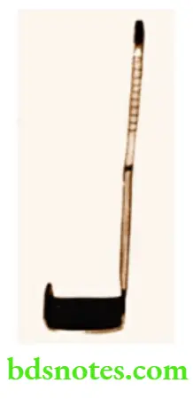
Langenbeck Retractor
- This instrument has only one blade.
- The uses of this are similar to that of Czerny’s retractor.
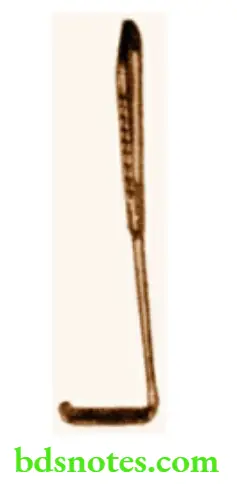
Kocher’s Thyroid Dissector
- This has a long handle and the operating end is small and blunt with an opening.
- Few longitudinal serrations are present at the tip.
- This was used to dissect the upper pole of the thyroid gland.
- This instrument can also be used to dissect the isthmus of the thyroid gland from the trachea.
- The silk thread can be fed into the opening so as to ligate the vascular pedicle or isthmus.
- With the availability of the right-angled forceps, this instrument is not in routine use nowadays.
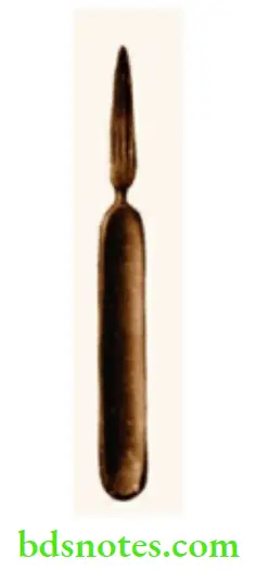
Aneurysm Needle
- This is a long instrument with an ‘EYE’ at the operating end.
- It is called an aneurysm needle because it is used to ligate the feeding artery in an aneurysm.
- This instrument, however, has limited use in present practice.
- During venesection or cut down, the silk suture can be threaded within the ‘eye’, passed around the vein and it is tied.
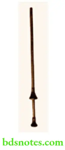
Trocar And Cannula
- This has two parts. The inner sharp part is the trocar and the outer blunt part is the cannula.
- It is used to drain hydrocoele fluid.
- Once the hydrocoele sac is delivered, it is punctured with a trocar and cannula, the trocar removed and the fluid drained.
- Make sure that the trocar and cannula are matched. Otherwise, injury to the deeper structures (testis) can occur.
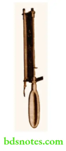
Humbly’s Knife
- This instrument has a handle and a long sheath.
- When in use, a disposable blade can be attached to it.
- The instrument is used to take skin grafts. Hence, it is also called a skin grafting knife.
- To facilitate the exact thickness of the skin to be removed, there is a screw at the operating end, with which, prior adjustment is done.
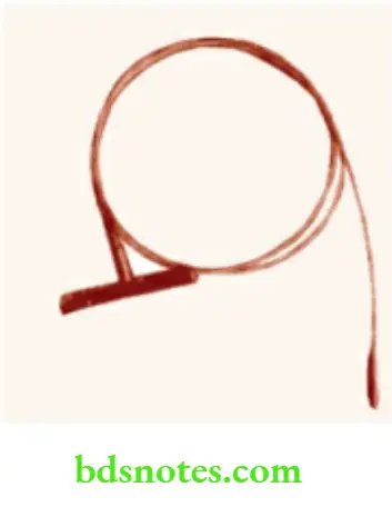
Myer’s Metal Stripper
- This is a long metallic chain or a stripper used in varicose vein surgery.
- It has a handle that is T-shaped and a blunt ‘advancing’ end that enters the vein. Once this end comes out of the cut end of the vein, a medium sized head is connected to it.
- With gentle force, (traction) exerted on the handle, the varicose vein can be stripped.
- Hence, it is also called a vein stripper.
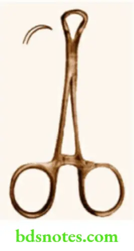
Towel Clip
- This instrument has a ratchet and the operating end is sharp.
- This is available in different sizes.
- Once the part is cleaned and draped, the clips are used to hold the towels in place.
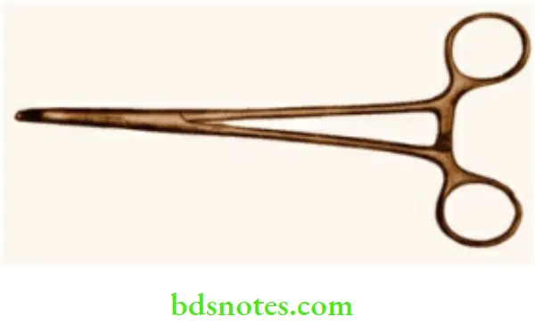
Right-Angled Forceps—Lahey’s Forceps
- This is a long instrument with a right angle at the operating end.
- This instrument is extremely useful in ligating the major vascular pedicles, e.g. superior thyroid pedicle—Thyroidectomy
- Cystic artery—Cholecystectomy
- Lumbar veins—Lumbar sympathectomy.
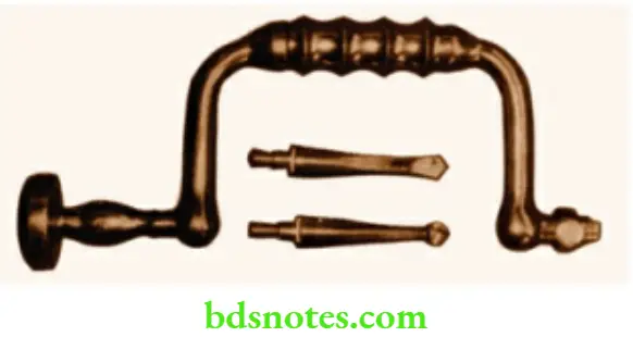
Hudson’S Brace And The Burr
- This is a heavy instrument with a brace and a burr (drill).
- This is used to create openings in the cranium so as to get access to the structures within.
- Thus once a ‘burr’ is made, drainage of blood, fluid, or pus can be done.
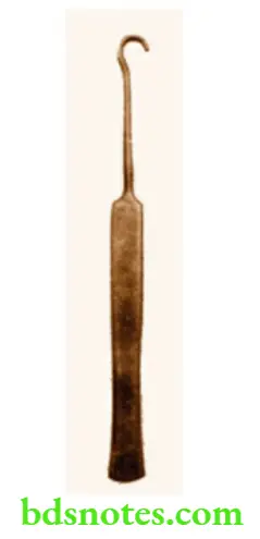
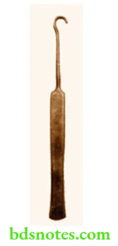
Cricoid Hook
- This has a broad handle and a thin shaft with a hook at the operating end.
- This is used to stabilize the trachea by hooking the cricoid cartilage ‘up’.
- This step is essential in children wherein veins are very superficial and can get injured easily when a child moves the head and neck. By stabilizing the trachea, it is easy to incise the trachea, without injuring vessels.
Tracheal Dilator
- This is an instrument with no ratchet at the non-operating end.
- The operating end is blunt and curved.
- The peculiarity of this instrument is that when the handle is opened, the operating end is closed and when the handle is closed, the operating end is opened.
- The tracheal dilator is used in the post-tracheostomy period when the tube has to be changed due to blockage. In such situations, once the tube is removed, the tracheal dilator is introduced, the opening in the trachea is kept open, and the new tube is introduced. However, once the track is formed, the tracheal dilator need not be used.
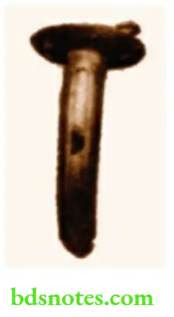
Metal Tracheostomy Tube
- This has two tubes, the inner long and the outer short tube.
- This has no cuff.
- Once the tube is introduced, the tape is passed around the neck, passed through the opening, and tied so as to keep the tube in place.
- If this tube is blocked, the inner tube can be removed, cleaned, and reintroduced.
- Metal tracheostomy tubes are useful as permanent tracheostomy tubes.
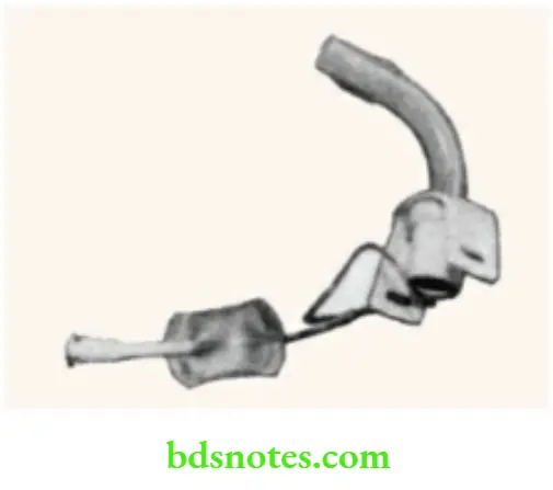
Cuffed Tracheostomy Tube
- This is made of polyvinyl chloride. It is a single tube.
- Once the tube is introduced within the trachea, the cuff is inflated by using 5–8 ml of air.
- The cuff prevents leakage of air and prevents acid aspiration syndrome—(Mendelson’s syndrome).
- If this tube is blocked, it is an emergency. In such cases, the tube has to be cleaned and mucus plugs have to be removed. Otherwise, the tube is removed, the tracheal opening is kept open with the help of a tracheal dilator, and a new tube is introduced. Alternatively, endotracheal intubation may need to be done to ensure patency of the airway.
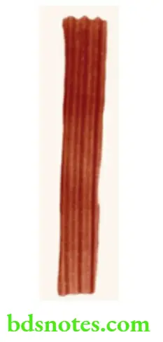
Corrugated Red Rubber Drain
- It is made of red rubber. It has corrugations on both sides. Whenever a major surgery is done, some amount of blood loss or anastomotic leakage is expected. This drain is used so that fluid can escape freely outside.
- Thus, it is used after thyroidectomy, gastrectomy, cholecystectomy, etc. The drain is removed after it stops draining.
- Usually, it takes about 3–5 days. After laparotomy for peritonitis, these drains are used to prevent residual abscesses in the postoperative period.
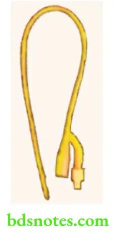
Foley’s Self-Retaining Urinary Catheter
- This is made of latex with silicon coating. At the tip, there is a bulb, the capacity of which is written at the other end.
- Before inflating the bulb, one must make sure that the catheter is in the urinary bladder, not in the urethra. This is assessed by the free flow of urine.
- After introducing the catheter, the bulb is inflated using saline. Thus, it becomes self-retaining. After usage, it is removed by deflating the bulb. It can also be used to drain the peritoneal cavity as in biliary peritonitis. An inflated bulb compresses the prostatic bed and controls bleeding after prostatectomy.
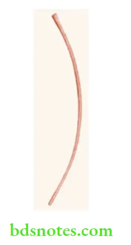
Red Rubber Catheter
This is used to drain urine temporarily. It causes urethritis if it is left long in the urinary bladder. Once the urine is emptied, it is removed. It is not a self-retaining catheter. Not routinely used nowadays because of the availability of Foley catheters. It is stiffer than Foley’s catheter.
Hence, in cases of stricture urethra where a Foley catheter cannot be passed, a red rubber catheter may be used.
Procedure of Urinary Catheterisation
- First, wash your hands with soap and running water. Spirit can be applied. Then, wear sterile gloves.
- External genitalia are cleaned with mild antiseptic agents such as Savlon. Do not use strong iodine-containing solutions. In males, the prepuce is retracted and smegma is removed if there is any.
- Identify the external urinary meatus. In males about 5 ml of lignocaine jelly is injected into the penile urethra, slowly. Wait for 2 minutes.
- A sterile catheter is taken out from the pack. It is lubricated with lignocaine jelly and slowly introduced into the penile urethra and advanced further into the urinary bladder.
- In females, once an external urinary opening is identified, the catheter is lubricated and introduced into the urethra.
- Once the catheter reaches the bladder, there is a free flow of urine.
- Now the bulb is inflated with 10 ml of air to make it self-retaining.
- The catheter is connected to a sterile bag for continuous drainage.
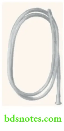
Ryle’s Tube (Nasogastric Tube)
- This is also called a nasogastric tube. At the end of this tube, there are lead shots. After introduction within the stomach, its position is confirmed by pushing 5–10 ml of air and auscultating in the epigastrium or aspirating gastric juice. It is a long tube having 3 marks.
- When the tube is passed upto the 1st mark, it enters the stomach. Usually, it is passed upto the 2nd mark.
- The life-saving use of Ryle’s tube is in acute gastric dilatation. In the volvulus of the stomach, it is impossible to pass Ryle’s tube.
- Ryle’s tube is used to aspirate gastric contents as in intestinal obstruction or pyloric stenosis, to diagnose GI hemorrhage, and also to provide enteral feeding in patients who cannot naturally, e.g. comatose or tetanus patients.

Leave a Reply