Spinal Cord
Question 1: What is the spinal cord? List its functions.
Answer. The spinal cord is a long lower cylindrical part of the central nervous system. It is about 45 cm long and lies in the upper two-thirds of the vertebral canal. It extends from the lower border of the medulla oblongata to the lower border of the L1 vertebra. It encloses the central canal of the spinal cord which contains CSF. It gives off 31 pairs of spinal nerves.
Functions Of Spinal Cord
- Transmission of sensory information from most of the body to the brain
- Transmission of motor information from the brain to the body
- Execution of simple reflexes
Question 2: Enumerate the main ascending and descending tracts present within the spinal cord.
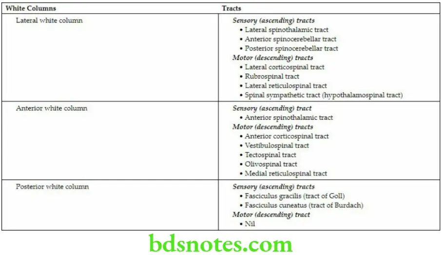
Answer. In each half of the spinal cord, the white matter is divided into three regions called white columns.
The important ascending and descending tracts in each white column of the spinal cord.
Ascending and Descending Tracts in White Columns
The main ascending and descending tracts as seen in the transverse section of the spinal cord.
Question 3: Enumerate the arteries supplying the spinal cord.
Answer. The spinal cord is supplied by the following arteries.
Anterior Spinal Artery
It is the artery formed by the union of anterior spinal branches of the vertebral arteries. It descends on the front of the spinal cord in the anterior median fissure.
Posterior Spinal Arteries
These are two posterior spinal arteries, one on each side. They are the branches of vertebral arteries. Each posterior spinal artery divides into two branches, which descend one on either side of the dorsal nerve roots of the corresponding side in the posterolateral sulcus.
Radicular Arteries (segmental arteries)
They run along the spinal nerve roots to reach the spinal cord. Their sources of origin vary in the different regions. From above downwards, they are branches of the deep cervical and ascending cervical in the cervical region, posterior intercostals in the thoracic region, and subcostal and upper lumbar arteries.
The segmental/radicular arteries at the T1 and T11 spinal segment levels are large and are termed arteria radicularis magna.
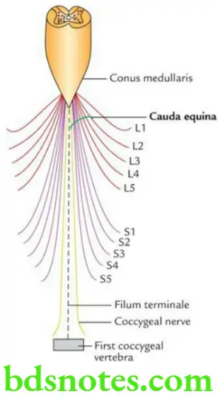
Question 4: Write a short note on cauda equina.
Answer. It is a leash of nerve roots of the lumbar (except L1), and sacral and coccygeal nerves around the filum terminale. It is called cauda equina because of its fancied resemblance to the tail of a horse (cauda = tail, equina = horse).
Question 5: Write a short note on the lateral spinothalamic tract.
Answer. It is a part of the spinothalamo-cortical pathway. It carries pain and temperature sensations from the opposite side of the body to the brain.
- First-order neurons are found in the dorsal root ganglia. Their central processes (axons) enter the spinal cord through the lateral division of the dorsal root of the spinal nerve and relay in the posterior horn of the spinal cord.
- Second-order neurons are found in the posterior horn. Their axons cross to the opposite side in the anterior white commissure and ascend in the contralateral white column (lateral spinothalamic tract). These axons terminate in the thalamus (ventral posterolateral [VPL] nucleus).
- Third-order neurons are found in the VPL nucleus of the thalamus. They project through the posterior limb of the internal capsule to the primary somatosensory cortex (Brodmann areas 3, 1, and 2).
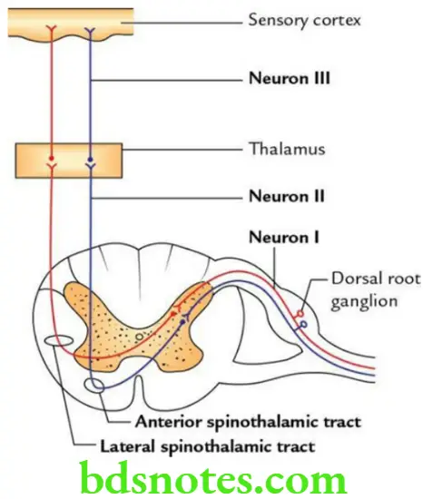
Applied anatomy The damage of the lateral spinothalamic tract results in contralateral loss of pain, touch (simple/crude), and temperature sensations.
Question 6: Write a short note on the ventral spinothalamic tract.
Answer. The ventral spinothalamic tract carries light/simple touch (crude touch), pressure, and itching sensations from the opposite half of the body.
The course of this tract is the same as that of the lateral spinothalamic tract, except that the second-order neurons after crossing to the opposite side ascend in the contralateral ventral white column of the spinal cord.
Ventral Spinothalamic Tract Applied Anatomy The damage of the ventral spinothalamic tract leads to loss of light touch (crude touch) and pressure on the opposite side of the body below the level of the lesion.
Question 7: Write a short note on the dorsal column–medial lemniscal pathway.
Answer. The dorsal column–medial lemniscal pathway carries proprioceptive sensations (e.g. muscle and joint sense, fine touch, vibration sensations) from the opposite side of the body.
- First-order neurons are located in the dorsal root ganglia. Their axons enter the spinal cord through the medial root of the spinal nerves, ascend in the ipsilateral dorsal white column as fasciculus gracilis and fasciculus cuneatus, and terminate in gracile and cuneate nuclei, respectively, located in the caudal part of the medulla.
- Second-order neurons are located in the gracile and cuneate nuclei of the medulla. Their axons (internal arcuate fibers) decussate with those of opposite sides in the midline. After decussation, they form a compact fiber bundle (medial lemniscus), which ascends in the contralateral half of the brainstem and terminates in the ventral posterolateral (VPL) nucleus of the thalamus.
- Third-order neurons are located in the VPL nucleus of the thalamus. Their axons project to the primary somatosensory cortex.
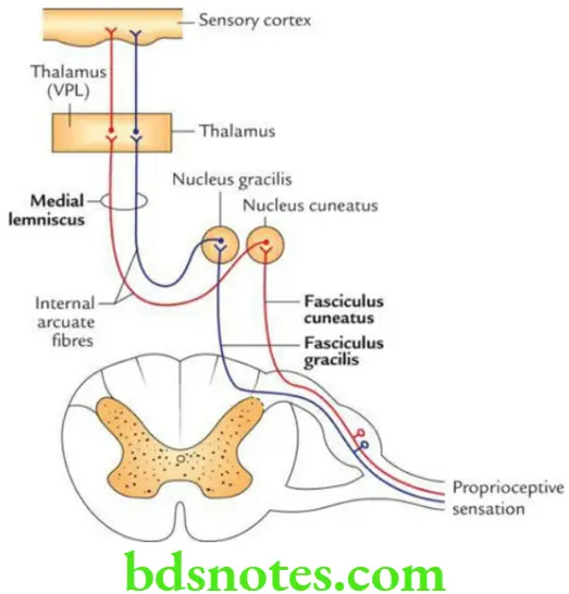
Applied Anatomy
The damage of the dorsal column–-medial lemniscal pathway above the sensory decussation causes contralateral loss of proprioceptive sensations, while below the sensory decussation, it causes ipsilateral loss of proprioceptive sensations.
Question 8: Describe the corticospinal (pyramidal) tract in brief and discuss its applied anatomy.
Answer. The pyramidal tract is a motor tract consisting of both corticospinal and corticonuclear tracts. However, conventionally it refers to only the corticospinal tract.
Origin, Course, And Termination
- Most of the fibers of the pyramidal tract arise from pyramidal cells of motor and premotor areas (areas 4 and 6) of the cerebral cortex.
- These fibers descend and traverse the following parts of the CNS in succession, viz. corona radiata, internal capsule (anterior two-thirds of the posterior limb and genu), crus cerebri (middle three-fifth), basilar part of pons and pyramid of the medulla.
Read And Learn More: Selective Anatomy Notes And Question And Answers
Note that after emerging from pons, they condense to form pyramid-shaped bundles in the upper part of the medulla oblongata.
In the lower part of the medulla oblongata, about 70%–80% of fibers of the pyramidal tract cross to the opposite side and then descend in the lateral white column of the spinal cord on the opposite side as lateral corticospinal/crossed pyramidal tract and terminate on the anterior horn cells.
About 20%–30% of fibers of the pyramidal tract do not cross to the opposite side and descend in an uncrossed pyramidal tract/anterior corticospinal tract in the anterior white column of the spinal cord of the same side. These fibers finally also cross to the opposite side and terminate on the anterior horn cells.
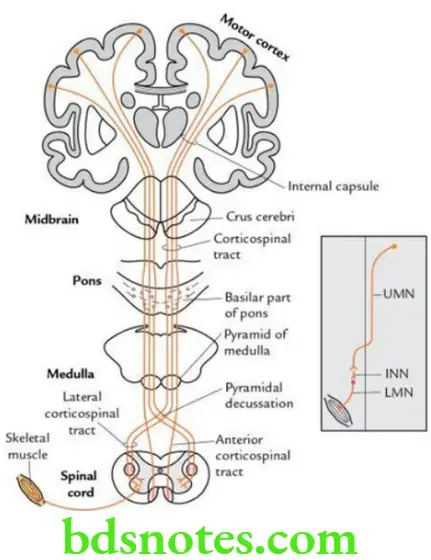
Corticospinal Tract Function
The pyramidal tract is concerned with voluntary movements of the body.
Corticospinal Tract Applied anatomy
Lesion of pyramidal tract: It produces upper motor neuron (UMN) type of paralysis.
If the lesion is above the level of motor decussation, it causes spastic paralysis on the opposite side of the body, i.e. contralateral hemiplegia; while if the lesion is below the level of motor decussation, it leads to ipsilateral hemiplegia.
- Effects of upper and lower motor neuron type of paralysis: The lesions of upper motor neurons (UMNs) lead to:
- Spasticity paralysis
- Increased muscle tone
- Exaggeration of tendon reflexes
- No wasting of muscles except disuse atrophy.
The above signs and symptoms occur due to the hyperactivity of LMNs, as the control of UMNs on LMNs is lost.
The lesions of lower motor neurons (LMNs) lead to:
- Flaccid paralysis
- Decreased muscle tone
- Loss of tendon reflexes
- Wasting of muscle i.e. muscle atrophy
All these signs and symptoms occur due to the loss of nerve supply of the muscle.

Leave a Reply