Skin Tumors
Question.1. Write short note on basal cell carcinoma.
Answer. It is the most common malignant skin tumor.
- It is a slow-growing neoplasm.
- It arises from basal cell of the pilosebaceous adnexa and occurs only on the skin.
Basal Cell Carcinoma Common Sites
- Majority of lesions are found on the face.
- Inner canthus and outer canthus of eyes.
- Eyelids.
- Bridge of the nose.
- Around nasolabial fold.
- These sites are the area where the tears roll down; hence,it is also called ‘Tear cancer’.
- Nodular: Common on face.
- Cystic/Nodulocystic
- Ulcerative
- Multiple: Associated with syndromes and other malignancies.
- Pigmented basal cell carcinoma: It mimics melanoma
- Geographical or forest fie basal cell carcinoma: Involve wide area with central scabbing and peripheral active proliferating edge.
- Basio-squamous: Behave-like squamous cell carcinoma and spread in lymph nodes
Read And Learn More: General Surgery Question And Answers
Basal Cell Carcinoma Etiology
- Ultraviolet rays are the main factor for its development.
- Fair skin is vulnerable for development of it.
- Prolonged administration of arsenic in the form of arsenical ointments.
Basal Cell Carcinoma Clinical Features
- It is common in males in middle aged and elderly
- The most common clinical presentation is an ulcer that never heals.
- It is common on the region of face, i.e. above the line drawn between angle of mouth and ear lobe.
- It can also present as a painless, fim, nodule.
- It is pigmented with fie blood vessels on its surface.
- The ulcer has raised and *beaded edge, induration may be present, and bleed on touch.
Basal Cell Carcinoma Spread
- It spread by local invasion.
- It slowly penetrates deep inside, destroying the underlying tissue-like bone, cartilage or even eyeball, hence, the name “rodent ulcer”.
Basal Cell Carcinoma Treatment
- It is radiosensitive. lf lesion is away from vital structure then curative radiotherapy can be given.
Radiotherapy is not given, once it erodes cartilage or bone. - Surgery: Wide excision (l cm clearance) with skin grafting,primary suturing or flp (Z plasty, rhomboid flp, rotation flp) is the procedure of choice.
- Laser surgery, photodymamic therapy, 5-florouracil local application.
- Cryosurgery.
- Microscopically Oriented Histographic Surgery (MOHS):
- It is useful to get a clearance margin and in conditions such as basal cell carcinoma close to eyes, nose or ear, to preserve more tissues.
- MOHS is becoming popular in basal cell carcinoma. Procedure is done by dermatological surgeon along with a histotechnician/histologist.
- Under local anesthesia, a saucerized excision of the primary tumour is done and quadrants of the specimen are mapped with diffrent colors.
- Specimen is sectioned by histotechnician from margin and depth and it is stained using eosin and hematoxylin. lt is studied by MOHS surgeon or histologist.
- Residual tumor from relevant mapped area is excised and procedure is repeated until clear margin and clear depth are achieved.
- Clearance must be complete and proper in basal cell carcinoma.
Question.2. Write short note on epithelioma.
Answer. It is also called “squamous cell carcinoma”.
- It is the second common malignant tumor of skin after basal cell carcinoma.
- It arises from prickle cell layer of the skin.
- It usually affcts elderly males.
Epithelioma Clinical Features
- It is an ulcerative or cauliflwer-like lesion.
- Edges are *everted and *indurated.
- Base is indurated and it may be subcutaneous tissue,muscle or bone.
- Floor contains cancerous tissue, which look like granulation tissue.
- It is pale, *friable, bleed easily on touch.
- Surrounding area is also indurated
- Mobility is usually restricted.
Epithelioma Investigations
- Wedge biopsy from edge.
- FNAC from lymph node
- USG/CT scan to identify the nodal disease
- MRI to identify local extension.
Epithelioma Treatment
- Radiotherapy using radiation needles, moulds, etc. is given.
- Wide excision, 2 cm clearance followed by skin grafting or flps.
- Wide excision should show clearance both at margin as well as in the depth.
- If muscle, fascia, cartilage are involved, it should be cleared.
- Reconstruction is usually done by primary split skin grafting. Delayed skin grafting can also be done once wound granulates well.
- Often flops of different type are needed depending on the site of lesion.
- Amputation with one joint above.
- For lymph nodes, block dissection of the regional lymph nodes is done.
- Curative radiotherapy is also useful in tumors which are not adherent to deeper planes or cartilage as squamous cell carcinoma is radiosensitive.
- It is also useful in recurrent squamous cell carcinoma and in patients who are not fi for surgery.
- A dose of 6000 cGy units over 6 weeks; 200 units/day is used. Recurrence after radiotherapy is treated by surgical-wide excision.
- In advanced cases with fied lymph nodes, palliative external radiotherapy is given to palliate pain, function and bleeding.
- Chemotherapy is given using methotrexate, vincristine, bleomycin.
- Field therapy using cryoprobe or topical florouracil or electrodesiccation.
Question.3. Write short note on neurofibroma.
Answer. Neurofibroma is a benign tumor arising from connective tissue of a nerve containing ectodermal neural and mesodermal connective tissue components.
Types of Neurofiroma
Nodular:
- Single, smooth, fim, tender swelling which moves horizontally or perpendicular to direction of nerve.
Plexiform:
- Occurs along distribution of trigeminal nerve in skin of face.
- Attain enormous size with thickening of skin which hang downwards.
Von-Recklinghausen’s Disease:
- Inherited disease with multiple neurofiromas in body.
- It can be cranial, spinal or peripheral
- Associated with pigmented spots on skin, i.e. Caféau-lait spots.
Elephantiasis:
- Origin is congenital and involve limbs.
- Skin of limb is thickened, dry and coarse.
Cutaneous:
- Small, multiple, firm/hard nodules arising from terminal ends of dermal nerves
- It can be pedunculated or sessile.
Neurofibroma Clinical Features
- Mild pain or painless swellings seen in subcutaneous and cutaneous plane with tingling, numbness and paresthesia.
- Most commonly affected sites are trunk, face and extremities
- Sessile or pedunculated elevated small nodules of various sizes
- Majority ofpatients have asymmetric areas ofpigmentation known as Café-au-lait spots. They are smooth-edge dark brown macules.
- Lisch nodules are present which are translucent brown pigmentation on iris.
- Crowe’s sign is present, i.e. axillary freckling, brown spot on skin.
Neurofibroma Complications
- Cystic degeneration is present.
- Spinal and cranial neurofiromas can cause neurological defiits.
- Spine dumb-bell tumor lead to compression of spinal cord and paralysis of limb.
- Hemorrhage in tissues.
Neurofibroma Treatment
Excision is done.
Question.4. Describe features of mole turning into melanocarcinoma.
Answer.
Following are the Features of mole turning into Melanocarcinoma.
- Lesion show superfiial radial growth pattrn.
- Lesion becomes ulcerated and growing day by day in size.
- Fungating growth is present associated with the bleeding.
- It leads to destruction of the underlying bone
- Lesion is fim on palpation
- Borders of lesion are erythematous.
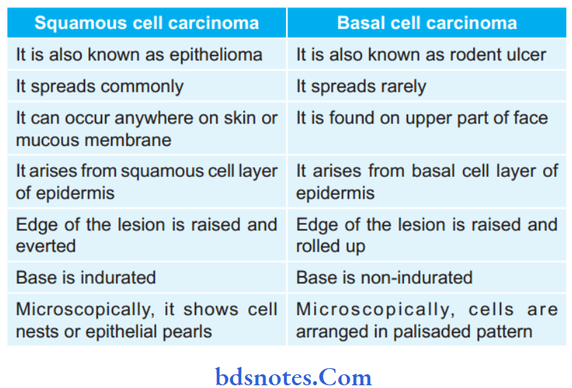
Question.5. Write difference between squamous cell carcinoma and basal cell carcinoma.
Answer. Following are the differences between squamous cell carcinoma and basal cell carcinoma:


Leave a Reply