Skeletal Maturity Indicators
Question 1. Write short note on Fishman’s skeletal maturity indicators.
Answer. Leonard S Fishman in 1982 had proposed this system.
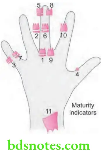
- Fishman had used four anatomical sites, i.e. location on thumb, third figer, fit figer and radius.
- Eleven discrete adolescent skeletal maturity indicators covering entire period of adolescent development are described.
- Fishman system of interpretation uses four stages of bone maturation which are:
- Epiphysis equal in width to diaphysis
- Appearance of adductor sesamoid of thumb
- Capping of epiphysis
- Fusion of epiphysis.
Read And Learn More: Orthodontics Question And Answers
- Following are the Fishman’s skeletal maturity indicators:
-
- S.M.I 1: The third finger proximal phalanx shows equal width of epiphysis and diaphysis.
- S.M.I 2: Width of epiphysis is equal to that of diaphysis in the middle phalanx of third finger.
- S.M.I 3: Width of epiphysis is equal to that of diaphysis in the middle phalanx of fit finger.
- S.M.I 4: Appearance of adductor sesamoid of the thumb
- S.M.I 5: Capping of epiphysis seen in distal phalanx of third finger.
- S.M.I 6: Capping of epiphysis seen in middle phalanx of third finger.
- S.M.I 7: Capping of epiphysis seen in middle phalanx of fit finger.
- S.M.I 8: Fusion of epiphysis and diaphysis in the distal phalanx of third figer.
- S.M.I 9: Fusion of epiphysis and diaphysis in the proximal phalanx of third figer.
- S.M.I 10: Fusion of epiphysis and diaphysis in the middle phalanx of third figer.
- S.M.I 11: Fusion of epiphysis and diaphysis is seen in the radius.
S.M.I denotes skeletal maturity indicator.

Question 2. Write short note on CVMI.
Or
Write short answer on CVMI, (Cervical vertebral maturation indicator)
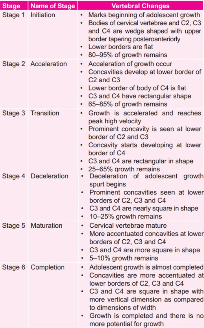
Answer. Full form of CVMI is cervical vertebrae maturity indicator.
- CVMI was given by Hassel and Farman.
- It is a skeletal maturity indicator.
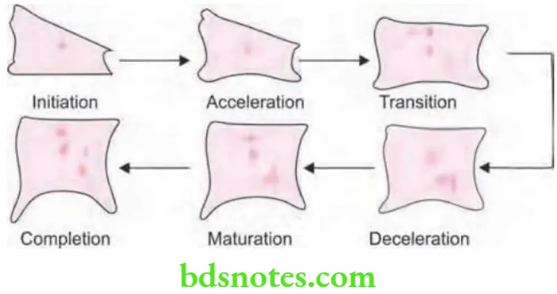
- Shapes of cervical vertebrae shows difference during each level of skeletal development.
- Six stages are described in the vertebral development which are:


Question 3. Write short note on carpal index.(Oct 2015, 5 Marks)
Answer. Carpal index denotes skeletal maturity.
- Carpal bones were fist named by Lyser in 1683.
- Carpals are the bones in hand-wrist region.
- Carpals are the multiple small bones in hand-wrist region. Carplals follow a pattern in ossification along with union of epiphysis and diaphysis.
- For carpal index left hand-wrist region is used.
- PA view is taken to record the hand-wrist region.
- Each hand-wrist region has 8 carpals, 5 metacarpals, 14 phalanges which form total 27 bones.
- Carpal bones are arranged in distal and proximal rows. Distal row consists of trapezium, trapezoid, capitate, hamate; Proximal row consists of scaphoid, lunate, triquetral, pisiform.
- The above irregular bones lie between long bones of forearm and metacarpals.
- Each of the carpal bone ossifies from one primary center which appears in a particular predictable pattern.
Question 4. Write short note on hand-wrist radiograph.
Answer. Hand-wrist region is made by multiple small bones which show a predictable and scheduled pattern of appearance, ossification and union from birth to maturity. So by merely comparing patient’s hand wrist radiograph with standard radiographs which represent different skeletal ages, one can be able to determine skeletal maturation status of that individual.
Most commonly used methods to access skeletal maturity by using hand-wrist radiographs are:
- Bjork, Grave and Brown method
- Hagg and Taranger method
- Fishman’s skeletal maturity indicator
- Atlas method by Greulich and Pyle
Bjork, Grave and Brown Method
They divided skeletal development in nine stages. All of these stages represent a level of skeletal maturity.
Appropriate chronological age for each of the stage was given by Schopf
Following are the stages:
- Stage 1 (Males 10.6 years, Females 8.1 years): Both epiphysis and the diaphysis of proximal phalanx of the index finger are equal. This occurs approximately 3 years before the peak of pubertal growth spurt.
- Stage 2 (Males 12.0 years, Females 8.1 years): Epiphysis and diaphysis of middle phalanx of the index finger are equal. It is noticed before the peak of pubertal growth spurt.
- Stage 3 (Males 12.6 years, Females 9.6 years): It is characterized by presence of three areas of ossification i.e. hamular process of hamate exhibits ossification, ossification of the pisiform, epiphysis and diaphysis of radius are equal.
- Stage 4 (Males 13 years, Females 10.6 years): This stage marks the beginning of pubertal growth spurt which is characterized by the initial mineralization of ulnar sesamoid of thumb and increase in ossification of hamular process of hamate bone.
- Stage 5 (Males 14 years, Females 1 year): It heralds the peak of pubertal growth spurt. Capping of the diaphysis by epiphysis is seen in middle phalanx of third finger, proximal phalanx of thumb and radius.
- Stage 6 (Males 15 years, Females 13 years): It signifies the end of pubertal growth spurt. It is characterized by union between epiphysis and diaphysis of distal phalanx of middle finger.
- Stage 7 (Males 15.9 years, Females 13.3 years): Union of both epiphysis and diaphysis of proximal phalanx of litt
- e finger occur. This is seen a year after growth spurt.
- Stage 8 (Males 15.9 years, Females 13.9 years): It shows fusion between epiphysis and diaphysis of middle phalanx of middle finger.
- Stage 9 (Males 18.5 years, Females 16.0 years): It is the last stage and signifies end of skeletal growth. This is characterized by fusion of epiphysis and diaphysis of radius.
Maturation Assessment by Hagg and Taranger
Development of skeleton in hand and wrist is analyzed by annual radiographs which are taken between ages of 6 and 18 years, by assessing the ossifiation of ulnar sesamoid of metacarpophalangeal joint of fist figer (S) and certain specific stages of three epiphyseal bones i.e. middle and distal phalanges of third figer (MP3 and DP3) and distal epiphysis of radius (R)
Sesamoid
It is attained during the acceleration period of the pubertal growth spurt. i.e. the onset of peak height velocity.
Third Finger Middle Phalanx (MP3)
- Stage 6: Here epiphysis as wide as metaphysis. 40% of person attains this stage before the onset of peak height velocity. At peak height velocity many of the persons attain this.
- Stage 6: Here epiphysis is wide as metaphysis and there is distinct medial and/or lateral border of epiphysis which forms the line of demarcation at right angle to the distal border. About 90% of persons attain this stage 1 year before peak height velocity.
- Stage 7: Sides of epiphysis are thickened and there is capping of the metaphysis which form the sharp edge distally at either one end or at both the ends. About 90% of persons attain it at or 1 year after peak height velocity.
- Stage 8: Fusion of the epiphysis and metaphysis has begun and is attained after peak height velocity 90% of girls and all the boys attain it after peak height velocity but before end of pubertal growth spurt.
- Stage 9: Fusion of both the epiphysis and metaphysis is completed. All persons except few girls have ended pubertal growth spurt.
Third Finger Distal Phalanx
DP3 – I: Completion of fusion of epiphysis and metaphysis. It signifies fusion of epiphysis and metaphysis and is attained at the deceleration period of pubertal growth spurt by all persons.
Radius
- R – I: Beginning of fusion of epiphysis and metaphysis. It is attained one year before or at end of growth spurt by about 80% of girls and about 90% of boys.
- R – IJ At this stage fusion is almost completed but there is still a small gap at either one or at both the margins.
- R – J: It is characterized by the fusion of epiphysis and metaphysis
Above stages were not attained before end of peak height velocity by any of the person.
Atlas Method by Greulich and Pyle
- Gruelich and pile publish an atlas containing pictures of wrist – hand for different chronological ages for both males and females.
- Radiographs of patient’s are matched with one of the photograph in the atlas which is representative of particular skeletal age.
Indications of Hand-wrist Radiograph
- In cases with major discrepancies between chronological and dental age.
- In cases where prediction of pubertal growth spurt is required.
- In cases with skeletal Class II and Class III malocclusion before beginning their treatment so that their growth potential is assessed.
- For skeletal age assessment to study growth of an individual.
- Indicated in research studies to make out the effect of hereditary and environment on dentofacial growth.
- In patients with skeletal malocclusion and need orthognathic surgery.
Question 5. Write short note on skeletal maturity indicator.
Answer. Various skeletal maturity indicators are:
- Hand wrist radiographs: For details refer to Ans 4 of same chapter
- Skeletal maturation evaluation by using cervical vertebrae:
- Tooth mineralization as an indicator of skeletal maturity
Tooth Mineralization as an Indicator of Skeletal Maturity
Dental development is widely investigated as the potential predictor of skeletal maturity level.
Ability to assess skeletal maturity by developmental stage of dentition via the examination of panoramic radiograph offer various advantages over conventional hand – wrist radiographic method
Demerjian, Goldstein and Tanner describe tooth mineralization as an indicator of skeletal maturity.
Dental Calcification Stages Using Demerjian Index (DI)
- Stage A: Calcification of the single occlusal point without fusion of different calcifications.
- Stage B: Fusion of the mineralization points, now contour of occlusal surface is recognizable.
- Stage C: Formation of enamel is completed at occlusal surface, dentine formation is commenced. Pulp chamber is curved and no pulp horns are visible.
- Stage D: Completion of crown formation to the level of cementoenamel junction. Commencement of root formation. Pulp horns begin to differentiate, walls of pulp chamber remains curved.
- Stage E: Length of the root remains shorter as compared to height of crown. Walls of pulp chamber are straight and pulp horns become more differentiated than in previous stages. In molars the root bifurcation starts to calcify.
- Stage F: Walls of the pulp chamber form an isosceles triangle, and the root length is equal to or greater than crown height. Development of bifurcation in molars sufficiently to give roots a distinct form.
- Stage G: Walls of root canal are now parallel, apical end is partially open. In molars only distal root is rated.
- Stage H: Complete closure of root apex. Periodontal membrane surrounds the root and apex is uniform in width throughout.

Leave a Reply