Scalp Temple And Face
Question 1. Describe the scalp under the following headings:
- Definition,
- Layers,
- Arterial supply,
- Venous drainage,
- Nerve supply and
- Applied anatomy.
Answer.
“Early warning signs of gaps in understanding scalp, temple, and face basics: Common questions”
The soft-tissue structures covering the vault of the skull form the scalp. The boundaries of the scalp are as follows.
Anteriorly: Superciliary arches of the frontal bone.
Posteriorly: Superior nuchal lines of the occipital bone.
On each side: Superior temporal line.
“Understanding the layers and structures of the scalp, temple, and face through FAQs: Q&A explained”
Read And Learn More: Selective Anatomy Notes And Question And Answers
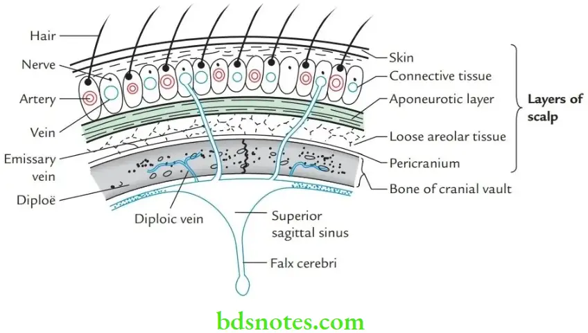
Layers of scalp The scalp consists of five layers, which can be easily remembered by the initial letter of each layer, i.e. SCALP.
- S: Skin
- C: Connective tissue
- A: Aponeurosis (epicranial aponeurosis)
- L: Loose areolar tissue
- P: Pericranium (outer periosteum)
“Importance of studying scalp, temple, and face anatomy for medical and dental students: Questions explained”
Mnemonic: SCALP
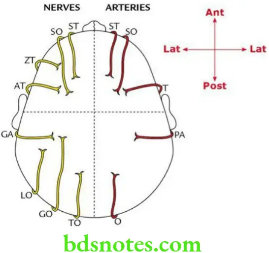
Scalp Temple Arterial supply Each lateral half of the scalp is supplied by five arteries:
“Common challenges in understanding scalp, temple, and face anatomy effectively: FAQs provided”
Venous drainage Each lateral half of the scalp is supplied by five veins:
- Three in front of the auricle
- Supratrochlear vein
- Supraorbital vein
- Superficial temporal vein
- Two behind the auricle
- Posterior auricular vein
- Occipital vein
Nerve supply Sensory Each lateral half of the scalp is supplied by eight nerves:
- Four in front of the auricle (i.e. anterior quadrant)
- Four behind the auricle (i.e. posterior quadrant)
- Great auricular, GA (C2, C3)
- Lesser occipital, LO (C2, C3)
- Greater occipital, GO (C2)
- Third occipital, TO (C3)
“Factors influencing success with scalp, temple, and face anatomy knowledge: Q&A”
Motor Each lateral half of the scalp is supplied by two nerves – one in front of the auricle and one behind the auricle.
- In front of the auricle
- Temporal branch of the facial nerve
- Behind the auricle
- Posterior auricular branch of the facial nerve
Scalp Temple Applied anatomy Wounds of the scalp bleed profusely because:
“Steps to explain the anatomy of the scalp, temple, and face: Layers vs blood supply vs innervation: Q&A guide”
- Walls of torn vessels fail to retract because they adhere to the dense connective tissue.
- It is profusely supplied with blood.
- Wounds of the scalp heal quickly because of profuse blood supply.
- The scalp is the most common site of sebaceous cysts because it contains a maximum number of sebaceous glands as compared to anywhere else in the body.
“Role of the temporal artery in supplying blood to the temple region: Questions answered”
- A dangerous layer of the scalp: The layer of loose areolar tissue (i.e., the fourth layer of the scalp) is called the dangerous layer of the scalp because if the pus collects in this layer, the infection from here may travel through emissary’s veins into the intracranial dural venous sinuses causing their thrombosis and associated meningitis.
- Black eye: If blood collects in the fourth layer of the scalp due to trauma on the head. It tracks anteriorly in the subcutaneous tissue of eyelids for a couple of days where it clots. As a result, the eyelids appear black (black eye).
Leave a Reply