Salivary Glands
Question 1. Classify salivary glands and write about the functions of saliva.
Answer:
Salivary Glands Classification:
1. Based on the size of the glands:
- Major glands:
- Parotid gland.
- Submandibular gland.
- Sublingual gland.
- Minor glands:
- Glossopalatine.
- Palatine.
- Labial and lingual glands.
- Posterior lingual glands.
- Von Ebner’s glands.
Read And Learn More: BDS Previous Examination Question And Answers
2. Based on the location of the glands:
- Labial glands.
- Lingual glands.
3. Based on the nature of secretion:
- Serous glands:
- Parotid gland.
- Von Ebner’s gland.
- Mucous gland:
- All minor glands except von Ebner’s gland
- Mixed:
- Submandibular gland.
- Sublingual gland.
Four Functions Of Saliva
1. Functions of Saliva Protection of oral cavity:
- Flushes away nonadherent bacteria and other debris
- Clears away sugars thereby reducing the occurrence of acidogenic bacteria.
- Mucin in saliva.
- Lubricate the oral tissue.
- Prevents their adherence to each other.
- Forms barrier against noxious stimuli, microbial toxins, and minor trauma.
- Protects the oral tissue from chemical and thermal insults by reducing the concentration.
2. Functions of Saliva Buffering action:
- The components of saliva that help in buffering action are.
- Bicarbonate
- Phosphate
- Salivary proteins and
- Peptides.
- These protect the teeth from demineralization caused by acid produced by bacteria.
- Metabolism of salivary proteins and peptides increases the pH of salvia by providing urea and ammonia.
- In this increased pH, acidogenic bacteria cannot survive or ferment carbohydrates.
- Thus, they become unable to cause tooth decay.
3. Functions of Saliva Pellicle formation:
- Along with salivary glycoproteins and certain proline-rich proteins bind to the tooth surface, forming a thin film, the salivary pellicle.
4. Functions of Saliva Maintenance of tooth integrity:
- Saliva is supersaturated with calcium and phosphate ions.
- This reduces dissolution and increases the surface hardness of enamel, and resistance to demineralization.
- Remineralization is enhanced by fluorides in saliva.
5. Functions of Saliva Antimicrobial action:
- Salivary proteins like lysozyme, lactoferrin, and peroxidase have antimicrobial activity.
- Lysozyme hydrolyzes the polysaccharide of bacterial cell walls resulting in cell lysis.
- Antifungal action is exerted by statins, chromogranin A, and immunoglobulins.
- Mucins aggregate microorganisms.
- Major salivary immunoglobulin IgA (secretory) causes agglutination of specific microorganisms, preventing their adherence to oral tissues and forming clumps that are swallowed.
6. Functions of Saliva Tissue repair:
- Growth factors, peptides, and proteins present in saliva promote tissue growth, differentiation, and wound healing.
7. Functions of Saliva Digestion:
- Salivary enzymes such as amylase and lipase begin the digestive process by solubilization of food substances.
- Amylase act on ingested carbohydrates and produce glucose and maltose.
- Lingual lipase initiates the digestion of dietary lipids, hydrolyzing triglycerides to monoglycerides and diglycerides, and fatty acids.
- The moistening and lubricative properties of saliva allow the formation and swallowing of a food bolus.
8. Functions of Saliva Taste:
- Saliva solubilizes food substances so that they can be sensed by taste receptors located in taste buds.
- Saliva helps in maintaining taste receptors.
9. Functions of Saliva Speech:
- Saliva keeps the oral tissue moist and well-lubricated which facilitates speech.
- It helps in vocalization and communication ability.
10.Functions of Saliva Mastication and deglutition:
- Allows the formation of bolus and facilitate deglutition.
- Saliva not only moistens the dry food but also reduces the temperature of the hot foods.
11. Functions of Saliva Excretion:
- Many substances from blood reach the saliva as a route of excretion.
- The low molecular weight serum constituents can be demonstrated in saliva.
- Infective agents from blood can reach the saliva.
- The nitrates in the food reach the saliva and are reduced to nitrites by microorganisms.
Question 2. Describe the composition, functions, and formation of saliva.
Answer:
Composition of saliva:
- Saliva contains 99% water and 1% organic and inorganic substances.
1. Composition of saliva – Inorganic substances:
- Main electrolytes Na, K, Ca, CI, HCO3, and HPO4
- Electrolytes in small
2. Composition of saliva – Organic substances:
- Secretory proteins.
- Amylase, proline-rich proteins, mucins, histatin, cystatin, peroxidase, lysozyme, lactoferrin.
- Enzymes ribonuclease, kallikrein, acid phosphatase.
- Serum constituents.
- Albumin, blood clot factors, immunoglobulins IgA, IgM, IgG
3. Composition of saliva – Small organic molecules:
- Glucose, amino acids, urea, uric acid, lipids, hormones.
4. Composition of saliva – Other components:
- EGF, insulin, cyclic adenosine monophosphate.
Functions of saliva:
1. Protection of oral cavity:
- Provides washing action by flushing nonadherent bacteria and other debris.
- Mucins lubricate the oral tissues and protect against thermal and chemical insult.
2. Buffering action:
- Saliva maintains its pH through its bicarbonate and phosphate ions.
- It neutralizes the acid produced by bacteria.
3. Pellicle formation:
- Salivary proteins bind to the tooth surface and form a salivary pellicle.
4. Antimicrobial action:
- Salivary proteins, mucins, and immunoglobulins in saliva contribute to its antimicrobial action.
5. Tissue repair:
- Growth factors, peptides, and proteins promote tissue growth, differentiation, and wound healing.
6. Digestion:
- Water and mucin help in bolus formation which facilitates swallowing.
7. Taste:
- Saliva solubilizes food substances that are sensed by taste receptors.
8. Speech:
- It lubricates the oral tissue which facilitates speech.
9. Excretion:
- Many substances from blood reach saliva and are used as a route of excretion.
10. Maintenance of tooth integrity:
- Saliva reduces dissolution, increases the surface hardness of enamel, and increases resistance to demineralization.
Mechanism Of Saliva Formation
Formation of Saliva:
- The formation of saliva occurs in two stages.
1. Formation of Saliva First stage:
- Cells of the secretory end pieces and intercalated ducts produce primary saliva, which is an isotonic fluid containing most of the organic components and all of the water that is secreted by the salivary glands.
2. Formation of Saliva Second stage:
- Primary saliva is modified as it passes through the striated and excretory ducts mainly by reabsorption and secretion of electrolytes.
- The final saliva that reaches the oral cavity is hypotonic.
Question 3. Describe briefly the ductal system of a salivary gland.
Answer:
Ductal system of salivary gland:
- The ductal system of salivary glands consists of hollow tubes.
- It is a varied network of tubules that progressively increase in diameter, beginning at the secretory end pieces and extending to the oral cavity,
- Three classes of ducts are present.
- Intercalated intralobular.
- Striated intralobular
- Excretory interlobular.
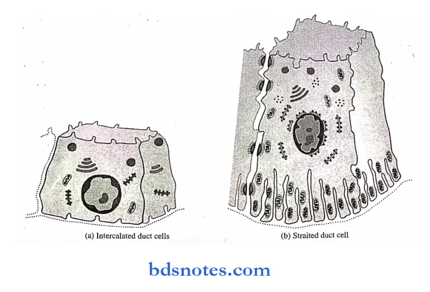
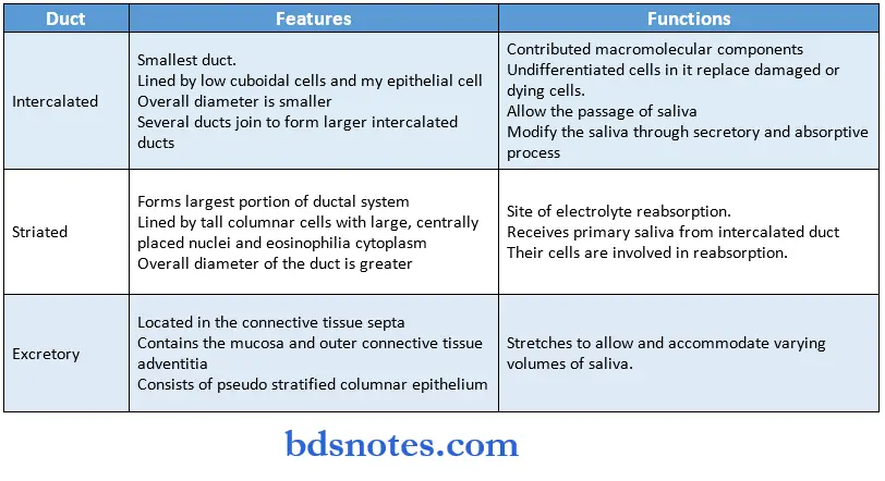
1. Intercalated duct:
- They are the smallest duct connecting secretory units to the striated duct.
- They are lined by a single layer of flow cuboidal cells and myoepithelial cell bodies.
- The overall diameter is smaller.
- Several ducts join to form larger intercalated ducts.
- Its length differs in major and minor salivary glands.
Ductal System of a Salivary Gland Functions:
- The intercalated ducts contributed macromolecular components stored in their secretory granules.
- Undifferentiated cells present in it may proliferate and undergo differentiation to replace damaged or dying cells.
- Allow the passage of saliva.
- Modify the saliva through the secretory and absorptive processes.
Mechanism Of Saliva Formation
2. Striated duct:
- Forms the largest portion of the ductal system.
- Receives primary saliva from the intercalated duct.
- They are lined by tall columnar cells with large, centrally placed nuclei and eosinophilic cytoplasm.
- The overall diameter of the duct is greater than that of the secretory end pieces and intercalated ducts.
- The combination of in-foldings and mitochondria accounts for the striations.
Striated Duct Functions:
- They are sites of electrolyte reabsorption, especially of sodium and chloride, and secretion of potassium and bicarbonate.
- Their cells are involved in active reabsorption.
3. Excretory ducts:
- They are located in the connective tissue septa between the lobules of the gland.
- It contains 2 layers.
- The mucosa
- Outer connective tissue adventitia.
- They consist of pseudostratified columnar epithelium which finally merges with the epithelium of the oral cavity.
- Cells seen in the excretory duct are:
- Basal cells with tonofilaments.
- Tuft cells with microvilli.
- Cells with pale cytoplasm and dense nuclear chromatin.
- Lymphocytes and macrophages.
- Dendritic cells; antigen-presenting cells.
Excretory Ducts Functions:
- The excretory duct stretches passively and allows and accommodates varying volumes of saliva.
- Intercalated duct cells
- Striated duct cell

Question 4. Classify salivary glands and describe the parotid gland briefly.
Answer:
Classification of salivary glands:
1. Based on the size of the glands:
- Major glands:
- Parotid gland
- Submandibular gland Sublingual gland
- Minor glands:
- Glossopalatine.
- Palatine
- Von Ebner’s gland
- Labial and lingual gland Posterior lingual gland.
2. Based on the location of the glands:
- Labial glands
- Lingual glands.
3. Based on the nature of secretion:
- Serous glands:
- Parotid gland
- Von Ebner’s gland
- Mucous glands:
- All minor glands except von Ebner’s gland.
- Mixed.
- Submandibular gland
- Sublingual gland.
Parotid gland:
Anatomy:
- It is the largest major salivary gland.
Parotid gland Parts:
- Superficial part.
- Located subcutaneously in front of the external ear.
- Deeper portion.
- Lies behind the ramus of the mandible.
Parotid gland Size:
- 5.8 cm craniocaudally.
- 3.4 cm ventrodorsally.
- Weight: Between 14 and 28 grams.
- Duct: Stensen’s duct.
- Runs forward across the masseter muscle, and turns inward at the anterior border of the masseter.
- It opens into the oral cavity at a papilla opposite the maxillary second molar.
- Accessory gland:
- Located just anterior to the superficial portion of the parotid gland.
Blood supply:
- From branches of the external carotid artery.
Nerve supply:
- Parasympathetic nerve supply from the glossopharyngeal nerve.
- Preganglionic fibers synapse in the otic ganglion.
- Postganglionic fibers reach the gland through the auriculotemporal nerve.
- The sympathetic innervations from the superior cervical ganglion.
Nerve supply Histology:
- Secretory end prices are spherical and serious.
- Acinar cells.
- Pyramidal shaped.
- Have a basally situated nucleus
- The nucleus is surrounded by a small, central lumen.
- The basal cytoplasm is basophilic.
- Secretory granules are acidophilic.
- Fat cell spaces are seen.
Ducts:
- Intercalated duct.
- Numerous and long.
- Slightly acidophilic.
- Consist of a simple columnar epithelium with round, centrally placed nuclei.
- Faint striations representation infoldings and mitochondria may be visible below the nucleus.
Question 5. Write briefly about the Sublingual Salivary Gland.
Answer:
- Sublingual salivary gland.
Sublingual Salivary Gland Anatomy:
- It is the smallest of the paired salivary gland.
Sublingual Salivary Gland Weight:
- Approximately 2 grams.
Sublingual Salivary Gland Location:
- In the anterior part of the floor of the mouth between the mucosa and mylohyoid muscle.
Sublingual Salivary Gland Ducts and their opening:
- Ducts of previous, small ducts open along the sublingual fold.
- Bartholin’s duct, a large duct opens at the sublingual caruncle.
Sublingual Salivary Gland Blood supply:
- Sublingual and submental arteries.
Sublingual Salivary Gland Nerve supply:
- Parasympathetic from VII cranial nerve, facial nerve.
- It reaches the gland via the lingual nerve after synapsing in the submandibular ganglion.
Sublingual Salivary Gland Lymphatic drainage:
- Submandibular lymph nodes.
Sublingual Salivary Gland Histology:
- It is a mixed gland, but mucous secretory units are more than the serous units.
- Serous end pieces are rare.
Sublingual Salivary Gland Acinar cells:
- Mucous cells are arranged in a tubular pattern
- Serous cell and pathway of synthesis, storage, and exocytosis of secretory protein.
- Rought endoplasmic reticulum synthesizing protein, 2Golgi complex transfer protein to transfuse, Immature granules, 4Mature granules with concentrated protein, Exocytosis
- Mucous cell and pathway of synthesis and exocytosis of mucous.
- Rough endoplasmic reticulum synthesizing mucous protein,
- Golgi complex transfer protein to transfuse,
- Formation of the mucous pool,
- Exocytosis of mucous
Question 6. Write briefly about the sublingual salivary gland.
Answer:
- Sublingual salivary gland.
Sublingual Salivary Gland Anatomy:
- It is the smallest of the paired salivary gland.
Sublingual Salivary Gland Weight:
- Approximately 2 grams.
Sublingual Salivary Gland Location:
- In the anterior part of the floor of the mouth between the mucosa and mylohyoid muscle.
Sublingual Salivary Gland Ducts and their opening:
- Ducts of previous, small ducts open along the sublingual fold.
- Bartholin’s duct, a large duct opens at the sublingual caruncle.
Sublingual Salivary Gland Blood supply:
- Sublingual and submental arteries.
Sublingual Salivary Gland Nerve supply:
- Parasympathetic from VII cranial nerve, facial nerve.
- It reaches the gland via the lingual nerve after synapsing in the submandibular ganglion.
Sublingual Salivary Gland Lymphatic drainage:
- Submandibular lymph nodes.
Sublingual Salivary Gland Histology:
- It is a mixed gland, but mucous secretory units are more than the serous units.
- Serous end pieces are rare.
Sublingual Salivary Gland Acinar cells:
- Mucous cells are arranged in a tubular pattern
- Pure serous acini are rare or absent.
- Serous demilunes may be present at the blind ends of the tubules.
Sublingual Salivary Gland Ducts:
- Intercalated and striated ducts are poorly developed.
- Intercalated ducts are short.
- Interlobular ducts are few in number.
- Ducts are lined by cuboidal or columnar cells.
- They lack the infolded basolateral membranes.
Question 7. Ductal system of major salivary glands.
Answer:
- The ductal system of salivary glands. consist of hollow tubes.
- Three classes of ducts are preset.
- Intercalated duct cells
- Striated duct cell

Question 8. Serous cell.
Answer:
Serous cell Structure:
- Typically spherical
- Consists of 812 cells surrounding a central lumen.
- Lumen has a finger-like extension located between adjacent cells called intercellular canaliculi.
- The nucleus is spherical and placed basally.
- The apical cytoplasm shows the accumulation of secretory granules.
- The basal cytoplasm contains numerous cisternae of rough endoplasmic reticulum, a large Golgi complex.
- The cell contains all typical cell organelles
- The secretory granules are 1 mm in diameter.
- The granules are zymogen granules and are formed by glycosylated proteins.
- Cell shows enzymatic activity.
- The mature granule stored at the apex of the cell is emptied into the lumen by exocytosis.
- The Golgi apparatus consists of 4 6 smooth surface saccules located apically and laterally to the cell nucleus.
- Cells also contain a good number of mitochondria, a powerhouse of the cells.
- Lysosomes are seen with hydrolytic enzymes, which help to destroy foreign material and warn out cell organelles.
- Bundles of tonofilaments, associated with desmosomes and microfilaments, may be seen in the cytoplasm.
Serous cell Functions:
- They are specialized for the synthesis, storage, and secretion of proteins. They secrete glycoproteins.
- Rought endoplasmic reticulum synthesizing protein, 2Golgi complex transfer protein to transfats,
- Immature granules,
- Mature granules with concentrated protein,
- Exocytosis


Question 9. Histology of mucous salivary gland
Answer:
- The sublingual salivary gland is a mucous salivary gland
- It contains predominately mucous secretory endpieces
- Mucous cells
- They are arranged in tubules
- Joined to each other by intracellular junctions
- Large amounts of secretory products are present at the apical cytoplasm
- The nucleus is oval or flattened in shape
- Located just above the basal membrane
- Serous demilunes
- These are crescent-shaped serous cells covering mucous cell
- Present at the blind end of tubules
- Serous cells
- Absent or rare
- Ducts
- Striated and intercalated ducts are poorly developed
- Mucous tubules open directly into ducts
Question 10. Histology of minor salivary gland
Answer:
- Located within the submucosa of the oral mucosa
- 12 mm in diameter
- Not encapsulated by connective tissue instead mix with it
- Contains a number of acini connected in a tiny lobule
- It may have a common excretory duct with another gland or may have its own excretory duct
- All minor salivary glands are mucous except von Ebner which is serious

Question 11. Serous and mucous cells
Answer:
- They are darkly stained
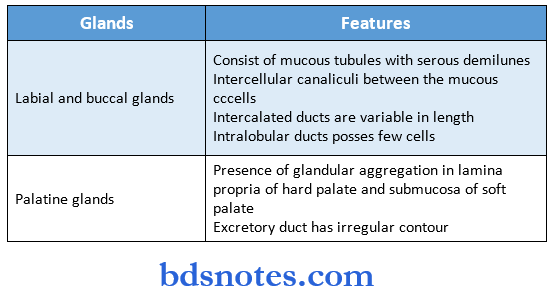
- Intercellular canaliculi between the mucous cells
- Intercalated ducts are variable in length
- Intralobular ducts possess few cells
- Presence of glandular aggregation in lamina propria of the hard palate and submucosa of the soft palate
- The excretory duct has an irregular contour
- Have rounded nuclei placed toward the base
- They are arranged in the form of rounded acini or roughly pyramid
- The base is towards the basement membrane and the apex is towards the lumen
- Apex shows microvilli and pinocytotic vesicles
- Lumen has intercellular secretory canaliculi
- Cytoplasm contains
- Small and homogenous secretory granules
- Prominent Golgi apparatus
- Abundant rough endoplasmic reticulum
- Mitochondria
- Lysosomes
- Microfilaments
Mucous cells:
- They are lightly stained
- The nucleus is flattened and present towards the basement membrane
- Cells are arranged in the form of tubules
- Cells lining the tubules are columnar
- Secretory granules are large and ill-defined
Question 12. Myoepithelial cell
Answer:
- They are stellate or spider-like cells with flattened nuclei and scanty cytoplasm
- Also known as basket cell
- The structure is similar to smooth muscle and contains actin and myosin
- They expel secretion by contraction.
- Present in intercalated and terminal ducts.
Myoepithelial cell Functions:
- Contractile process
- Accelerate outflow of saliva from acini
- Shorten and widen intercalated ducts
- Supports parenchyma
- Contribute to secretory pressure in the acini or duct
Question 13. Stenson’s duct
Answer:
- It is the main excretory duct of the parotid salivary gland
Stenson’s duct Course:
- Crosses masseter muscle
- Turns medially
- Penetrates buccinator muscle
Stenson’s duct Opening:
- At the papilla at the buccal mucosa opposite to the maxillary second molar
Stenson’s duct Size:
- 46 cm in length, 5 mm in diameter
- A small portion of the gland is accompanied by duct-forming accessory gland
Question 14. Striated duct
Answer:
- Striated ducts form the largest portion of the ductal system
- Receives primary saliva from intercalated duct
- They are lined by tall columnar cells with large, centrally placed nuclei and eosinophilic cytoplasm
- The overall diameter of the duct is greater than that of the secretory end pieces and intercalated ducts
- The combination of in-foldings and mitochondria accounts for the striations
Striated duct Functions:
- They are sites of electrolyte reabsorption, especially of sodium and chloride and secretion of potassium and bicarbonate
- Their cells are involved in active reabsorption
Question 15. Intercalated duct
Answer:
- Intercalated ducts are the smallest ducts connecting secretory units to striated duct
- They are lined by a layer of low cuboidal cells and myoepithelial cells
- Its overall diameter is small
- Its length varies in major and minor salivary glands
- Several ducts join to form larger intercalated duct
Intercalated duct Functions:
- The intercalated ducts contribute macromolecular components stored in their secretory granules
- Undifferentiated cells present in it may proliferate and undergo differentiation to replace damaged or dying cells
- Allow the passage of saliva
- Modify the saliva through the secretory and absorptive process
Question 16. Von Ebner glands
Answer:
- Minor salivary gland
Von Ebner glands Site:
- Posterior lingual serous gland
Von Ebner glands Functions:
- Their secretions wash out the trough of papillae and ready the taste receptors for new stimulus
- Has antibacterial enzymes peroxidase and lysozyme
- Contains lingual lipase for digestion of lipids
- Its arrangement provides a continuous flow of saliva
Von Ebner glands Opening:
- Trough of vallate papilla and at foliate papilla on sides of the tongue
Question 17. Serous cells.
Answer:
- Specialized cells for synthesis, storage, and secretion of glycoproteins.
- They are spherical cells with spherical nuclei located basally.
- It contains all typical cell organelles.
- Cytoplasm shows the accumulation of zymogen secretory granules.
- Cells show enzymatic activity.
- Bundles of tonofilaments, associated with desmosomes and microfilaments may be seen in the cytoplasm.
Question 18. Mucous cell.
Answer:
- It is a tubular cell with an oval or flat nucleus located just above the basal plasma membrane.
- Its main product of it is mucin which lubricates and forms a barrier on surfaces.
- A cell has a central lumen of large size.
- They have large Golgi complex and other organelles in the basal cytoplasm.
- The cells are joined by a variety of intercellular junctions.
- They have little or no enzymatic activity.
Question 19. Myoepithelial cell.
Answer:
- They are stellate or spiderlike cells with flattened nuclei and scanty cytoplasm.
- They contain keratin intermediate filaments and contractile actin filaments.
- Its structure is similar to smooth muscles.
- It is located between the basal lámina and the secretory or duct cells.
- They are joined to adjacent cells by desmosomes.
Myoepithelial cell Functions:
- Provide support for the secretory end pieces.
- Expel the primary saliva from the end pieces into the duct system.
- Maintains the potency of ducts.
- Reduce luminal volume.
- Support underlying parenchyma.
- Maintains cell polarity.
- Provide a barrier against invasive epithelial neoplasms.
Question 20. Functions of saliva.
Answer:
- Protects the oral cavity.
- The components of saliva help in buffering action.
- Salivary glycoprotein forms a salivary pellicle.
- It maintains tooth integrity.
- Salivary proteins have antimicrobial action.
- Salivary proteins are also involved in tissue repair.
- Salivary enzymes help in the digestion of food constituents.
- Saliva helps in maintaining taste receptors.
- It helps in vocalization and communication.
- It facilitates deglutition.
- Many substances from blood reach the saliva as a route of excretion.
Question 21. Salivary enzymes.
Answer:
- Enzyme
- Amylase inactivated by acid pH 8 proteolysis.
- Lingual lipase
- Function
- Carbohydrate digestion Produces glucose and maltose
- Digestion of dietary lipids
- Hydrolytic enzyme
- Hydrolyzes triglycerides and fatty acids Not yet established
Question 22. Whartons’ duct.
Answer:
- It is the main excretory duct of the submandibular gland.
Whartons’ duct Course:
- It runs forward above the mylohyoid muscle lying just below the mucosa of the floor of the mouth in its terminal portion.
Whartons’ duct Opening:
- It opens at the sublingual papillae called caruncula sublingual, lateral to the lingual frenum.
Question 23. Minor salivary glands.
Answer:
- They are small, discrete aggregates of secretory tissue present in the submucosa throughout most of the oral cavity except the gingiva and the anterior part of the hard palate or the anterior two-thirds of the dorsum of the tongue.
- They lack a distinct capsule.
- They are classified according to their anatomic location.
Question 24. Von-Ebner’s glands.
Answer:
- They are posterior lingual serous minor salivary glands.
Von-Ebner’s glands Location:
- Between the muscle fibers of the tongue below the vallate papillae.
Von-Ebner’s glands Opening:
- Into the trough of the validated papillae
- At rudimentary foliate papillae on the sides of the tongue.
Von-Ebner’s glands Functions:
- Wash out the trough of the papillae.
- Have protective and digestive functions.
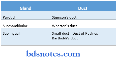
Question 25. Name ducts of major salivary glands.
Answer:

Question 26. Stensen’s duct.
Answer:
- It is the main excretory duct of the parotid gland.
- Stensen’s duct Wharton’s duct
Stensen’s duct Duct:
- Small duct Duct of Rivinus Bartholin’s duct
Stensen’s duct Size:
- 46 cm in length.
- 5 mm diameter.
Stensen’s duct Course:
- Crosses the masseter muscle.
- Turns inward at the anterior border of the masseter.
Stensen’s duct Opening:
- At the papilla opposite to the maxillary second molar.
Question 27. Ptyalin.
Answer:
- It is an alpha form of amylase that hydrolyses polysaccharides to produce glucose and maltose.
- It is most prominent in saliva and maltose.
- It is most prominent in saliva and pancreatic juice.
- It is inactivated in the stomach by gastric acid.
- Overall, the ptyalin degrades only a small amount of the total starch, rest is done by pancreatic amylase.
Question 28. Glands of Blandin and Nuhn
Answer:
- They are anterior lingual glands
Glands of Blandin and Nuhn Location:
- Near the apex of the tongue
Glands of Blandin and Nuhn Nature:
- Mucous
Glands of Blandin and Nuhn Opening:
- Ventral surface of the tongue near the lingual frenum
Question 29. Serous demilunes
Answer:
- These are mucous cells covered by serous cells
- They are present at the ends of mucous tubules
Question 30. Basket cell
Answer:
- Myoepithelial cells are also called basket cells due to their appearance of basket cradling the secretory unit
- They are stellate or spider-like cells with flattened nuclei and scanty cytoplasm
- The structure is similar to smooth muscle and contains actin and myosin
- They expel secretion by contraction.
- Present in intercalated and terminal ducts.
Basket cell Size:
- 46 cm in length.
- 5 mm diameter.
Basket cell Course:
- Crosses the masseter muscle.
- Turns inward at the anterior border of the masseter.
Basket cell Opening:
- At the papilla opposite to the maxillary second molar.
Question 31. Ptyalin.
Answer:
- It is an alpha form of amylase that hydrolyses polysaccharides to produce glucose and maltose.
- It is most prominent in saliva and maltose.
- It is most prominent in saliva and pancreatic juice.
- It is inactivated in the stomach by gastric acid.
- Overall, the ptyalin degrades only a small amount of the total starch, rest is done by pancreatic amylase.
Question 32. Glands of Blandin and Nuhn
Answer:
- They are anterior lingual glands
Glands of Blandin and Nuhn Location:
- Near the apex of the tongue
Glands of Blandin and Nuhn Nature:
- Mucous
Glands of Blandin and Nuhn Opening:
- Ventral surface of the tongue near the lingual frenum
Question 33. Serous demilunes
Answer:
- These are mucous cells covered by serous cells
- They are present at the ends of mucous tubules
Question 34. Basket cell
Answer:
- Myoepithelial cells are also called basket cells due to their appearance of basket cradling the secretory unit
- They are stellate or spider-like cells with flattened nuclei and scanty cytoplasm
- The structure is similar to smooth muscle and contains actin and myosin
- They expel secretion by contraction.
- Present in intercalated and terminal ducts.

Leave a Reply