The Permanent Maxillary Premolars
Question 1. Describe in detail the morphology of the maxillary first premolar. (or) The occlusal surface of the permanent first premolar (or) Mesial surface of the permanent maxillary first premolar
Answer:
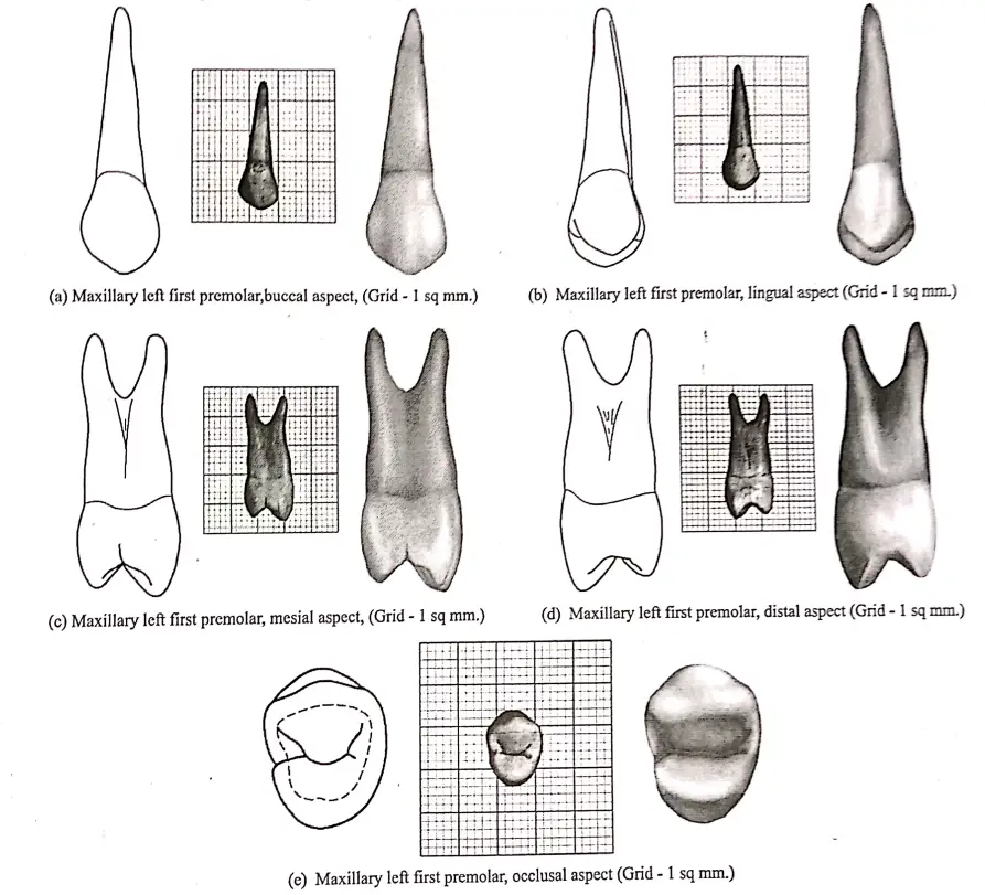
Buccal aspect:
- The crown is roughly trapezoid
- The crown exhibits little curvature at the cervical line and its crest is present near the center of the root.
- The mesial outline is slightly concave while the distal outline is straight.
- The mesial slope of the buccal cusp is straight and longer while the distal slope is shorter and more curved.
- It has a broader contact area
- It has a convex surface with a longer buccal cusp buccal ridge extending from the cusp tip to the cervical margin on the buccal surface of the crown.
- Mesial and distal to the buccal ridge, developmental depression are seen.
Read And Learn More: BDS Previous Examination Question And Answers
Lingual aspect:
- The crown tapers lingually
- The lingual cusp is smooth and spheroidal.
- The cusp tip is pointed.
- The mesial and distal slopes meet an angle of 90 degrees.
- The lingual ridge starts from the crest of the smooth lingual portion up to a point over the lingual cusp.
- The mesial and distal outlines are convex.
- The cervical line is regular, curving apically the lingual portion of the root is smooth and convex with a blunt apex.
Read And Learn More: BDS Previous Examination Question And Answers
Mesial aspect:
The crown appears trapezoidal
- The cusp’s tip is within the root trunk.
- The cervical line may be regular or irregular.
- The buccal outline of the crown curves outwards below the cervical line.
- The lingual outline of the crown curves outwards below the cervical line.
- The lingual outline of the crown is a smoothly curved line starting from the cervical line up to the lingual cusp tip.
- The lingual cusp tip is along with the lingual border of the lingual root.
- The mesial development depression is bordered buccally and lingually by the mesiobuccal and mesiolingual line angles.
- A well-defined development groove is seen in the enamel of the mesial marginal ridge.
- This is continuous with the central groove.
- The buccal outline of the buccal root and the lingual outline of the lingual root is straight.
- The root surface is smoothly convex, ending in a blunt apex.
Distal aspect:
- The crown surface is convex with less curvature of the cervical line.
- The developmental groove is shallow and insignificant.
- It has a flat root trunk.
- The bifurcation of the roots is abrupt near the apical third.
Occlusal aspect:
- It resembles a hexagonal figure with sides mesiobuccal, mesial, mesiolingual, distolingual distal, and distobuccal.
- The two buccal sides are equal.
- The mesial side is shorter than the distal side.
- The mesiolingual side is shorter than the distolingual marginal ridges.
- The angle formed by the mesiobuccal cusp ridge and the mesial marginal ridge is 90 degrees.
- The angle formed by the distobuccal cusp ridge and the distal marginal ridge is acute.
- The crown is wider buccal.
- The distance from the buccal crest to the mesial crest is longer than that from the buccal crest to the distal crest.
- The distance from the mesial crest to the lingual crest is shorter than that from the distal crest to the lingual crest.
Grooves:
- Central developmental groove
- Extends from the mesial of the distal marginal ridge to the mesial marginal ridge.
- Divides the occlusal surface buccolingually.
- Mesial marginal developmental groove.
- Continuation of the central groove.
- It crosses the mesial marginal ridge.
- Mesiobuccal and distobuccal developmental groove.
- These are collateral developmental grooves joining the central groove.
Fossa:
- Mesial triangular fossa.
- Located distal to mesial marginal ridge.
- Harbors the mesiobuccal developmental groove.
- Distal triangular fossa.
- Located just mesial to the distal marginal ridge.
- Ridges: Arise near the center of the central groove.
- Buccal triangular ridge.
- More prominent
- Converge with buccal cusp tip.
- Lingual triangular ridge.
- Less prominent
- Converge with the tip of the lingual cusp.

Measurements:
- Cervo-occlusal crown length -8.5 mm
- Root length – 14 mm
- Mesiodistal crown diameter at cervix -7 mm
- Bucco-lingual crown diameter -5 mm
- Bucco-lingual crown diameter at cervix -9 mm
- Curvature of cervical line – mesial – 8 mm
- The curvature of the cervical line – distal – 1 mm
Question 2. Define the morphology of the maxillary first premolar and enumerate the difference between the maxillary first and second premolar.
Answer:
Morphology of maxillary first premolar:

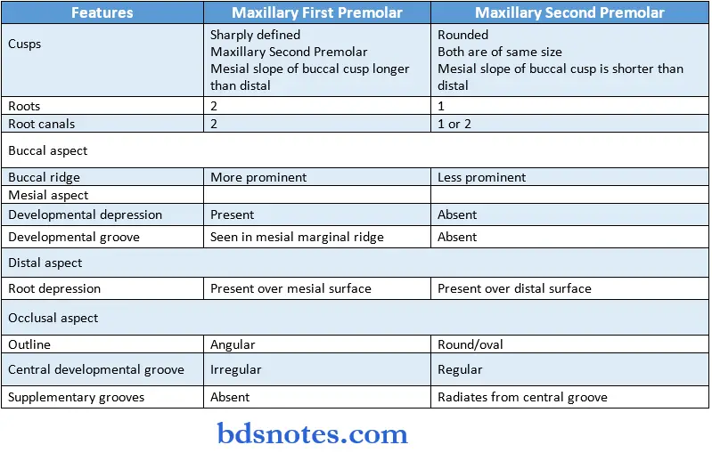
Differences between the maxillary first and second premolar:

Question 3. Differentiate between permanent maxillary 1 premolar and maxillary 2nd premolar
Answer:

Question 4. Chronology of permanent maxillary first premolar.
Answer:
- The first evidence of calcification -11%-1% year
- Enamel completed -5-6 years
- Eruption -10-11 year
- Root completed -12-13 years.

Leave a Reply