Periodontal Pocket
Question 1. Describe Classification, clinical feature, pathogenesis and histopathology of periodontal pocket.
Or
Write short note on Classification of pocket.
Or
Classify periodontal pocket. Write in detail the pathogenesis of periodontal pockets.
Or
Define periodontal pocket. Discuss various Classifications. Write pathogenesis of periodontal pocket in detail.
Or
Define periodontal pocket. Write in detail about pathogenesis and classification of pocket.
Or
Define periodontal pocket. Write its Classification and clinical features.
Or
Write short note on periodontal pocket.
Or
Define and classify periodontal pockets. Write in detail about the etiopathogenesis, histopathology and clinical features of periodontal pockets.
Or
Define and classify periodontal pockets and write in detail etiopathogenesis of periodontal pockets.
Or
Classify periodontal pocket.
Or
Define and classify periodontal pocket.
Answer. Periodontal pocket can be defined as pathological deepening of gingival sulcus.
Classification of Periodontal Pocket
Depending upon its morphology:
- Gingival/false pocket: Deepening of gingival sulcus mainly owing to increase in size of gingiva without any appreciable loss of underlying tissue or apical migration of junctional epithelium. It is known as false pocket.
- Periodontal/true pocket: It is formed by pathological deepening of gingival sulcus due to destruction of supporting tissues of periodontium and apical proliferation of epithelial attachment.
- Combined pocket.
Depending upon its relationship to crestal bone:
- Supra-bony/Supra-alveolar pocket: Base of the pocket is coronal to the crest of alveolar bone. Bone loss is horizontal
- Infra-bony/Intra-alveolar pocket: Base of the pocket is apical to the crest of alveolar bone. Bone loss is vertical.
Depending upon the number of surface involved:
- Simple pocket: It involves one tooth surface
- Compound pocket: This type of pocket involves two or more tooth surfaces. Base of the pocket is in direct contact with gingival margin along each of involved surface.
- Complex pocket: It originates on one tooth surface and twists around the tooth to involve one or more additional surfaces. Only communication with gingival margin is at surface where pocket originates. It is common in furcation areas.
Read And Learn More: Periodontics Question And Answers
Depending upon the nature of soft tissue wall of the pocket:
- Edematous pocket: It contains more cellular exudates and inflammatory infitrate and pocket wall appears bluish red, soft friable with smooth shiny surface.
- Fibrotic pocket: It contains more connective tissue fibers and pocket wall appears pink and relatively firm.
Depending upon the disease activity:
- Active pocket: It consists of more of inflammatory content and is marked by spontaneous bleeding or bleeding on slight provocation.
- Inactive pocket: It consists of less inflammatory contents and show no signs of attachment loss or bone loss.
Periodontal Pocket Clinical Features
Clinical Signs
- Enlarge bluish red marginal gingiva with a rolled edge separated from tooth surface.
- Shiny discolored and puffy gingiva associated with exposed tooth surface.
- Gingival bleeding, purulent exudate from the gingival margin.
- Mobility, extrusion and migration of teeth.
- Development of diastema may present.
Clinical Symptoms
- Foul taste in localized area.
- Tendency of material to stick in inter proximal space.
- Feeling of itching in gums.
- Sensitivity to heat and cold.
- Radiating pain.
- Sensation of pressure in gingiva after eating which gradually diminish.
- Symptom of localized pain is present.
Pathogenesis/Etiopathogenesis
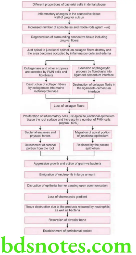
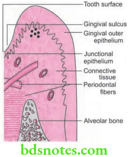
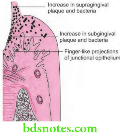
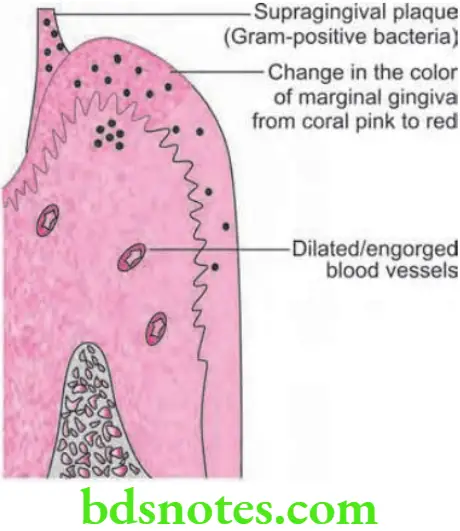
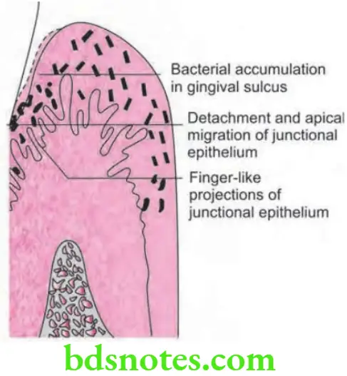
- Pocket formation starts with the laying down of gram-positive bacteria over the supragingival tooth surface and its extension into the gingival sulcus.
- Periodontal pockets are caused by microorganisms as well as their products, which produce pathologic changes that lead to the deepening of the gingival sulcus.
- Since there is an occurrence of inflammation, following changes are observed in the junctional epithelium:
- Junctional epithelium starts proliferating along the root which is in the form of finger-like projections.
- Coronal portion of junctional epithelium detaches from the root as the apical migration takes place. The epithelial cell detachment can take place due to:
- Enzymes produced by the bacteria.
- Physical force applied by speedly growing bacteria.
- Bacteria can also interfere with growth as well as synthetic activities of junctional epithelial cells and also with the normal maintenance of the attachment lamina.
- Exudate which is associated with the advancing bacteria can also be important. Now the sulcus base shift apically and this will be replaced by pocket epithelium.
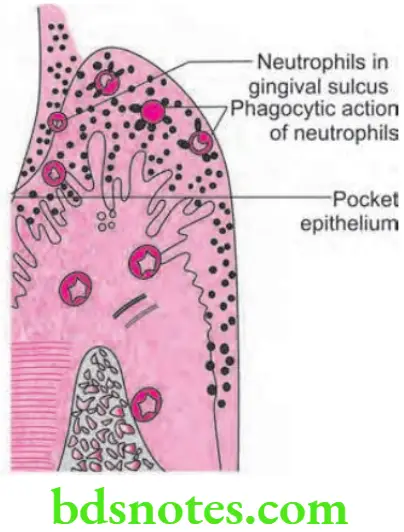
- In normal circumstances, the neutrophils emigrates from the vessels of the gingival plexus via the junctional epithelium inside the gingival sulcus and oral cavity. Most of the bacteria produce substances which attact the neutrophils. Chemotactic agents occur across normal, intact junctional epithelium and connective tissue. Neutrophils which leave the blood vessels are guided by chemical gradient toward the gingival margin and inside the gingival sulcus. In normal circumstances, the transmigrating cells leave no trace of their passage and produce no damage. Neutrophils are the primary and fist line of defense around the teeth while the epithelial barrier is the second.
- As plaque extends subgingivally this causes an increase in the transmigrating neutrophils which can be due to the increased concentration of chemotactic factors and other inflammation-induced substances derived from the bacteria. Such substances lead to vasculitis. Neutrophils get adhere to the endothelial lining and migrate inside the connective tissue, but they still do not accumulate there and they rapidly pass through the junctional or pocket epithelium and form a thick layer which covers the surface of the subgingival plaque. On arrival at the plaque surface, neutrophils are viable partly, and not become completely functional. Neutrophils limit further extension and spread of bacteria by phagocytosis and killing. This can be inhibited by lipopolysaccharides and leukotoxin. The normal neutrophil activities are sufficient to limit the extension of plaque, however, increasing growth rate of bacteria, swamps the neutrophil system and permits tissue destruction to occur.
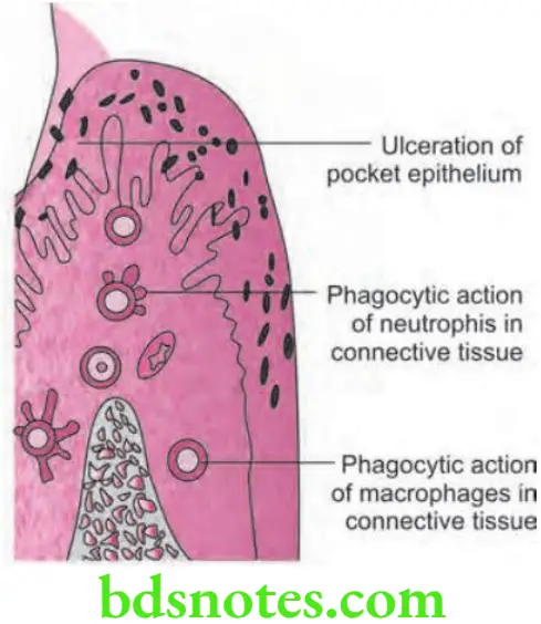
- As the pocket formation proceed either due to aggressive growth and action of bacteria or due to ever increasing number of neutrophils which transmigrate through the junctional epithelium and pocket epithelium leads to open communication between the pocket and connective tissue. Occurrence of ulceration of this type is the second major event in pocket formation. As the epithelial barrier gets breached, the gradient of chemotactic agents produced by pocket bacteria can be disrupted. Now the neutrophils have no longer a guidance system for directing them from the blood vessels through tissues inside the pocket. Neutrophils remain in connective tissue and move randomly. Due to rupture of epithelial barrier, connective tissue gets flooded with the bacterial substances and bacteria can now enter the connective tissue.
- Neutrophils within the connective tissue encounter these substances. Neutrophils become activated and undergo phagocytosis with release of lysosomal enzymes, collagenases and other substances prostaglandin E2 which lead to high tissue damage. As the bacterial substances enter the connective tissue, many systems other than neutrophils are activated such as macrophages, lymphocytes and complement system.
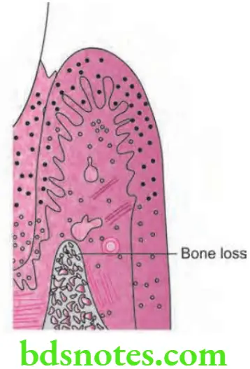
- As epithelial barrier again gets re-established the chemotactic gradient is created again and the destructive process subsides. If epithelial barrier does not get re-established the tissue destruction continues and alveolar bone is resorbed. A periodontal pocket is established.
Histopathological Features
Changes in Soft Tissue Wall
- Blood vessels are engorged and are dilated.
- Connective tissue become edematous and is densely infitrated with plasma cells, lymphocytes and a scattring of PMN leucocytes.
- Junctional epithelium at base of the pocket is shorter than the normal sulcus.
- Most of the degenerative changes occur along the lateral wall of the pocket. Following are the changes in lateral wall of the pocket:
- Epithelium at the lateral wall of the pocket show striking proliferative and degenerative changes.
- Epithelial projection extend deep in connective tissue and extend further apically than junctional epithelium.
- Epithelium is infitrated with leucocytes and various other inflammatory cells.
- Degeneration and necrosis of epithelium causes ulceration of epithelium and exposure of underlying connective tissue.
- Bacterial invasion is present along lateral and apical areas of pocket. Some bacteria traverse basement membrane and invade subepithelial connective tissue.
Changes in Root Surface Wall
- Structural changes:
- Presence of pathologic granules
- Areas of increased mineralization
- Areas of demineralization
- Chemical changes: Mineral content of exposed cementum is increased
- Cytotoxic changes: Bacterial penetration in cementum is found as deep as cementodentinal junction. Bacterial products such as endotoxins are also seen.
Question 2. Write short note on periodontal pockets and its contents.
Or
Write short note on contents of a pocket.
Or
Write short answer on contents of periodontal pocket.
Answer.
Contents of Periodontal Pockets
- Debris consist of:
- Microorganisms and their products (enzymes, endotoxins and metabolites).
- Dental plaque.
- Gingival fluid.
- Food remmants.
- Salivary mucin.
- Desquamated epithelial cells.
- Leucocytes.
- If purulent exudates is in amount:
- Degenerative and necrotic leukocytes.
- Dead and living bacteria.
- Serum and scanty amount of firm.
Question 3. Define and classify pockets. Write about the pathophysiology of pocket formation.
Or
Define and classify periodontal pocket. Write in detail about pathophysiology of pocket.
Answer. Periodontal pocket can be defined as pathological deepening of gingival sulcus.
Classification of Periodontal Pocket
- Depending upon its morphology:
- Gingival/false pocket.
- Periodontal/true pocket.
- Combined pocket.
- Depending upon its relationship to crestal bone:
- Suprabony/Supra-alveolar pocket.
- Infrabony/Intra-alveolar pocket.
- Depending upon the number of surface involved:
- Simple pocket.
- Compound pocket.
- Complex pocket.
- Depending upon the nature of soft tissue wall of the pocket:
- Edematous pocket.
- Fibrotic pocket.
- Depending upon the disease activity:
- Active pocket.
- Inactive pocket.
Pathophysiology of Pocket Formation
Question 4. Define and classify periodontal pocket. Write in detail about clinical features and pathogenesis of periodontal pocket. Add a brief note on periodontal disease activity.
Answer.
Periodontal Disease Activity
- As per this concept periodontal pockets undergo multiple periods of exacerbation and quiescence due to episodic bursts of activity which is followed by period of remission.
- Period of quiescence shows reduced inflammatory response and very litte or no loss of bone as well as connective tissue attachment.
- Bunch of unattached plaque with gram-negative, motile and anaerobic bacteria starts period of exacerbation which is characterized by loss of bone and connective tissue attachments and deepening of pocket.
- Period of exacerbation may last for several days, certain weeks or months and is followed by a period of remission or quiescence in which gram-positive bacteria proliferate and stable condition is achieved.
- Bone loss in untreated periodontal disease occurs in episodic manner. These periods of quiescence and exacerbation are called as periods of inactivity and periods of activity.
- Clinically active periods represent bleeding either spontaneously or on probing and presence of greater amounts of gingival exudates.
Question 5. Write short note on absolute periodontal pocket.
Answer. It is also known as true periodontal pocket.
- In true periodontal pocket, destruction of supporting periodontium is seen.
- It is associated with apical migration of junctional epithelium.
- There is presence of progressive pocket deepening which leads to destruction of periodontium. Due to which there is loosening and exfoliation of teeth.
- To have a true periodontal pocket, a probing measurement of 4 mm or more must be clinically evidenced. In this state, much of the gingival fiers that initially attached the gingival tissue to the tooth have been irreversibly destroyed.
Question 6. Define and classify periodontal pocket. Write in detail about clinical features and pathogenesis of periodontal pocket and treatment.
Answer.
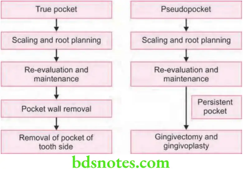
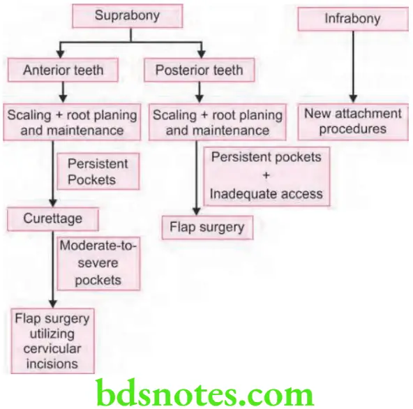
Treatment of Periodontal Pocket
Treatment Depending on the Type of Pocket
Treatment of Supra Bony and Infra Bony Pockets
Treatment of pockets classified under three main headings
- New attachment technique: Gingiva is reunited to the tooth at a position coronal to base of pre-existing pocket. Techniques are non-graft associated, graft associated and combined techniques.
- Removal of pocket wall:
This is done by:- Scaling and root planning which leads to retraction and shrinkage.
- Gingivectomy or undisplaced flap
- Apical displacement of pocket wall by apically displaced flap
- Tooth extraction or partial tooth extraction, i.e. hemisection or root resection can cause removal of tooth side of pocket.
Question 7. Describe the various nonsurgical procedures for the pocket eradication.
Answer.
Nonsurgical Procedures for the Pocket Eradication
- Scaling and root planning
- Use of chemotherapeutic agents
- Laser therapy
- Hyperbaric oxygen therapy
- Photodynamic therapy
Scaling and Root Planing
- Primary objective of scaling and root planning is to regain gingival health by completely removing elements that are responsible for the gingival inflammation (i.e., plaque, calculus, and endotoxins) in the oral environment.
- Both hand instruments and ultrasonic instruments are capable of dramatically reducing the numbers of subgingival microorganisms.
- Scaling and root planning procedure resolve the inflammatory process and gingiva shrinks which reduces the pocket depth.
- After scaling and root planning, maintenance and recall visits are necessary.
Uses of Chemotherapeutic Agents
- Effects of mechanical therapy might be augmented using antimicrobial agents which further suppress the remaining pathogens.
- Many chemotherapeutic agents are now available treating periodontal diseases. Systemic anti-infective therapy (oral antibiotics) and local anti-infective therapy (placing anti infective agents directly into the periodontal pocket) can reduce the bacterial challenge to the periodontium.
Four generations of antiseptics that includes:
- 1 generation: Antibiotics, phenols, quaternary ammonium compounds, and sanguinarine
- 2 generation: Bisbiguanides, bipyridines, quaternary ammonium compounds, phenolic compounds, metalions, halogens, enzymes, surfactants, oxygenating agents, natural products, urea, amino alcohols, salifuor, and agents that increases the redox potentials
- 3 generation: Effective against specific periodontogenic organisms
- 4 generation: Probiotics are incorporated in mouthwashes.
- Combination of metronidazole and amoxicillin has been found to produce more pocket depth reduction than control medication.
- Most commonly used antibiotics for periodontal organisms are metronidazole, amoxicillin, tetracycline, clindamycin, azithromycin, ciprofloxacin, and augmentin.
- Two daily rinses with 10 mL of 0.2% aqueous solution of chlorhexidine digluconate almost completely inhibit the development of dental plaque, calculus and gingivitis. This helps in elimination of periodontal pocket.
Laser Therapy
- Laser irradiation has been reported to exhibit bactericidal, and detoxification effects without producing a smear layer and root surface treated with laser may, therefore, provide favorable conditions for the attachment of periodontal tissue.
- Although there is no clear evidence to date that laser applications improve clinical outcome due to the action of curettge, laser treatment has a potential advantage of accomplishing soft tissue wall treatment effectively along with root surface debridement, and should be further investigated.
Hyperbaric Oxygen Therapy
- This method of administering pure oxygen at greater than atmospheric pressure to a patient to improve or correct conditions.
- Hyperbaric oxygen therapy should be used to compliment conventional therapies and treatments.
- It is showed that hyperbaric oxygen therapy combined with supragingival and subgingival scaling therapy had synergistic action on periodontitis.
- Hyperbaric oxygen therapy had good therapeutic effects on human severe periodontitis, the effects can keep more than 1 year.
Photodynamic Therapy
It is found that scaling and root planning combined with photodisinfection leads to signifiant improvements of the investigated parameters over the use of scaling and root planning alone and this helps in elimination of periodontal pockets.
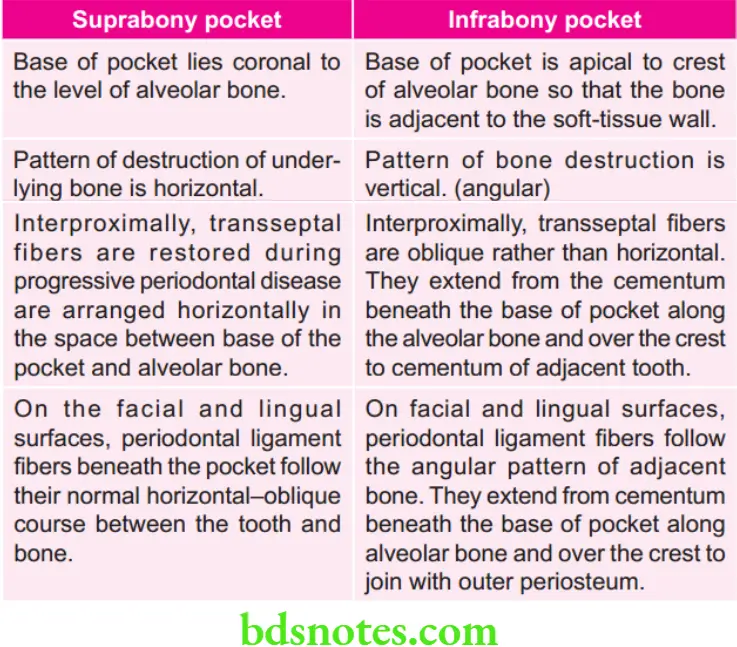
Question 8. Write differences between suprabony and infrabony periodontal pocket.
Answer.

Leave a Reply