Periodontal Ligament
Question 1. Describe in detail the structure of the periodontal ligament, (or) Discuss the cells and fibers of the periodontal ligament.
Answer:
Cells of periodontal ligament:
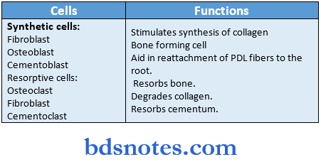
1. Synthetic cells:
- Fibroblasts:
- It is the predominant cell of PDL.
- Fibroblasts Origin:
- Ectomesenchyme of investing layer of the dental papilla.
- Dental follicle.
- Fibroblasts Origin:
- Fibroblasts Features:
- These are large cells with extensive cytoplasm and abundant organelles, associated with protein synthesis and secretion.
- The nucleus occupies a large volume of cells and contains one or more nucleoli.
- They have cilia, a well-developed cytoskeleton with a prominent actin network.
- They show frequent cell-to-cell contact between the adherents and the gap junction types.
- Fibroblasts Function:
- Remodeling of collagen.
- Fibroblasts produce growth factors and cytokines.
- Stimulates the synthesis of collagen and inhibits the synthesis of collagenase.
- The cilia of fibroblast may be associated with control of the cell cycle or inhibition of centriolar activity.
- It is the predominant cell of PDL.
- Osteoblasts:
- They are bone-forming cells lining the tooth socket.
- They are cuboidal with prominent round nuclei.
- They contain abundant rough endoplasmic reticulum, mitochondria, and vesicles.
- These cells contact each other through desmosomes and tight junctions.
- They contact with underlying osteocytes through the cytoplasmic process.
- Osteoblasts Function:
- Constant apposition of bone-
- Osteoblasts Function:
- Cementoblasts:
- They are almost cuboidal, with large vesicular nuclei, one or more nucleoli and abundant
- cytoplasm lining the surface of the cementum,
- They have an irregular outline,
- Cementoblasts Function:
- Aid in the reattachment of PDL fibers to root by forming fresh cementum whenever required,
- Cementoblasts Function:
Read And Learn More: BDS Previous Examination Question And Answers
2. Resorplive cells:
- Osteoclasts:
- They may be large and multinucleated or small and mononuclear,
- They are formed by the fusion of precursor cells.
- Sometimes appear to occupy Howships lacunae.
- The plasma membrane lying adjacent to the bone being resorbed has a folding called a ruffled border.
- Osteoclasts Function:
- They resorb bone.
- Fibroblasts:
- They contain organelles associated with the degradation of collagen.
- They show the rapid generation of collagen by fibroblast phagocytosis.
- Cementoclasts:
- They resemble osteoclasts.
- They are often located in Howship’s lacunae but during resorption, they are found on the surface of the cementum.
3. Progenitor cells:
- They have a perivascular location.
- They tend to have small, close-faced nuclei and very little cytoplasm.
Progenitor cells Functions:
- They divide to give rise to daughter cells which differentiate into functional cells.
- They replace differentiated cells at the end of their lifespan.
4. Epithelial rests of molasses:
- They are remnants of HERS
- They occur close to the cementum as clusters or strands of cells.
- They are abundant in the furcation areas.
- They are cuboidal, with a prominent nucleus and scanty cytoplasm.
- Tight junctions are seen between the cells.
- These cells may proliferate to form cysts and tumors or may also undergo calcification to become cementicles.
5. Defense cells:
- Mast cells:
- They are round or oval cells, with numerous cytoplasmic granules and small, round nuclei.
- Mast cells Functions:
- Plays a role in the inflammatory reaction through the release of histamine.
- Causes the proliferation of endothelial cells and mesenchymal cells.
- Regulate endothelial and fibroblast cell population.
- Macrophages:
- Macrophages Site: Predominantly located close to blood vessels.
- Derived: from monocytes.
- Macrophages Functions:
- Phagocytosing dead cells.
- Secreting growth factors.
- Regulate the proliferation of adjacent fibroblasts.
- Enhance the growth of fibroblasts and endothelial cells.
- Macrophages Functions:
- Eosinophils:
- Consist of one or more crystalloid structures.
- Capable of phagocytosis.
Extracellular substance:
1. Ground substance:
- It binds tissue and fluids which leads to diffusion of gases and metabolic substances.
- It consists of glycoproteins and proteoglycans.
- It helps in tire transportation of materials from and to the cells.
2. Interstitial tissue:
- It is composed of blood vessels, nerves, and lymphatics.
- Blood supply is through branches of superior and inferior alveolar arteries.
- Types of nerve endings in PDL are free nerve endings, Ruffini corpuscles, knob type, and spindle type.
- A network of lymphatics follows the blood vessels.
Fibers of PDL:
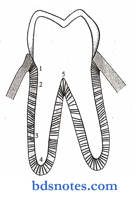
- Principal fibers:

1. Alveolar crest group:
- Attached to the cementum just below the CEJ.
- Inserted into the rim of the alveolus.
2. Horizontal group:
- Located just apical to the alveolar crest group.
- They ran at right angles to the long axis of the tooth from cementum to bone just below the alveolar crest.
3. Oblique group:
- Run from the cementum in an oblique direction and insert into the bone coronally.
4. Apical group:
- Radiate from the cementum around the apex of the root to the bone.
5. Interradicular group:
- Found between the roots of multirooted teeth running from the cementum into the bone.
Fiber bundles composing ligament:
1. Dentogingival group:
- Extends from cervical cementum to lamina propria of the free and attached gingiva.
2. Alveologingival group:
- Extends from the bone of the alveolar crest to lamina propria of the free and attached gingiva.
3. Circular group:
- It forms a band around the neck of the tooth.
4. Dentoperiosteal group:
- Runs apically from the cementum up to the alveolar process.
5. Transseptal fiber:
- Run interdentally from the cementum of one tooth to the cementum of the adjacent tooth.
Elastic Fibers:
Elastic Fibers Types:
Elastin fibers:
- Observed only in walls of afferent blood vessels.
Oxytalanfibers:
- They are numerous and dense in the cervical region of the ligament.
- They run parallel to the gingival group of collagen fibers.
- They regulate vascular flow in relation to tooth function.
Elauninfibers:
- They are found within fibers of the gingival ligament.
Reticular fibers:
- They are composed of type 3 collagen.
Secondary fibers:
- They are located between and among the principal fibers and are randomly oriented.
Indifferent fiber plexus:
- These are small fibers running in all directions forming a plexus called indifferent fiber plexus.
Question 2. Describe the histology and functions of PDL.
Answer:
Functions of PDL:
1. Supportive:
- When a tooth is moved in its socket due to the force of mastication or orthodontic force, part of the periodontal space is narrowed while the other is widened.
- The periodontal ligament in the narrow periodontal space is compressed.
- The collagen fibers in this area act as a cushion for the displaced tooth.
2. Sensory:
- When teeth move in their sockets, they distort receptors in the PDL and trigger a response.
- PDL carries tactile sensation from teeth and hence helps in the localization of pain.
- PDL contributes to the sensation of touch and pressure.
3. Nutritive:
- The blood vessel within the PDL provides nutrition to the centrocytes of the PDL and osteocytes of the alveolar bone.
- The blood vessel also helps in the removal of the catabolites from the cells.
4. Homeostatic:
- The cells of the PDL have the capability to synthesize and resorb the extracellular substance of the connective tissue of the ligament.
- If the balance between synthesis and resorption is disturbed, the quality of the tissue is changed.
- This gradually leads to loss of attachment which results in tooth loss.
- In all areas of PDL, there is continual cell death which is replaced by new cells produced by the division of progenitor cells.
5. Eruptive:
- PDL components enable teeth to adjust their position.
- PDL provides space and acts as a medium for cellular remodeling and hence continued eruption occurs.
6. Physical:
- PDL protects vessels and nerves from mechanical forces.
- It offers resistance to impact from occlusal forces.
- Acts as a shock absorber to transmit occlusal forces to the bone.
7. Pormative/Resorptive:
- Cementoblast and osteoblast form cementum and bone respectively.
- Cementoclast and osteoclast resorb cementum and bone respectively.
Question 3. Cell rests of Malassez.
Answer:
- They were first described by Malassez in 1884.
- They are remnants of HERS.
Cell rests of Malassez Site:
- Found close to the cementum.
- Abundant in the furcation areas.
Cell rests of Malassez Features:
1. Cell-cuboidal, closely packed.
2. Cytoplasm.
- Scanty.
- Contains tonofibrils inserted into desmosomes and hemidesmosomes.
3. Nucleus
- Prominent and deeply stained.
4. Cell organelles.
- Mitochondria are distributed throughout the cytoplasm.
- Poorly developed rough endoplasmic reticulum and approx
- Less – in older individuals.
- More- in children.
2. In position.
Apical region – during the second decade of life
Cervically – later life.
Cell rests of Malassez Fate:
pathological conditions, they undergo rapid proliferation and produce a variety of cysts and tumors or may also undergo calcification to become clementines.
Question 4. Functions of PDL
Answer:
1. Supportive
- When a tooth is moved in its socket due to the force of mastication or orthodontic force, part of the periodontal space is narrowed while the other is widened
- The periodontal ligament in the narrow periodontal space is compressed
- The collagen fibers in this area act as a cushion for the displaced tooth
2. Sensory
- When teeth move in their sockets, they distort receptors in the PDL and trigger a response
- PDL carries tactile sensation from teeth and hence helps in the localization of pain
- PDL contributes to the sensation of touch and pressure
3. Nutritive
- The blood vessel within the PDL provides nutrition to the cementocytes of the PDL and osteocytes of the alveolar bone
- The blood vessel also help in the removal of the catabolites from the cells
4. Homeostatic
- The cells of the PDL have the capability to synthesize and resorb the extracellular substance of the connective tissue of the ligament
- If the balance between synthesis and resorption is disturbed, the quality of the tissue is changed
- This gradually leads to loss of attachment which results in tooth loss
- In all areas of PDL, there is continual cell death which is replaced by new cells produced by the division of progenitor cells
5. Eruptive
- PDL components enable teeth to adjust their position
- PDL provides space and acts as a medium for cellular remodeling and hence continued eruptions occur
6. Physical
- PDL protects vessels and nerves from mechanical forces
- It offers resistance to impact from occlusal forces
- Acts as a shock absorber to transmit occlusal forces to the bone
7. Formative/Resorptive
- Cementoblast and osteoblast forms cementum and bone respectively
- Cementoclast and osteoclast resorbs cementum and bone respectively
Question 5. Resorptive cells
Answer:
- Osteoclast
- Multinucleated giant cells
- Lies adjacent to the bone
- Undergoes resorption of bone
- Formed by monocytes
- Fibroblast
- Contains fragments of collagen
- These undergoes digestion
- Results in resorption of bone
- Cementoclast
- Located in Howship’s Lacunae
- Causes resorption of cementum
Question 6. Periodontium.
Answer:
- It is defined as those tissues supporting and investing in the tooth.
- It attaches the teeth to the bones of the jaws and supports the teeth.
It consists of:
- Cementum.
- Periodontal ligament.
- Bone lining the socket.
- Part of the gingiva facing the tooth.
- It is attached to the dentin of the root by cementum and to the bone of the jaws by alveolar bone,
- The periodontium loss can be repaired by the cells regulating the formation, maintenance, and regeneration of the periodontium.
Question 7. Cell rest of Malassez.
Answer:
- They are remnants of HERS.
- They are found close to the cementum and are found abundant in furcation areas.
- They consist of closely packed cuboidal cells with prominent nuclei and scanty cytoplasm.
- They persist as strands, islands, or tubelike structures close to the cementum.
- They may show variations in number and site with respect to age.
Question 8. Oxytalin fibers.
Answer:
- They are a type of immature elastic fibers.
- They consist of microfibrillar components only.
- Size: 0.5 – 2.5 pm in diameter (approx.)
- They are not susceptible to acid hydrolysis.
- Fibers run in the axial direction.
- One end is embedded in cementum/bone and the other is in the wall of blood vessels.
- They play a role in tooth support and regulate vascular flow.
- They are numerous and dense in the cervical region of the ligament.
Question 9. Fibroblasts.
Answer:
- It is the predominant cell of PDL.
- They originate from the ectomesenchyme of the investing layer of the dental papilla.
- They are spindle-shaped large cells with extensive cytoplasm and abundant organelles with large nuclei.
- They consist of cilia which are associated with the control of the cell cycle.
- They play an important role in the remodeling of collagen which includes collagen synthesis and . degradation of collagen.
Question 10. Periodontal ligament.
Answer:
- It is soft, specialized connective tissue situated between the cementum covering the root of the tire tooth and the bone forming the socket wall.
- Width: 0.15 – 0.38 mm.
Periodontal ligament Cells:
1. Synthetic cells
- Osteoblasts
- Fibroblast
- Cementoblast.
2. Resorptive cells.
- Osteoclast
- Fibroblast
- Cementoclast
3. Progenitor cell.
4. Epithelial rest of malassez.
5. Defense cells.
- Macrophages
- Eosinophils.
Periodontal ligament Functions:
- Supportive
- Sensory
- Nutritive
- Homeostatic
- Eruptive
- Physical
Question 11. Cells of periodontal ligament.
Answer:


Question 12. Sharpey’s fibers.
Answer:
- These are collagen fibers that are embedded into the cementum on one side and into the alveolar bone on another side.
- Fibers in primary acellular cementum are fully mineralized while those in cellular cementum and bone are partly mineralized.
- Their mineralized part appears as a projecting covered with mineral clusters.
- Few of them pass uninterrupted through the alveolar bone to continue as principal fibers of PDL.
- It passes through alveolar bone only when it consists entirely of compact bone.
- It consists of noncollagenous proteins like osteopontin and bone sialoprotein.
Question 13. Alveolar crest group of fibers.
Answer:
- They radiate from the crest of the alveolar process and attach themselves to the cervical part of the cementum.
- They are located beneath the junction epithelium.
Alveolar crest group of fibers Function:
- They resist tilting, intrusive, extra use, and rotational forces.
Question 14. Synthetic cells of PDL.
Answer:

Question 15. Transseptal fibers.
Answer:
- These fibers run interdentally from the cementum apical to the junctional epithelium of one tooth over the alveolar crest to a similar region of the adjacent tooth.
- By these, all tire teeth are connected in an arch.
- They are responsible for post-retention relapse of orthodontic treatment.
- They are capable of turnover and remodeling under normal physiologic conditions and therapeutic tooth movement.
- They ensure clinical stability of tooth position.
Question 16. Bundle fibers of the periodontal membrane.
Answer:
Fiber bundles composing ligament:
1. Dentogingival group:
- Extends from cervical cementum to lamina propria of the free and attached gingiva.
2. Alveologingival group:
- Extends from the bone of the alveolar crest to lamina propria of the free and attached gingiva.
3. Circular group:
- It forms a band around the neck of the tooth.
4. Dentoperiosteal group:
- Runs apically from the cementum up to the alveolar process.
5. Transseptal fiber:
- Run interdentally from the cementum of one tooth to the cementum of the adjacent tooth.
Question 17. Age changes in the periodontal ligament.
Answer:
- The cell number and activity decrease with age
- PDL fibers become attached to the scalloping ends of the alveolar bone.
- PDL activity decreases.
- Destructive changes occur due to the presence of gingival and periodontal diseases in old age.
- Some of the teeth become non-functional.
- PDL width decreases.
Question 18. Birbeck’s granules
Answer:
- They are named after its discoverer Michael Stanley Clive Birbeck
- It is a rod-shaped cytoplasmic organelle
- Found in Langerhans cells
Birbeck’s granules Functions:
- They migrate to the periphery of Langerhans cells and release its contents into the extracellular matrix
- Act as receptor-mediated endocytosis
Question 19. Principal fibers
Answer:
- Trans-septal fibers
- Connects cementum of one tooth with that of other
- Alveolar crest
- Extends from cementum to alveolar crest
Principal fibers Functions:
-
- Retains tooth in the socket
- Retains lateral tooth movement
- Horizontal Group
- Extends from cementum to alveolar bone
- Oblique group
- Extends coronally from the cementum to the bone
- Periodontal Ligament
Functions:
- Resist axially directed forces
- Apical group
- Extends from the cementum to the bone of the alveolar fundus
Functions:
- Prevents tipping movement
- Resists luxation
- Inter-radicular fibers
- It is present between the cementum of multi-rooted teeth
Functions:
- Resists luxation Resists tipping and torquing
Question 20. The protective function of PDL
Answer:
- The periodontal ligament protects vessels and nerves from mechanical forces
- It offers resistance to impact from occlusal forces
- Acts as a shock absorber to transmit occlusal forces to the bone.

Leave a Reply