Leukemia
Question 1. Discuss in brief acute myeloid leukemias.
Or
Write notes on acute myeloid leukemia.
Or
Write a short note on acute myeloid leukemia.
Answer:
Acute myeloid leukemia is a heterogeneous disease characterized by the infiltration of malignant myeloid cells in the blood, bone marrow, and other tissues.
Acute myeloid leukemia Classification
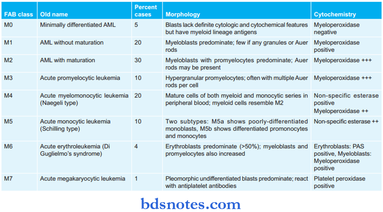
Revised FAB (French American British) Classification for Acute myeloid leukemia

Acute myeloid leukemia Clinical Features
- Due to bone marrow failure
- Anemia produces pallor, lethargy, and dyspnea.
- Bleeding manifestations due to thrombocytopenia cause petechiae and bleeding from the gums.
- Infections of the mouth, throat, skin, respiratory and other sites are common.
- Fever is attributed to infections in acute leukemias.
Read And Learn More: Pathology Question And Answers
- Due to organ infiltration
- Pain and tenderness in bones.
- Presence of gum hypertrophy
- Lymphadenopathy and enlargement of tonsils may occur.
- Moderate splenomegaly, splenic infarction, subcapsular hemorrhages
- Presence of hepatomegaly
- Leukemic infiltration of the kidney
- Chloroma and granulocytic sarcoma is a localized tumor-forming masses occurring on the skin or orbit.
- Meningeal involvement manifested by raised intracranial pressure, headache, nausea and vomiting, blurring of vision, and diplopia are seen.
- Other organ infiltrations include testicular swelling and mediastinal compression.
Acute myeloid leukemia Laboratory Findings
1. Blood picture
Anemia
- It is generally severe, progressive, and normochromic in type.
- Moderate reticulocytosis up to 5% and few nucleated red cells may be present.
Thrombocytopenia
- The platelet count is less than 50 000/µL.
- Whenplateletcountis below20 000/µL serious spontaneous hemorrhagic episodes develop.
- Acute promyelocytic leukemia (M3) may be associated with a serious coagulation abnormality called disseminated intravascular coagulation.
White Blood Cells
- In advanced cases, the WBC count is more than 100 000/µL.
- The majority of leucocytes in the peripheral blood blasts and there is often neutropenia due to marrow infiltration by leukemic cells.
- Some patients of myelodysplastic syndrome present with pancytopenia and have a few blasts labeled sub-leukemic leukemia or have no blasts labeled as aleukemic leukemia.
2. Bone Marrow Examination
- Cellularity: Typically the marrow is hypercellular with a predominance of myeloblasts and promyelocytes. A dry tap may also occur due to pancytopenia or the adhesive nature of leukemic cells which are enmeshed in reticulin fibers.
- Leukemic cells: Diagnosis of the type of leukemic cells is done by routing Romanowsky stains and cytochemical stains. The presence of at least 30% blasts in the bone marrow is the essential criterion for the diagnosis of acute leukemia.
- Erythropoiesis: Erythropoietic cells are reduced. Dyserythropoiesis, megaloblastic features, and ring sideroblasts are common.
- Megakaryocytes: They are usually reduced or absent.
- Cytogenetics: 75% of cases show karyotypic abnormalities in the dividing leukemic cells.
The common chromosomal abnormalities inAML are as follows:
- Aneuploidy: Hypo and hyperdiploid cell lines are found with equal frequency in AML.
- Philadelphia chromosome: 25-30% of cases of AML in adults shows the Philadelphia chromosome. It is associated with poor prognosis.
3. Cytochemistry
- Myeloperoxidase: It is positive in immature myeloid cells containing granules and Auer rods, i.e. all forms of AML from mL to M6 but negative in M0 myeloblasts.
- Sudan black: Positive in immature cells in AML.
- Periodic acid-SchiffPAS): Positive in erythroleukemia (M6)
- Nonspecific esterase: Positive in monocytic series (M4 and M5)
- Acid phosphatase: Diffse reaction in monocytic cells (M4 and M5).
4. Biochemical Investigations
- Serum muramidase: Serum levels of lysozyme, i.e. muramidase are elevated in myelomonocytic (M4) and monocytic (M5) leukemia.
- Serum uric acid: Serum uric acid level is frequently increased because of a rapidly growing number of leukemic cells.
Question 2. Describe leukemia.
Answer:
Leukemia is caused by the mutation of bone marrow pluripotent or most primitive cells.
- The proliferation of leukemic cells takes place primarily in the bone marrow and in certain forms in the lymphoid tissues.
- Leukemias are classified on the basis of cell types into myeloid and lymphoid and on the basis of the natural history of the disease, into chronic and acute. Thus the main types of leukemia are:
-
- Acute myeloblastic leukemia.
- Acute lymphoblastic leukemia
- Chronic myeloid leukemia
- Chronic lymphocytic leukemia
Chronic Lymphocytic Leukemia
It constitutes about 25% of all leukemias and is a disease of the elderly with a male predisposition.
Leukemia Clinical Features
- Features of anemia such as weakness, fatigue, and dyspnea.
- Lymph nodes are symmetrically enlarged, discrete and non-tender
- Splenomegaly and hepatomegaly are present
- Thrombocytopenia is present which leads to hemorrhagic tendencies.
Leukemia Laboratory Findings
- Blood picture:
- Anemia: It is of normocytic normochromic type.
- WBCs: Marked leukocytosis is present, i.e. 50,000 to 200,000/µl.
- Platelet count: Thrombocytopenia is present.
- Bone marrow findings:
- Increased lymphocyte count is present.
- Reduced myeloid and erythroid precursors.
Question 3. Write a short note on leukemoid reaction.
Answer:
Leukemoid reactions are characterized by an increase in total leucocyte count beyond 25000/µl.
- The clinical features of leukemia such as splenomegaly, lymphadenopathy, and hemorrhages are usually absent
- The leukemoid reaction may be myeloid or lymphoid.
Myeloid Leukemoid Reaction
Her total WBC count is morbidly increased with a predominance of cells of myeloid series including occasional immature cells.
Etiology
- Infection: Staphylococcal pneumonia, Disseminated tuberculosis, Meningitis, diphtheria, endocarditis, etc.
- Intoxication: Mercury poisoning and burns
- Malignant diseases such as multiple myeloma, Hodgkin’s disease, and bone metastasis.
- Severe hemorrhage and severe hemolysis.
Laboratory Findings
- Leucocytosis is present, i.e. beyond 25,000/ml.
- Immature cells are mild to moderate and comprised of metamyelocytes, myelocytes, and blasts, blood picture simulates chronic myeloid leukemia.
- Infective cases show Dohle bodies in the cytoplasm of neutrophils.
- Neutrophil alkaline phosphatase levels are high.
Lymphoid Leukemoid Reaction
Etiology
Infections: Infectious mononucleosis, whooping cough, chickenpox, measles, and tuberculosis.
- Chronic lymphocytic leukemia
- Carcinoma.
Laboratory Diagnosis
- Leucocytosis is present, i.e. beyond 25,000/ml
- Differential leucocyte count reveals mature lymphocytes simulating the blood picture found in chronic lymphocytic leukemia.
Question 4. Write a short note on acute leukemia.
Answer:
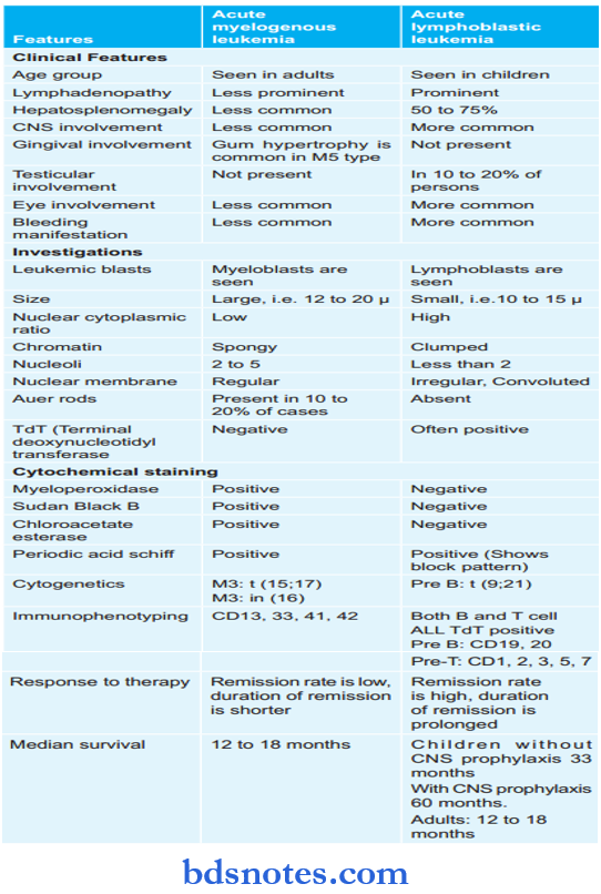
Acute leukemias are of two types, i.e. acute myelogenous leukemia and acute lymphoblastic leukemia.
Both the leukemias are explained on a comparison basis:

Question 5. Write a short note on chronic myeloid leukemia.
Answer:
Chronic myeloid leukemia is a hematological malignancy in which myeloid leukemia cells are predominant.
- Chronic myeloid leukemia comprises about 20% of all leukemias.
- More in 3rd and 4th decade of life.
Chronic myeloid leukemia Clinical Features
- Features of anemia are seen, i.e. weakness, pallor, dyspnea, and tachycardia.
- Symptoms due to hypermetabolism-weight loss, lassitude, anorexia, and night sweats.
- Splenomegaly is almost always present and is frequently massive.
- Bleeding tendencies: Easy bruising, epistaxis, menorrhagia, and hematomas.
Features of gout, visual disturbances, neurological manifestations, and priapism less commonly, lymph node enlargement, frequent infections, and facial rash in juvenile CML.
Chronic myeloid leukemia Lab Findings
- Blood picture
- Anemia: It is normocytic and normochromic
- WBCs: Leucocytosis is present in 10,000 cells/ml
- Platelets: Platelet count is normal but can be raised.
- Bone marrow findings
- Cellularity: Hypercellularity with total or partial replacement of fat spaces by proliferating myeloid cells
- Myeloid cells: They predominate in bone marrow with an increased myeloid-erythroid ratio
- Erythropoiesis: It is normoblasts but the reduction in erythropoietic cells
- Megakaryocytes: Smaller in size than normal.
- There is an increase in number of phagocytes.
- Cytogenetics: Characteristic chromosomal abnormality called the Philadelphia chromosome formed by a reciprocal translocation between the part of the long arm of chromosome 22 and that of chromosome 9 is usually seen in 70 to 90% of cases.
Chronic myeloid leukemia Treatment
- Chronic phase: Respond favorably to chemotherapy.
-
- The main chemotherapeutic agents used are Busalphan, hydroxyurea, and cyclophosphamide.
- Others splenectomy, leucopheresis, and bone marrow transplantation.
- Blast crisis: Remission of the actual blastic phase of chronic myeloid leukemia can be obtained by vincristine and prednisone or other combination chemotherapy regimes.
Question 6. Write a short note on the classification of acute leukemia.
Answer:
Acute leukemia Classification
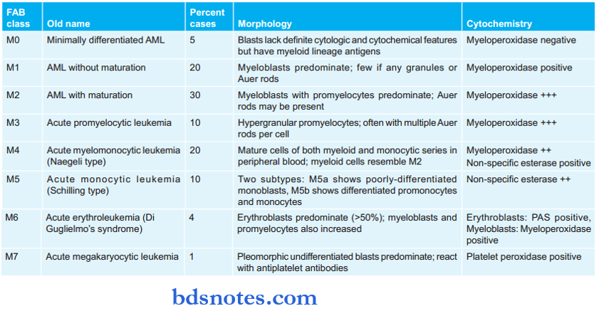
Revised FAB ( French – American – British ) Classification for Acute Myeloid Leukemia

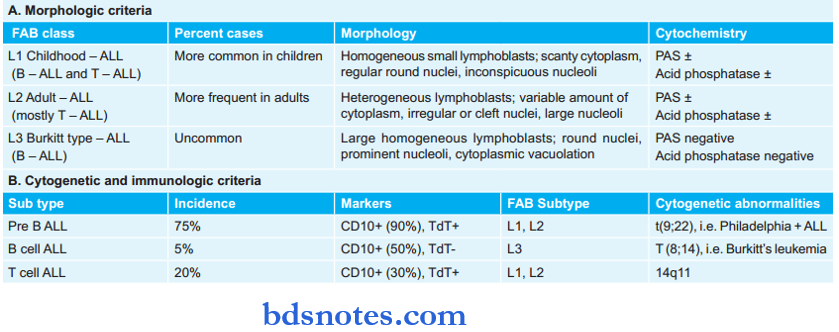
Revised FAB (French – American – British) Classification for acute Lymphocytic leukemia

World Health Organization (WHO) Classification of Acute Leukemias
Blast count for diagnosis of acute leukemia ≥ 20% in peripheral blood or bone marrow (in FAB classification the cut of is 30%; it has been demonstrated that the survival pattern of patients with 20-30% blasts is similar to those with a count of >30%).
1. WHO classification of AML
- AML with recurrent genetic abnormalities
- AML with t(8; 2l) (q22; q22); AMLl/ETO
- AML with abnormal bone marrow eosinophils inv(l6) (p l3; q22) or t (16; 16) (p l3; q 22); (CBFβ/MYH 1l)
- Acute promyelocytic leukemia AML with t(l5; l7)(q 22; q l2) (PML/RARα) and variants
- AML with 11q23(MLL) abnormalities
- AML with multilineage dysplasia
- Following a myelodysplastic syndrome
- Without antecedent myelodysplastic syndrome
- AML and myelodysplastic syndromes therapy related
- Alkylating agent related
- Topoisomerase Type 2 inhibitor-related
- Other types
- AML not otherwise characterized/specified
- AML minimally differentiated
- AML without maturation
- AML with maturation
- Acute myelomonocytic leukemia
- Acute mono blastic and monocytic leukemia
- Acute erythroid leukemia
- Acute megakaryoblastic leukemia
- Acute basophilic leukemia
- Acute pan melodies with myelofibrosis
- Myeloid sarcoma
- Myeloid proliferations related to Down’s syndrome
- Blastic plasmacytoid dendritic cell neoplasms
2. Classifiation of ALL


Leave a Reply