Panoramic Radiography Or Orthopantomography (OPG)
Question 1. Write short note on orthopantomography.
or
Write in detail about OPG.
Answer.
Orthopantomography
It is an extraoral radiography technique which images maxilla and mandible in a single film.
Orthopantomography is also known as panoramic radiography.
Read And Learn More: Oral Radiology Question And Answers
Purpose of orthopantomography
It provides the overall view of maxilla and mandible with their supporting structures, it helps in evaluation of following:
- Fractures of jaw
- Impacted, unerupted and supernumerary teeth
- Eruption pattern of teeth
- Cyst and tumors of jaw
- Various endocrine, nutritional and blood disorders in jaws
- Developmental anomalies affecting teeth and jaws
- Temporomandibular joint and maxillary sinus.
Principle of orthopantomography
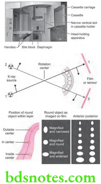
- If the film moves at the speed, which follows the moving projection of a certain point, this point will always be projected on the same spot on film and will not appear unsharp.
- In OPG, the film is attached to a rotating system and moves in opposite direction to the beam. The film is given correct speed by apposing this movement with contrary movement relative to the beam.
Orthopantomography Procedure
- Explain the procedure to the patient.
- Make the patient wear a lead apron and remove all objects from the head which will interfere with film exposure, e.g. ear rings, necklace, nose rings.
- Special attntion must be paid for proper positioning of patient along focal trough area.
- Patient is instructed to look straight rather than following movement of tube.
- Patient is positioned such that dental arches are located in middle of focal trough area.
- Mid-sagittl plane is kept perpendicular to flor.
- Patient back and spine is adjusted in erect position and the occlusal plane adjusted so that Frankfurt plane is parallel to flor done by asking the patient to place central incisor into a notched incisal device using a bite block.
- Mark on fim: Lef and right with lead marker.
- Center the lower border of mandible on the chin rest and is equidistant.
- Instruct the patient to position the tongue on the palate.
- After the exposure is complete, than film is subjected to routine processing.
Orthopantomography Advantages
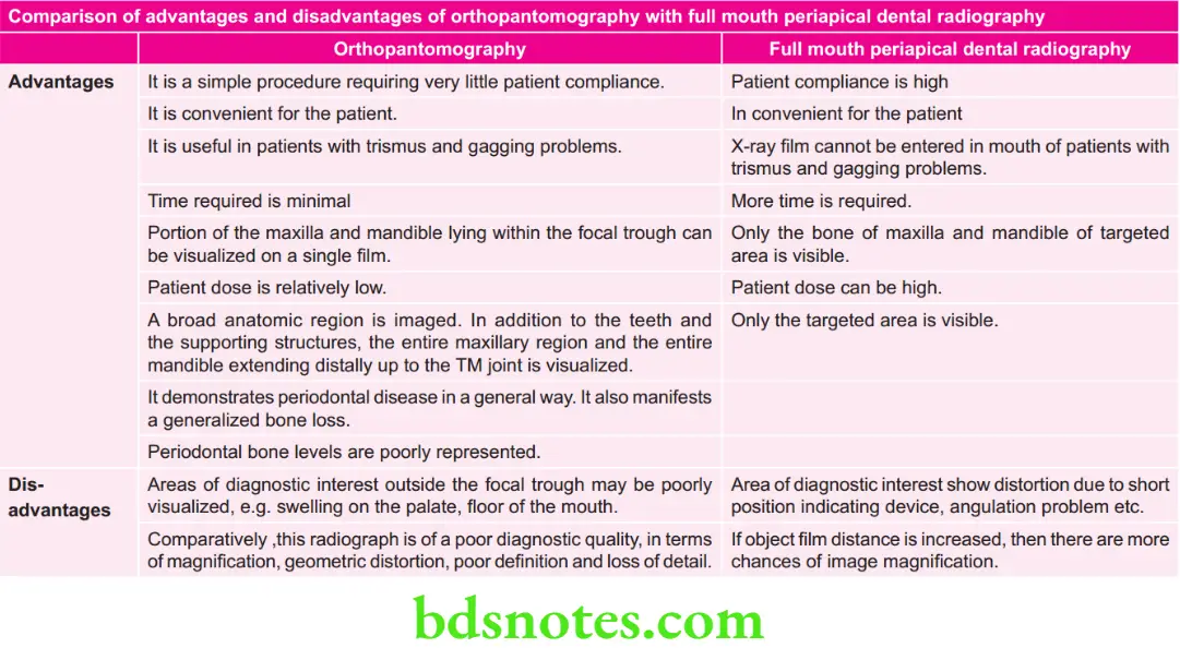
- It is a simple procedure requiring very little patient compliance.
- It is convenient for the patient.
- It is useful in patients with trismus and gagging problems.
- Time required is minimal compared to a full mouth intraoral periapical radiographs.
- Portion of the maxilla and mandible lying within the focal trough can be visualized on a single fim.
- Patient dose is relatively low.
- Panoramic radiographs taken for diagnostic purpose are valuable visual aid in patient education.
- Abroad anatomic region is imaged. In addition to the teeth and the supporting structures, the entire maxillary region and the entire mandible extending distally up to the TM joint is visualized.
- Anatomical structures are most identifible and the teeth are oriented in their correct relationship to the adjacent structures and to each other.
- It allows for the assessment of the presence and position of unerupted teeth in orthodontic treatment.
- It demonstrates periodontal disease in a general way. It also manifests a generalized bone loss.
- All the parameters are standardized and repetitive images can be taken, on recall visits for comparative and research purposes.
- It is very useful for mass screening.
- This view helps in localization of objects/pathology in conjunction with a topographic occlusal view or an intraoral periapical radiograph.
Orthopantomography Disadvantages
- Areas of diagnostic interest outside the focal trough may be poorly visualized, e.g. swelling on the palate, flor of the mouth.
- Comparatively, this radiograph is of a poor diagnostic quality, in terms of magnifiation, geometric distortion, poor defiition and loss of detail.
- There is an overlapping of the teeth in the bicuspid area of the maxilla and the mandible.
- In cases of pronounced inclination, the anterior teeth are poorly registered.
- Density of the spine, especially in short-necked people can cause lack of clarity in the central portion of the fim.
- Number of radiopaque and radiolucent areas may be present due to the superimposition ofreal / double or ghost images and because of sof tissue shadows and airspaces.
- Due to prescribed rotation, patient with facial asymmetry or patients who do not conform to the rotation curvature, cannot be X-rayed with any degree of satisfaction.
- If the patient positioning is improper, the amount of vertical and horizontal distortion will vary from one part of the fim to another part of the fim.
- Ease and convenience of obtaining an OPG may encourage careless evaluation of a patient’s specifi radiographic needs.
- Artifacts are easily misinterpreted and are more commonly seen, e.g. nose ring as a periapical radiopaque lesion, ear ring as a calcifiation in the maxillary sinus.
- OPG shows an oblique, rather than true lateral view of the condylar heads and hence, the joint space cannot be accurately assessed.
- Some patients do not conform to the shape of the focal trough and some structures will be out of focus.
- Cost of the machine is very high.
Question 2. Describe in detail technique of orthopantomography and also write its advantages and disadvantages comparing it with full mouth periapical dental radiography.
Answer. Synonyms: Panoramic imaging or rotational radiography or pantomography.
Definition: It is a technique for producing a single tomographic image of the facial structures that induces both the maxillary and mandible dental arches and their supporting structures.
Orthopantomography Procedure
- Explain the procedure to the patient.
- Make the patient wear a lead apron and remove all objects from the head which will interfere with film exposure, e.g. ear rings, necklace, nose rings.
- Special attntion must be paid for proper positioning of patient along focal trough area.
- Patient is instructed to look straight rather than following movement of tube.
- Patient is positioned such that dental arches are located in middle of focal trough area.
- Mid-sagittl plane is kept perpendicular to flor.
- Patient back and spine is adjusted in erect position and the occlusal plane adjusted so that Frankfurt plane is parallel to flor done by asking the patient to place central incisor into a notched incisal device using a bite block.
- Mark on fim: Lef and right with lead marker.
- Center the lower border of mandible on the chin rest and is equidistant.
- Instruct the patient to position the tongue on the palate.
- After the exposure is complete, then film is subjected to routine processing.


Question 3. Write short note on advantages of orthopantomography.
Answer.
Orthopantomography Advantages
- It is a simple procedure requiring very little patient compliance.
- It is convenient for the patient.
- It is useful in patients with trismus and gagging problems.
- Time required is minimal compared to a full mouth intraoral periapical radiographs.
- Portion of the maxilla and mandible lying within the focal trough can be visualized on a single fim.
- Patient dose is relatively low.
- Panoramic radiographs taken for diagnostic purpose are valuable visual aid in patient education.
- Abroad anatomic region is imaged. In addition to the teeth and the supporting structures, the entire maxillary region and the entire mandible extending distally up to the TM joint is visualized.
- Anatomical structures are most identifible and the teeth are oriented in their correct relationship to the adjacent structures and to each other.
- It allows for the assessment of the presence and position of unerupted teeth in orthodontic treatment.
- It demonstrates periodontal disease in a general way. It also manifests a generalized bone loss.
- All the parameters are standardized and repetitive images can be taken, on recall visits for comparative and research purposes.
- It is very useful for mass screening.
- This view helps in localization of objects/pathology in conjunction with a topographic occlusal view or an intraoral periapical radiograph.
Question 4. Enumerate the various specialized radiographic techniques. describe in detail about panoramic radiography.
or
Enumerate advances in radiography and describe in detail about all aspects of orthopantomography.
Answer. Various specialized radiographic techniques are:
- Tomography OPG (Panoramic radiography).
- Dentascan imaging.
- Three-dimensional CT.
- Cone beam radiography.
- Scanography.
- Magnetic resonance imaging (MRI).
- Nuclear medicine.
- Diagnostic ultrasound.
- Arthrography and arthroscopy.
- Xeroradiography.
- Sialography.
- Implant imaging.
Question 5. Describe indications, principle, advantages, disadvantages of panoramic radiography.
or
Describe in detail about the principles, advantages and disadvantages of panoramic radiography.
or
Discuss the philosophy, indications, advantages and disadvantages of panoramic radiography.
or
Write indications, advantages, disadvantages and principles of OPG.
Answer.
Principle/Philosophy
- If the film moves at the speed which follows the moving projection of a certain point, this point will always be projected on the same spot on film and will not appear unsharp.
- In OPG, the film is attached to a rotating system and moves in opposite direction to the beam. The film is given correct speed by apposing this movement with contrary movement relative to the beam.
Panoramic Indications
- As a substitute for full mouth intraoral periapical radiographs.
- For evaluation of tooth development for children, the mixed dentition and also the aged.
- To assist and assess the patient for and during orthodontic treatment.
- To establish the site and size oflesions such as cysts, tumors and developmental anomalies in the body and rami of the mandible.
- Prior to any surgical procedures such as extraction of impacted teeth, enucleation of a cyst, etc.
- For detection of fractures of the middle third and the mandible aftr facial trauma.
- For follow-up of treatment, progress of pathology or postoperative bony healing.
- Investigation of TM joint dysfunction.
- To study the antrum, especially to study the flor, posterior and anterior walls of the antrum.
- Periodontal disease—as an overall view of the alveolar bone levels.
- Assessment for underlying bone disease before constructing complete or partial dentures.
- Evaluation of developmental anomalies.
- Evaluation of the vertical height of the alveolar bone before inserting osseo-integrated implants.
Question 6. Discuss the principles, indications and contraindications of panoramic radiography.
or
Write short note on indications and contraindiacations of panoramic radiography.
Answer.
Panoramic Contraindications
- Younger children are on risk while taking panoramic radiographs.
- Where fie anatomic details are recorded.
- For detecting small carious lesions
- For detecting fie structures of marginal periodontium
- In periapical diseases
- For equal magnification
Question 7. Write various principles of panoramic radiography.
Answer.
- If the film moves at the speed, which follows the moving projection of a certain point, this point will always be projected on the same spot on film and will not appear unsharp.
- In OPG, the film is attached to a rotating system and moves in opposite direction to the beam. The fim is given correct speed by apposing this movement with contrary movement relative to the beam.
- During the exposure, the X-ray tube and the cassett carrier rotate around the patient in opposite direction and produce a section or an image layer which conforms to the shape of dental arches.
- Pivotal point or the axis around which X-ray tube and the cassett carrier rotate is known as rotational center.
- Depending on manufacturer of X-ray machine, rotational center can be double, triple or moving.
- There is difference in the number and location of the rotational centers which influence the size as well as shape of focal trough.
- Focal trough is a three-dimensional curved zone which is designed in panoramic machine to accommodate patient’s jaw for structures to appear well defied on the resultant radiograph.
Question 8. Draw a well-labeled diagram of OPG machine. discuss its principle of working.
or
Write in detail about principle of OPG machine
Answer.
Principle of Working of OPG Machine
- If the film moves at the speed, which follows the moving projection of a certain point, this point will always be projected on the same spot on film and will not appear unsharp.
- In OPG, the film is attached to a rotating system and moves in opposite direction to the beam. The film is given correct speed by apposing this movement with contrary movement relative to the beam.
The Focal trough or image Layer

- It is defied as that zone which contains those object points which are depicted with optimum resolution in other words, it is a three-dimensional curved zone in which structures lying within are clearly demonstrated on a panoramic radiograph.
- In the OPG, the arches should be placed within the image layer.
- Image layer thickness, depends upon the effective projection radius and the width of the beam. The size and shape of the focal trough varies according to the manufacturer. The closer the rotation center to the teeth, narrower is the focal trough.
- In most machines, the focal trough is narrow in the anterior region and wide in the posterior region. Since the jaws are not circular, a variety of movement patterns for the beam have been developed.
Rotation center
- In panoramic radiography, the film or cassette carrier and the tube head are connected and rotated simultaneously around a patient during exposure. The pivotal point or axis, around which the cassette carrier and X-ray tube head rotate is known as rotational center.
- Depending on the manufacturer, the number and location of the rotational centre differ:
- Single center of rotation: Dr. Paatero applied the principles of curved surface tomography, to relate to circular tomography, e.g. the rotagraph machine.
- Two centers of rotation: This follows the principle that, the individual lef and right sides of the arc formed by the teeth and jaws closely form a part of a circle. It was suggested that the center of rotation be positioned somewhat anteriorly to the location of the third molar opposite the side being examined. This double rotational principle was used in the Panorex machine.
- Three centers of rotation—Three centers of rotation system divided the arc of the jaws into three areas:
- A condyle to fist bicuspid posterior segment
- A cuspid to cuspid anterior segment
- A contralateral-posterior segment.
- These three curved segments have three different centers; two are bilaterally situated slightly posteromedial to the third molars, and the third is situated in the midline posterior to the incisors.
- The X-ray beam can be shifted from one center to the other without any interruption and a continuous image can be made from condyle to condyle, e.g. the orthopantomograph, panoram, panora.
- Moving Rotational Centers-Systems described so far have rotation centers or X-ray beams which were positioned at one or more fied locations during exposure. Pantomography is also achieved, if the beam rotates around a fied point or center.
- All these machines employ a moving rotational center that traces a path of the shape of an eclipse.
- Therefore, this system is also called “Ellipso pantomography”.
- In all cases, the center of rotation changes as the film and tube head rotate around the patient. The rotational change allows the image layer to conform to the elliptical shape of the dental arches. The location and number of rotational centers inflence the size and shape of the focal trough.

Leave a Reply