Miscellaneous Question And Answers
Question 1. Write short note on hemoptysis.
Answer. Hemoptysis is defined as coughing out of the blood which includes stained sputum.
Causes of hemoptysis
- Pulmonary infection: Tuberculosis, lung abscess, bronchiectasis, aspergilloma
- Mitral stenosis
- Tumor: Carcinoma, adenoma, endobronchial, metastasis.
- Pulmonary infarction.
- Trauma: Pulmonary contusion, transbronchial biopsy, transthoracic needle biopsy.
- Hemorrhagic diathesis: Purpura, leukemia, hemophilia.
- Pulmonary hemorrhage: Hemorrhagic fever, systemic lupus erythematous.
- Vascular abnormalities.
- Anticoagulants.
- Idiopathic: Symptoms and signs are of pulmonary and cardiac disease.
Read And Learn More: Oral Medicine Question And Answers
Investigations of hemoptysis
- Hemodynamic resuscitation and bronchoscopy is done.
- Chest radiograph for TB, pneumonia, tumor, pulmonary infarction.
- Full blood count and hematological tests.
- Bronchoscopy to exclude central bronchial carcinoma and to provide tissue diagnosis for the suspected.
- CT scan: For peripheral lesion investigation which are seen on chest radiograph.
Management of hemoptysis
- Establishing a diagnosis is a first priority.
- When hemoptysis is maintained, adequate gas exchange preventing blood from spleening into unaffected area of lung and avoiding asphyxiation are the highest priority.
- Keeping the patient at rest and partially suppressing cough are helpful to subside bleeding.
- If origin of blood is known and is limited to one lung, then bleeding lung should be placed in the dependent position so that blood is not aspirated to the affected lung.
- Endotracheal intubation and mechanical intubation are necessary to maintain the airways.
- Balloon catheters and inflattening balloon at the bleeding site are helpful in control of the bleeding.
- LASER phototherapy, embolotherapy and surgical resection of involved area of lung are the other methods. Surgical resection is done in life-threatening hemoptysis.
Question 2. Write short note on halitosis.
Answer. Halitosis is the term used to describe noticeably unpleasant odor exhaled in breathing.
Classification of Halitosis
- Genuine Halitosis: Physiologic halitosis.
- Oral
- Extraoral.
- Pseudohalitosis: Halitophobia.
Etiology
Causes for Physiologic Halitosis
- Mouth breathing.
- Medications.
- Aging and poor dental hygiene.
- Fasting/Starvation.
- Tobacco.
- Foods and alcohol.
Causes for Pathologic Halitosis
- Periodontal infection: Odor from subgingival dental biofilm. Specific diseases such as ANUG and pericoronitis.
- Tongue-coating harbors microorganisms.
- Stomatitis, xerostomia.
- Faulty restorations retaining food and bacteria.
- Unclean dentures.
- Oral pathologic lesions such as oral cancer, candidiasis.
- Parotitis, cleft palate.
- Aphthous ulcer, dental abscess.
Management of Halitosis
The treatment of halitosis is a step-by-step problem-solving procedure.
- The simplest way from distinguishing oral from non-oral origin is to compare smell from mouth and nose. If origin is from nose patient is referred to concerned specialist, and if origin is from mouth, patient is referred to the dentist for treatment.
- For genuine halitosis with oral causes the treatment is as follows:
- Reduction of anabolic load by improving oral hygiene and basic periodontal health by basic dental care, if necessary incorporate advanced oral hygiene methods including oral irrigation and sonic or ultrasonic tooth brushes.
- If halitosis persists in spite of adequate conventional oral hygiene, tongue brushing is advised.
- Use of chlorhexidine mouth rinses causes reduction of micro organisms which leads to reduction of halitosis.
- Conversion of volatile sulfur compounds by using various metal ions. Zinc is an ion which bond to twice negatively charged sulfur radicals to reduce expression of volatile sulfur compounds. Halita is a new solution containing 0.55 %, chlorhexidine, 0.05% cetylpyridinum chloride and 0.14 % zinc lactate with no alcohol is more efficient than 0.2 %. Chlorhexidine formulation in reducing volatile sulfur compounds.
- Reduction of anabolic load by improving oral hygiene and basic periodontal health by basic dental care, if necessary incorporate advanced oral hygiene methods including oral irrigation and sonic or ultrasonic tooth brushes.
Question 3. Write short note on osteoradionecrosis.
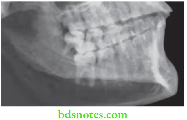
Answer. Osteoradionecrosis is a radiation-induced pathologic process characterized by the chronic and painful infection and necrosis is accompanied by the late sequestration and sometimes permanent deformity.
This is one of the most serious complications of radiation to head and neck seen frequently today because of better treatment modalities and prevention.
Factors Leading to Osteoradionecrosis
- Irradiation of an area of previous surgery before adequate healing had taken place.
- Irradiation of lesion in close proximity to bone.
- Prolong oral hygiene and continued use of irritants.
- Poor patient’s corporation in managing irradiated tissues.
- Surgery in irradiated area.
- Failure to prevent trauma to irradiated bony areas.

Osteoradionecrosis Clinical Features
- Osteoradionecrosis is the result of non healing dead bone.
- Mandible is affected more commonly than maxilla.
Osteoradionecrosis Treatment
- Debridement of necrotic tissue should be done along with removal of sequestrum.
- Administration of intravenous antibiotic and hyperbaric oxygen therapy are essential.
- Maintenance of oral hygiene is necessary.
Question 4. Write short note on Melkersson-Rosenthal syndrome.
Answer. Melkersson-Rosenthal syndrome is a rare neurological disorder.
- It consists of following three condition, i.e. cheilitis granulomatosa, facial paralysis and scrotal tongue.
- Onset is in childhood or early adolescence.
- After recurrent attacks (ranging from days to years in between), swelling may persist and increase, eventually becoming permanent.
- The lip may become hard, cracked, and fissured with a reddish-brown discoloration.
- The cause of Melkersson-Rosenthal syndrome is unknown, but there may be a genetic predisposition.
- It can be symptomatic of Crohn’s disease or sarcoidosis.
Treatment of Melkersson-Rosenthal syndrome
- Treatment is symptomatic and may include nonsteroidal anti-inflammatory drugs (NSAIDs) and corticosteroids to reduce swelling, antibiotics and immunosuppressants.
- Surgery may be indicated to relieve pressure on the facial nerves and reduce swelling, but its efficacy is uncertain.
- Massage and electrical stimulation may also be prescribed.
Question 5. Write short note on dental management in hypertensive patient.
Answer. Following steps are to be undertaken while treating the hypertensive patient:
- Take BP before and after injection of local anesthetics with vasoconstrictors.
- Defer elective care and provide only urgent care for patients with Stage 3 hypertension or those experiencing hypertensive signs and symptoms. Minimize or avoid the use of vasoconstrictors in these patients. Refer immediately or within one week to the appropriate medical provider depending on the clinical situation.
- Avoid long, stressful appointments.
- Nitrous oxide may be beneficial in controlling anxiety, however rebound hypertension may result if inadequate oxygenation (hypoxia) occurs.
- Minimize the use of local anesthesia with vasoconstrictor to 0.036–0.054 mg of epinephrine (2–3 cartridges of 2% lidocaine with 1:100,000 epinephrine) per visit in Stage 2,3, and Risk Group C patients.
- Avoid epinephrine impregnated retraction cords in Stages 1-3.
Question 6. Write short note on differential diagnosis of cervicofacial lymphadenopathy.
Answer. Following is the differential diagnosis of cervicofacial lymphadenopathy:
Cervicofacial Lymphadenopathy Infective Causes
- Acute lymphadenitis
- Chronic non-specific lymphadenitis
- Tuberculous lymphadenitis
- Infectious mononucleosis
- Toxoplasmosis
- Cat scratch fever.
Cervicofacial Lymphadenopathy Neoplastic Causes
- Metastatic lymph node enlargement
- Lymphomas i.e Hodgkin’s lymphoma, Non-Hodgkin’s lymphoma, Burkitt’s lymphoma
- Lymphosarcoma
- Chronic lymphatic leukemia
Cervicofacial Lymphadenopathy Other Causes
- Autoimmune diseases i.e. Systemic lupus erythematosus, rheumatoid arthritis
- HIV infection, immunosuppression.
Acute Suppurative Lymphadenitis
- It is a bacterial infection leading to acute inflammation and suppuration of lymph nodes caused by group A streptococci or staphylococci.
- There is presence of tender, enlarged, firm or soft palpable neck lymph nodes.
- Overlying skin may become red, hot and brawny.
Chronic Non-specific Lymphadenitis
- Infections such as chronic tonsillitis, recurrent dental infection can lead to chronic non-specific lymphadenitis.
- There is presence of firm, non-tender, multiple, bilateral lymph node enlargement in neck.
Tuberculous Lymphadenitis
- It is caused by Mycobacterium tuberculosis infection.
- Commonly affect deep cervical lymph nodes. Besides these mesenteric and axillary lymph nodes are also affected.
- Lymph nodes are matted, enlarged with cold abscess or sinus formation.
- Fever with chills, weight loss, anorexia and respiratory complaints may be present.
Infectious Mononucleosis
- Caused by Epstein-Barr virus
- Lymph nodes are enlarged, discrete and slightly tender affecting, especially the cervical and submandibular lymph nodes.
- Petechial rash can occur at junction of soft and hard palate on 4th day and can persist for 3 to 4 days.
- Monospot test is positive
Syphilitic Lymphadenitis
- Painless, firm, discrete and shotty glands which do not suppurate.
- In secondary syphilis, generalized lymphadenopathy is present.
Toxoplasmosis
- Caused by Toxoplasma gondii, protozoa through meat.
- Cervical lymphadenopathy is most common.
- Diagnosed by Sabin-Feldman dye test.
Cat Scratch Disease
- Caused by Bartonella henselae
- Lymphadenopathy is present.
- Diagnosed by lymph node biopsy with Warthin-Starry staining
Lymphomas
- In Hodgkin’s lymphoma, there is painless progressive enlargement of lymph nodes. Lymph nodes are smooth, firm, non-tender. In this, cervical lymph nodes, i.e. lower deep cervical group in posterior triangle are commonly enlarged. In this type, peripheral lymph node involvement is not common.
- In non-Hodgkin’s lymphoma any group of lymph node is involved. In this type, peripheral lymph node involvement is common.
Lymphosarcoma
- Commonly affects cervical lymph nodes which become enlarged, firm and fixed.
- Overlying skin is stretched and shiny with dilated blue veins under it.
- High malignant tumors grow rapidly and invade surrounding tissues.
Metastatic Lymph Node Enlargement
- Cervical and other lymph nodes become enlarged, irregular and fixed to structures including skin.
- Consistency of lymph nodes is stony hard.
- Patient may be cachectic and wasted.
HIV Associated Lymphadenopathy
- It is known as persistent generalized lymphadenopathy.
- It is seen in the stage of intermediate immune depletion following HIV infection.

Leave a Reply