Oral cavity
Question 1. Write a short note on oral cavity.
Answer.
Oral Cavity Boundaries
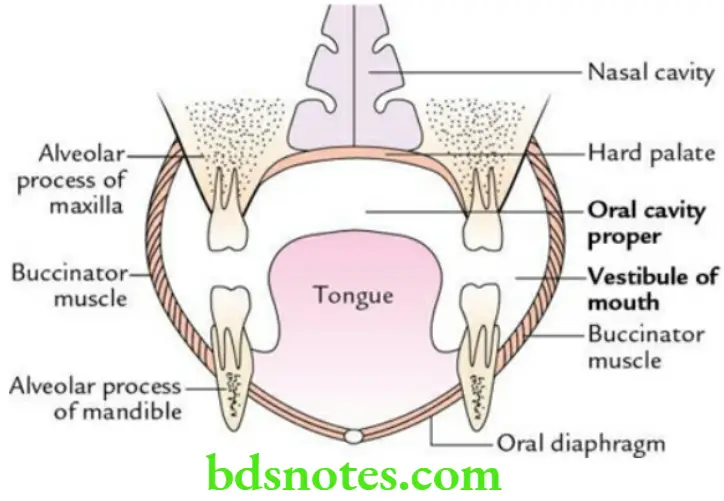
Oral Cavity Roof: Hard palate
Oral Cavity Floor: Oral diaphragm
Oral Cavity On either side: Cheek
Oral Cavity Communications
Anteriorly: To exterior through oral fissure guarded by the upper and lower lips.
Posteriorly: To oropharynx through oropharyngeal isthmus guarded on either side by the palatoglossal arch.
Structures present within the oral cavity
- Teeth and gums
- Tongue
- Soft palate
Question 2. What are the parts of the oral cavity? List their boundaries.
Answer.
The oral cavity (mouth) is divided into two parts:
- an outer smaller part called the vestibule of the mouth and
- an inner larger part called oral cavity proper.
Boundaries of vestibule of mouth
Oral Cavity Externally: Lips and cheeks
Oral Cavity Internally: Teeth and gums
Boundaries of oral cavity proper
Above (roof): Hard palate
Below (floor): Oral diaphragm formed by two mylohyoid muscles
On either side (lateral): Teeth and gums
Question 3. Enumerate the ducts that open in the oral cavity.
Answer.
Oral Cavity Parotid ducts One on either side, open in the vestibule of the mouth opposite the crown of the 2nd upper molar tooth.
Oral Cavity Submandibular ducts One on either side, open in the floor of the oral cavity proper on the summit of the sublingual papilla.
Oral Cavity Sublingual ducts About a dozen in number, on either side, open in the oral cavity proper on the sublingual fold in a row.
Question 4. Enumerate the layers of the cheek.
Answer.
From superficial to deep, the layers of the cheek are as follows:
- Skin
- Superficial fascia containing a buccal pad of fat
- Buccopharyngeal fascia
- Buccinator muscle
- Submucosa
- Buccal mucosa
Question 5. Briefly describe the parts of a tooth.
Answer.
Tooth Parts: Each tooth consists of three parts:
Tooth Parts Crown: A part that projects above the gum
Tooth Parts Neck: A part between crown and root, and surrounded by gum
Tooth Parts Root: A part that is embedded in the alveolar process of the jaw
Question 6. Write a short note on the structure of the tooth.
Answer.
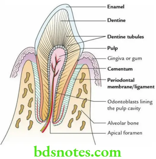
Structurally, each tooth is made up of five components:
- Pulp – an inner core of soft tissue containing blood vessels and nerves.
- Dentine – a calcified material surrounding the pulp/pulp cavity.
- Enamel – a densely calcified material covering the crown.
- Cementum – a thin bony covering over the dentine.
- Periodontal membrane – a fibrous membrane (akin to periosteum), connecting the root of the tooth with the alveolar socket.
Read And Learn More: Selective Anatomy Notes And Question And Answers
Question 7. Write a short note on the development of a tooth.
Answer.
The tooth develops from two sources:
- Ectodermal epithelial lining the alveolar process of the jaw and
- Underlying neural crest mesenchyme.
The details are summarized as follows:
Ectodermal epithelium lining of alveolar process → Dental lamina → Tooth buds/enamel organs → Ameloblasts → Enamel.
The structural components of the tooth are derived from these two sources.
Source of Development of Various Components of the Tooth

Question 8. Briefly describe the stages of tooth development.
Answer.
The stages in the development of a tooth are:
- Dental lamina
- Enamel organs
- Dental papilla
- Dental sac
Question 9. Briefly describe the eruption and shedding of teeth.
Answer.
Human beings are diphyodont animals, i.e. they have two sets of teeth, which develop at different times of life. The two sets are
- Deciduous teeth, develop first and shed off, and
- Permanent teeth, appear later after the shedding of deciduous teeth and do not shed off.
The usual time of eruption and shedding of the teeth.
Eruption and Shedding of Deciduous Teeth

Eruption and Shedding of Permanent Teeth

Question 10. Define tongue and list its functions.
Answer.
The tongue is a mobile, muscular organ situated on the floor of the mouth. It performs the following functions:
- Taste
- Speech
- Mastication
- Deglutition
Question 11. Enumerate the external features of the tongue.
Answer.
- The tongue has an apex, tip, and body.
- Body presents:
- Dorsal surface (also called dorsum)
- Ventral surface
- Right lateral margin
- Left lateral margin
Dorsum of Tongue
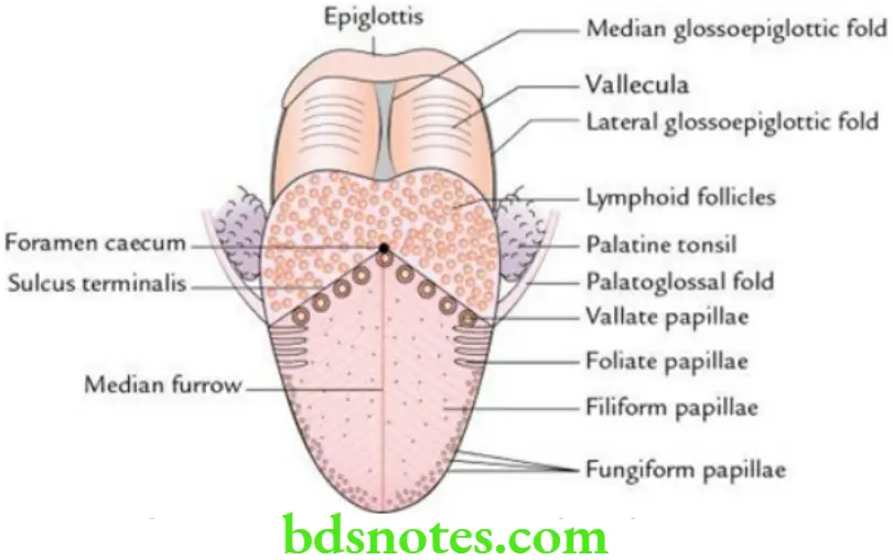
- Anatomically and developmentally, the dorsum of the tongue is divided into two parts: anterior two-thirds (oral part) and posterior one-third (pharyngeal part). The two parts are separated from each other by a V-shaped sulcus – the sulcus terminalis.
- A blind foramen at the apex of sulcus is called foramen caecum. The foramen caecum represents the site of development of the endodermal thyroglossal duct which grows down into the neck during embryonic development.
Features on the dorsal surface of Tongue
- Posterior one-third presents:
- A large number of lymphoid follicles, which together form lingual tonsils.
- A large number of openings of mucous and serous glands.
- Anterior two-thirds presents:
- A median furrow.
- A large number of papillae.
Features on the ventral surface of the tongue The ventral surface of the tongue presents:
- Frenulum linguae: A median fold of mucous membrane extending between the tongue and the floor of the mouth.
- Plica fimbriata: Two, fringed corrugated folds of mucous membrane, one on either side of frenulum linguae converging towards the tip of the tongue.
- Prominences of deep lingual veins: These are visible, one on either side, between frenulum linguae and plica fimbriata.
Question 12. Enumerate the features in the sublingual region.
Answer.
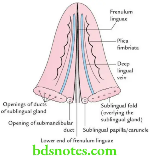
Sublingual papillae Two rounded elevations – one on either side of the root of frenulum linguae for the opening of the submandibular gland duct.
Sublingual folds
- Two elongated elevations – one on either side of frenulum linguae on the floor of the mouth produced by an underlying sublingual salivary gland.
- The sublingual ducts open on these folds.
Question 13. Enumerate the various types of papillae of the tongue.
Answer.
Vallate papillae They are of large size (1–2 mm in diameter) and are located in front of the sulcus terminalis. They are 8–12 in number and surrounded by a ditch (trench). The taste buds are found in the wall of the ditch.
Fungiform papillae They are numerous and located near the tip and margins of the tongue. They have a narrow pedicle and rounded head.
Filiform papillae These are the smallest and most numerous, and cover the dorsum of the anterior two – third of the tongue and give it a characteristic velvety appearance.
Foliate papillae These are transverse mucosal folds on the lateral margins of the tongue, in front of the palatoglossal arch. The papillae of the tongue.
Question 14. Describe the tongue under the following headings:
- Muscles of the tongue,
- Nerve supply,
- Blood supply,
- Lymphatic drainage and
- Applied anatomy.
Answer.
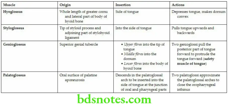
Muscles of tongue: The muscles of the tongue are paired and divided into two groups: intrinsic and extrinsic.
Intrinsic Muscles (arise and are inserted within the tongue). These are as follows:
- Superior longitudinal
- Inferior longitudinal
- Transverse
- Vertical
Extrinsic Muscles (arise outside the tongue but are inserted into the tongue).
These are as follows:
- Genioglossus
- Hyoglossus
- Styloglossus
- Palatoglossus
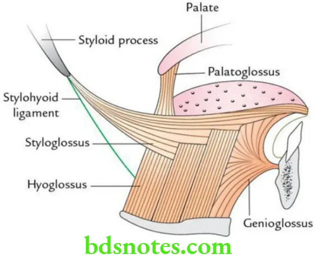
The origin, insertion, and actions of extrinsic muscles.
Origin, Insertion, and Actions of Extrinsic Muscles of the Tongue
The origin and insertion of extrinsic muscles of the tongue.
Nerve supply
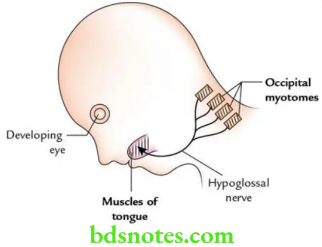
Motor Supply: All the intrinsic and extrinsic muscles of the tongue are supplied by the hypoglossal nerve, except palatoglossus which is supplied by the cranial root of accessory nerve via pharyngeal plexus.
Sensory supply:

Blood supply
- The lingual artery (chief artery of the tongue), a branch of the external carotid artery supplies the oral part of the tongue.
- Tonsillar and ascending palatine arteries, branches of the facial artery supply the pharyngeal part of the tongue.
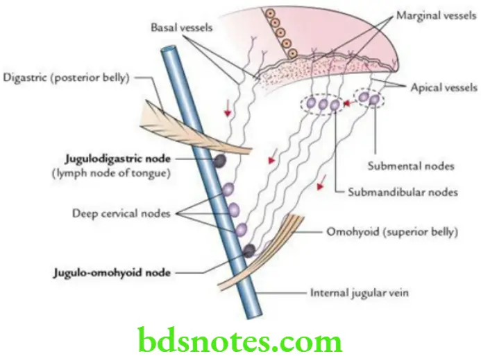
Lymphatic drainage The lymph from the tongue is drained by the following three sets of lymph vessels: marginal, central, and posterior.
Marginal Vessels:
- From the tip, drains bilaterally into the submental lymph nodes.
- From the margins and lateral part of the dorsum of the tongue, drains into the submandibular and jugulo-omohyoid lymph nodes.
Central Vessels: From the central region of the dorsum of the anterior two-thirds of the tongue descends between genioglossi muscles and drains bilaterally into the submandibular lymph nodes.
Posterior Vessels: From the posterior one-third of the tongue drains bilaterally into the deep cervical lymph nodes, principally into the jugulodigastric lymph node.
Applied anatomy
- Injury of the hypoglossal nerve causes paralysis of muscles of the tongue on the side of the lesion; hence, the protruded tongue deviates to the same side (i.e. the side of injury) due to unopposed action of muscles on the healthy side.
- In an unconscious patient, the tongue may fall backward into the oropharynx and obstruct the air passage to cause choking. This can be prevented by turning the head to one side and pulling the mandible forward.
- Carcinoma of the tongue most commonly occurs along the margin of the tongue. Cancer in the posterior one-third of the tongue has a bad prognosis because of bilateral lymphatic drainage.
Question 15. Describe the development of the tongue in brief and correlate the nerve supply of the tongue with its development.
Answer.
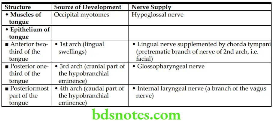
- The tongue develops in the floor of the primitive pharynx from the 1st, 2nd, 3rd, and 4th pharyngeal arches.
- The epithelium, muscles, and connective tissue of the tongue develop as follows.
Tongue Development Epithelium The epithelium of the tongue develops from four swellings:
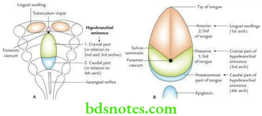
Correlation of Nerve Supply of Tongue with Its Development
The various parts of the tongue develop from above-mentioned four swellings as follows:
- Epithelium of the anterior two-thirds of the tongue develops from two lingual swellings and tuberculum impair derived from 1st arch. The contribution from tuberculum impact is insignificant.
- The epithelium of the posterior one-third of the tongue develops from the cranial (anterior) part of the hypobranchial eminence, derived from 3rd arch.
- The epithelium of the posteriormost part develops from the caudal (posterior) part of the hypobranchial eminence derived from the 4th arch.
Muscles The muscles of the tongue develop from occipital myotomes.
Connective tissue The connective tissue of the tongue develops from local mesenchyme.
Question 16. Give the embryological basis of tongue-tie.
Answer.
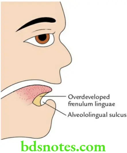
The tongue-tie (ankyloglossia) occurs when the frenulum linguae is overdeveloped and extends up to the tip of the tongue.
The overdevelopment of frenulum linguae occurs due to the incomplete separation of the tongue from the floor of the primitive mouth by alveololingual sulcus.
- Clinically it presents as:
- Disturbed speech, i.e. difficulty in speaking
- Restriction of tongue movements especially one which prevents protrusion

Leave a Reply