Nematodes
Question 1. Write a short note on the mode of infection by Ascaris lumbricoides. Name the definitive host.
Answer:
Mode of Infection
- Infective form: Embryonated eggs
- Portal of entry: Alimentary canal
- Site of location: Small Intestine
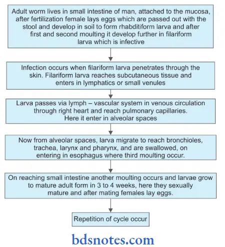
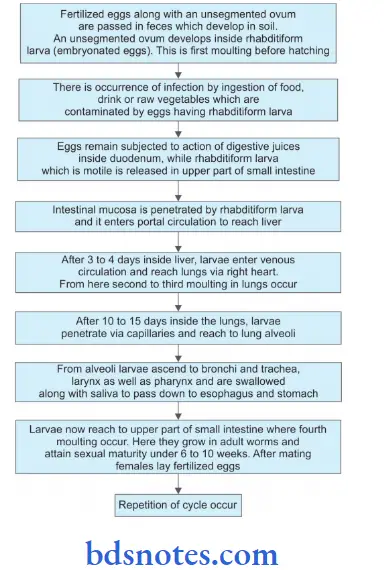
- The adult worm lives in the jejunum of man.
- Fertilized eggs containing unsegmented ova are passed in feces.
- Eggs are not infective to man.
- Eggs undergo development in soil and have a period of incubation in soil for acquiring infectivity.
- A rhabditiform larva is developed from an unsegmented ovum and undergoes fist molting within an eggshell in 10–40 days.
- Eggs containing rhabditiform larvae are pathogenic to man and are known as embryonated eggs.
- In the upper part of the small intestine, rhabditiform larvae are liberated from embryonated eggs.
- These newly hatched larvae reach the mucous membrane of the small intestine and are carried by portal circulation to the liver where they reside for 3 to 4 days.
- Later on they pass out of the liver and via right heart enter pulmonary circulation.
- The definitive host is a human being.
“Understanding nematodes through FAQs: Q&A explained”
Read And Learn More: Microbiology Question And Answers

“How do nematodes cause diseases in humans? FAQ answered”
Question 2. Describe morphology, life cycle, pathogenicity, and lab diagnosis of Ancylostoma duodenale.
Answer:
Morphology
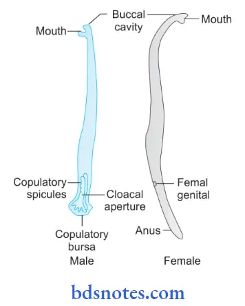
- Adult Worm:
- Its shape is cylindrical and is small. Its color is grayish-white.
- Its anterior end is bent dorsally.
- The freshly passed worm is usually reddish brown due to ingested blood in its intestine.
- The mouth of an adult worm consists of six teeth, which are four hook-like on the ventral surface and two knob-like over the dorsal surface.
- Male worm: It is small and is usually 8 to 11 × 0.4 mm. The posterior end of the male worm consists of an umbrella-like ovulatory bursa with two ovulatory spicules.
- The bursa consists of three lobes, i.e. one dorsal and two lateral.
- These three lobes are supported by 13 chitinous rays, i.e. three surround the dorsal lobe and five surround the ventral lobe.
- Female worm: It is larger as compared to male. It is 10 to 13 × 0.6 mm. The posterior end of the female worm is tapering, and the bursa is absent.
- The vulva opens ventrally at the junction of the posterior and middle third of the body.
- The female worm is oviparous, i.e. a mature female can lay 10,000 to 25,000 eggs in a day.
- Egg:
- The shape of the eggs is oval or elliptical.
- The size of an egg is 60 × 40 µ.
- Eggs are colorless and are non-bile stained.
- Eggshell is thin, transparent hyaline.
- When the egg is kept in a saturated solution of common salt, it flats over it.
- Ovum is segmented with 4 to 8 blastomeres
“Importance of studying nematodes for parasitology students: Questions explained”
Life Cycle of Ancylostoma Duodenal
“Common challenges in understanding nematodes effectively: FAQs provided”
Pathogenicity
- Due to larva:
- Ground itch or dermatitis: It is characterized by pruritic maculopapular dermatitis during the entrance of larvae.
- Creeping eruption: It is characterized by red itchy papules along the path traveled by larvae. Larva wanders aimlessly via skin layers for several weeks and months, producing symptoms known as larva migrans. This also may lead to bronchitis or bronchopneumonia.
- Due to adult worms:
- In the early phase, the adult worm causes epigastric pain, vomiting, and diarrhea.
- There is also the presence of anemia, which is microcytic photochromic.
- This occurs because of the sucking of blood by adult worms via a wound created in intestinal mucosa at the time of its attachment.
- A single adult worm can suck 0.2 mL of blood/day. Sucking of blood occurs by pumping in the action of the esophagus of the worm, which is assisted by the secretion of anti-coagulant by the buccal capsule.
- Mainly iron-deficiency anemia is present because of a deficiency of iron and various hemopoietic substances in diet and blood loss due to heavy infection.
- There can also be the presence of microcytic anemia due to a deficiency of folic acid and vitamin B12. Dimorphic anemia can also result as there is a deficiency of iron, vitamin B12, or folic acid.
- Individuals who remain malnourished suffer from hypoproteinemia.
“Why is early learning of nematodes critical for global health? Answered”
Laboratory Diagnosis
- Stool examination for adult worm (naked eye).
- Microscopic examination of stool for ova by saline and iodine preparation.
- Study of duodenal contents for adult worm and ova by saline and iodine preparation.
- Egg counting to find out the intensity of infection. If there are more than 50 eggs per milligram of feces, this indicates a massive infection.
- Indirect evidence:
- Blood examination for anemia and eosinophilic.
- Stool examination for occult blood and Charcot-Leyden crystals.
“Factors influencing success with nematode knowledge: Q&A”
Question 3. Write a short note on common ova found in stool.
Answer:
The following ova are found in human faces:
1. Eggs of Ascaris lumbricoides: Its eggs are of two types, i.e. fertilized eggs and unfertilized egg
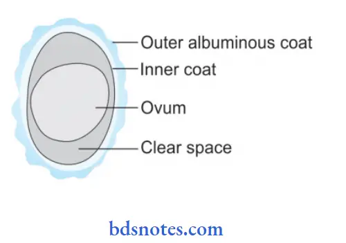
- Fertilized Egg:
- It is round to oval in shape.
- Its size is 60 to 70 µ × 40 to 50 µ.
- It is yellow-brown and bile stained.
- Its eggshell is thick, smooth, and translucent with an uneven outer albuminous coat known as rugosities or mammilliations. This albuminous coat is sometimes absent.
- The ovum is large and unsegmented.
- There is the presence of a clear, crescent area between the ovum and eggshell at each pole
- Egg flats on the saturated solution of common salt.
- Unfertilized egg:
- Female worm if remain unfertilized produces eggs.
- Its shape is narrow, long, and more elliptical as compared to a fertilized egg
- Its size is 80 to 90 µ × 45 to 55 µ
- Its color is brownish (bile stained)
- Eggshell of an unfertilized egg is thin with an irregular outer albuminous coat
- It has a small or atrophied ovum having a mass of disorganized and highly refractile granules of various sizes.
- The egg does not flat on the saturated solution of common salt.
“Steps to explain types of nematodes: Ascaris vs Ancylostoma vs Enterobius: Q&A guide”
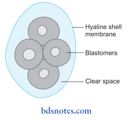
2. Eggs of Ancylostoma duodenal:
- They are oval or elliptical in shape.
- The size of an egg is 60 × 40 m
- They are colorless (not bile stained)
- They are surrounded by transparent hyaline shell membrane
- When the egg is kept in a saturated solution of common salt, it flats over it
- They contain a segmented ovum, usually four Blastomers.
“Role of nematode life cycles in disease transmission: Questions answered”
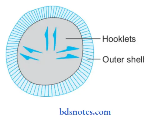
3. Eggs of Echinococcus granulosus:
- It is oval.
- Its size is 32 to 36 µ × 25 to 32 µ.
- It is brown in color when bile is stained.
- The outer layer surrounds inner embryophore
- The embryo consists of six hooklets, i.e. three pairs
- Eggs cause infection to humans, cattle, and sheep
- These eggs are excreted in the feces of canine animals.
- By single egg—one larvae consists of many scolices from which multiple adult worms are formed.
“How does Wuchereria bancrofti cause lymphatic filariasis? FAQ explained”
Question 4. Write a short note on the laboratory diagnosis of Ascaris lumbricoid.
Answer:
The following is the laboratory diagnosis of Ascaris lumbricoides:
- Stool examination: During stool examination, adult worms or eggs may be demonstrated. Eggs can be detected by direct microscopic examination of stool specimen or after concentration of same.
- Demonstration of adult worm: Adult worms may pass through the anus, mouth, and nose. Barium meal demonstrates
presence of adult worms in the small intestine. - Demonstration of larvae: Larvae may be detected in sputum during the stage of migration.
- Serodiagnosis: Antibodies against A. lumbricoides can be detected. These tests are used in the diagnosis of
intestinal ascariasis. - Blood examination: It demonstrates eosinophilia and is observed in the larval invasion stage.
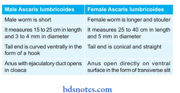
Question 5. Tabulate the difference between male and female Ascaris lumbricoides.
Answer:
“Early warning signs of gaps in understanding nematode basics: Common questions”
Question 6. Write briefly on hookworm infection.
Answer:
Human hookworms are Ancylostoma duodenale and Nector americanus.
Infections Caused by Hookworms
- Due to Larva:
- Ground itch or dermatitis: It is characterized by pruritic maculopapular dermatitis during the entrance of larvae.
- Creeping eruption: It is characterized by red itchy papules along the path traveled by larvae. Larva wanders aimlessly via skin layers for several weeks and months, producing symptoms known as larva migrans. This also may lead to bronchitis or bronchopneumonia.
- Due to Adult Worm:
- In the early phase, the adult worm causes epigastric pain, vomiting, and diarrhea.
- There is also the presence of anemia, which is microcytic hypochromic.
- This occurs because of the sucking of blood by adult worms via a wound created in intestinal mucosa at the time of its attachment.
- A single adult worm can suck 0.2 mL of blood/day. Sucking of blood occurs by the pumping action of the esophagus of the worm, which is assisted by the secretion of anti-coagulant by the buccal capsule.
- Mainly iron-deficiency anemia is present because of a deficiency of iron and various hemopoietic substances in diet and blood loss due to heavy infection.
- There can also be the presence of microcytic anemia due to a deficiency of folic acid and vitamin B 12. Dimorphic anemia can also result as there is a deficiency of iron, vitamin B12, or folic acid.
- Individuals who remain malnourished suffer from hypoproteinemia.
“Asymptomatic vs symptomatic effects of ignoring nematode principles: Q&A”
Question 7. Write a short note on the life cycle of Ascaris lumbricoides.
Answer:
Ascaris lumbricoides pass their life cycle only in a single host, i.e. man. Man is the only definitive host.
Leave a Reply