Neck Swelling
Question.1.Describe clinical features and treatment of carotid body tumor.
Answer. It is also called as chemodectoma or potato tumor.
Definition: It is a non-chromaffin paraganglioma.
It most commonly arises near the bifurcation of common carotid artery.
It is a benign tumor.
Carotid body tumor Clinical Features
- It is usually unilateral.
- More common in middle age.
- Swelling (75%) in the carotid region of the neck which is smooth, fim, pulsatile and moves only side to side but not in vertical direction.
- It can often compress over esophagus and larynx.
- Headache, neck pain, dysphagia, syncope are other presentations.
- It can present with unilateral vocal cord palsy; can cause Horner’s syndrome.
- Features of transient ischemic attcks due to compression over the carotids, “carotid body syncope. “
- Thrill may be felt and bruit may be heard.
- It is located at the level of hyoid bone deep to anterior edge of the sternomastoid muscle in anterior triangle, vertically placed, round, fim ‘potato-like swelling.
- Often tumor may extend into the cranial cavity along with internal carotid artery as dumbbell tumor.
Read And Learn More: General Surgery Question And Answers
Carotid body tumor Treatment
- If it is small, it can be excised easily as the tumor is situated in adventitia.
- When it is large, as commonly observed, complete excision has to be done followed by placing a vascular graft.
- During resection a temporary shunt is placed between common carotid below and internal carotid above to safeguard cerebral perfusion; external carotid artery is ligated. Venous or prosthetic graft is placed between common carotid and internal carotid arteries.
Question.2. Write in short torticollis.
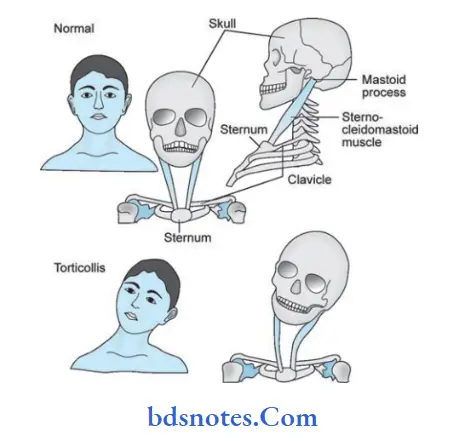
Answer. Torticollis or wryneck is a deformity in which the head is bent to one side with the chin point to the outer side.
In long standing cases there may be atrophy of the face on the affected side.
The different varieties of wry neck are:
- Congenital:
- The diagnosis is made by a history of diffilt labor, followed by the appearance of a sternomastoid tumor.
- The affected muscle feels fim and rigid.
- Traumatic: Fracture dislocation of the cervical spine.
- Rheumatic: Sudden appearance of wryneck after an exposure to cold or draught is suggestive.
- Inflammatory: For example, from inflamed cervical lymph node.
- Spasmodic: When the sternomastoid of the affcted side and the posterior cervical muscle ofthe opposite side are found in a state of spasm.
- Compensatory: For example, from scoliosis, defect in sight (ocular torticollis)
- From Potts disease of the cervical spine.
- From contracture: For example, after burns, ulcer, etc.
Torticollis Features
- Restricted neck movements
- Chin pointing towards opposite side
- Presence of squint
Torticollis Treatment
Botulinus toxins have been used to inhibit the spastic contraction of affcted muscle.

Question.3. Discuss the differential diagnosis of swelling in the lateral aspect of neck.
Answer. It is classified according to their location in three triangles of the neck:
1.Submandibular or digastric triangle
- Enlarged lymph node
- Enlarged submandibular salivary gland
- Calculus
- Chronic sialadenitis
- Cancer
- Chronic diseases-autoimmune.
2. Carotid triangle
- Aneurysm of carotid artery
- Carotid body tumor
- Branchial cyst
- Neurofiroma vagus
- Enlargement of thyroid gland
- Lymph node swelling (Cold abscess)
- Laryngocele.
- Sternomastoid tumor.
3.In posterior triangle
Solid swellings:
- Metastasis in lymph node
- Tuberculosis
- Lymphoma
- Lipoma
- Cervical rib
- Pancoast tumor.
Cystic swellings:
- Lymphangioma
- Hemangioma
- Cold abscess.
Pulsatile swellings:
- Subclavian artery aneurysm
- Vertebral artery aneurysm.
Sub-mandibular or digastric triangle
Enlarged Sub-mandibular lymph node
They form a nodular swelling which is deep to deep fascia.
They are palpable only in the neck.
The nodes can get enlarged due to following conditions:
- Acute lymphadenitis: Very often, poor oral hygiene or a caries tooth produces painful, tender, soft enlargement of these lymph nodes. Extraction of the tooth or with improvement of oral hygiene, lymph nodes regress.
- Chronic tuberculous lymphadenitis can affct these nodes along with upper deep cervical nodes.
The nodes are fim and matted. - Secondaries in the submandibular lymph nodes arise from carcinoma of the cheek, tongue, and palate.
The nodes are hard with or without fifty.
Non-Hodgkin’s lymphoma can involve submandibular lymph nodes along with horizontal group of nodes in the neck.
The nodes are fim or rubbery in consistency.
Submandibular Salivary Gland enlargement
The common causes are chronic sialadenitis with or without a stone, tumors of the salivary gland or enlargement due to autoimmune diseases.
They form irregular or nodular swelling.
The diagnosis is confimed by bidigital palpation of the gland.
Enlarged submandibular gland is bidigitally palpable because the deep lobe is deep to mylohyoid muscle.
Carotid triangle
- Branchial cyst: It is located in anterior triangle of neck.
It is soft, cystic, fluctuant, and transillumination negative. - Lymph node swelling (Cold abscess): Patient present with history of tuberculosis. Lymph nodes are fim and mattd.
Signs of inflmmation are absent. - Aneurysm of carotid artery: It is fim, flctuant and transillumination negative swelling with presence of expansile pulsations. Bruit/thrill can be heard.
- Carotid body tumor: It is has a typical location, i.e. located at level of hyoid bone in upper part of anterior triangle of neck beneath anterior edge of sternomastoid muscle.
On palpation it moves in transverse direction. Surface is smooth or lobulated, borders are round, oval in shape,vertically placed swelling. - Sternomastoid tumor: Swelling is present in infants or children.
It is tender, mobile sideways, medial and lateral borders are distinct.
Both superior and inferior borders are continuous with the swelling. - Laryngocele: It is a smooth, oval, boggy swelling which moves upwards on swallowing.
Expansile cough impulse is present. - Neurofibroma of vagus nerve: It produces swelling in carotid triangle in region of thyroid swelling.
It is a vertically placed oval swelling.
On pressure over the swelling dry cough occurs and in some cases bradycardia can occur.
Posterior triangle
Solid Swellings
- Metastasis in lymph nodes: Lymph nodes become enlarged and become fied to the underlying structures.
They become immobile and are stoney hard in consistency. - Tuberculosis: Lymph nodes become enlarged mainly cervical. The nodes are fim and mattd.
- Lymphoma: It can involve submandibular lymph nodes along with horizontal group of nodes in the neck. The nodes are fim or rubbery in consistency.
- Lipoma: It is a localized swelling with lobular surface,non-tender. It is semiflctuant and non-transilluminant. It is mobile with edges slipping between palpating figers.
- Cervical rib: It is an extra, rib present in the neck. A hard mass is visible or palpated in root of neck.
- Pancoast tumor: It is a tumor felt in the lower part of posterior triangle. It is hard in consistency, fied, irregular
and sometimes tender. Lower border of mass cannot be appreciated.
Cystic Swellings
- Lymphangioma: Skin vesicles contain watery or yellow flid. Bleeding in vesicle turn into brown or black.
Area is soft, spongy, often flctuant with flid thrill and translucency. It is non-compressible.
Vesicles will not fade on pressure.
Hemangioma: Swelling is warm and bluish in color, nonpulsatile, soft, flctuant, transillumination negative.
Compressibility is present.
When the swelling is compressed between figers blood diffuses under vascular spaces and when pressure is released it slowly fills up.
Cold abscess: The patient present with history of tuberculosis.
Lymph nodes are fim and mattd. Signs of inflmmation are absent.
Question.4. Write short note on swelling midline of neck.
Or
Write briefly on midline swellings of neck.
Answer. The midline swellings of neck are:
- Ludwig’s angina
- Enlarged sub-mental lymph node
- Sub-lingual dermoid cyst
- Thyroglossal cyst
- Sub-hyoid bursitis
- Goiter of thyroid, isthmus and pyramidal lobe
- Enlarged lymphnode and lipoma insubsternal space of burns
- Retrosternal goiter
- Thymic swelling
- Bony swelling arising from the manubrium sterni.
Swelling midline of neck ludwig’s angina
- This is an inflammatory edema of the flor of the mouth. It spreads to the submandibular region and sub-mental region.
- Tense, tender, browny edematous swelling in the submental region with putrid halitosis is characteristic of this condition.
Enlarged Submental Lymph Nodes
The three important causes ofenlargement:
1. Tuberculosis: Mattd submental nodes, fim in consistency,with enlarged upper deep cervical lymph nodes, with or without evening rise of temperature are suggestive of tuberculosis.
2. Non-Hodgkin’s lymphoma can present with sub-mental nodes along with other lymph nodes in the horizontal group of nodes such as submandibular, upper deep cervical, pre-auricular, post-auricular and occipital lymph nodes (external Waldeyer’s ring). Nodes are firm or rubbery, discrete without matting.
3. Secondaries in the submental lymph nodes can arise from carcinoma of the tip of the tongue, flor of the mouth,central portion of the lower lip.
The nodes are hard in consistency and sometimes, fied.
Sublingual dermoid Cyst
- It is a type of sequestration dermoid cyst which occurs due to sequestration of the surface ectoderm at the site of fusion of the two mandibular arches. Hence, such a cyst occurs in the midline, in the flor of the mouth.
- When they arise from 2nd branchial cleft, they are found lateral to the midline. Hence, lateral variety.
Subhyoid Bursitis
- Accumulation of inflammatory flid in the subhyoid bursa results in a swelling and is described as sub hyoid bursitis.
- Bursa is located below the hyoid bone and in front of the thyrohyoid membrane.
- It is swelling in front of neck in midline below hyoid bone.
- Swelling moves up with deglutition and is tender.
Thyroglossal Cyst
- It arises from thyroglossal tract or duct which extends from foramen cecum at base of tongue to isthumus of thyroid.
- It is common in females and is painless midline swelling.
Swelling is deviated to left side. - Thyroglossal cyst exhibits three types of mobilities, i.e.
1. It moves upwards with deglutition
2. Cyst moves with protrusion of tongue
3. Swelling move sideways but not vertically as it is tethered by the thyroglossal duct.
Enlarged Isthmus Of Thyroid Gland
Almostall the diseases ofthe thyroid gland result in enlargement of the isthmus. However, a solitary nodule and cysts can occur in relation to isthmus.
The swelling moves with deglutition.
However, it does not move on protrusion of the tongue.
Pretracheal and Prelaryngeal lymph nodes
These lymph nodes produce nodular swelling in the midline.
One or two discrete nodes are palpable. They can enlarge due to following conditions:
- Acute laryngitis: The nodes are tender, soft.
- Papillary carcinoma ofthyroid: The nodes are fim without mattng, with or without evidence of thyroid nodule.
- Carcinoma ofthe larynx: The nodes are hard in consistency.
Thymic Swelling
- It is caused by an aneurysm of innominate or subclavian artery.
- It is a pulsatile swelling.
lipoma in Suprasternal Space of Burns
- It is soft and lobular.
- Edge of lipoma slips under the palpating figer.
Question.5. Write short note on pulsatile swellings in neck.
Answer. Following are the pulsatile swellings in neck.
- Aneurysm
1.Arterial Hemangioma:
- An abnormal communication between artery and vein results in AV fitula.
- It is a soft, cystic, flctuant, transillumination negative, pulsatile swelling.
- A continuous bruit/murmur is characteristic.
- On compressing the feeding artery, venous return to heart diminishes which leads to fall in pulse rate and pulse pressure.
2. Cirsoid aneurysms:
- It is a rare variant of capillary hemangioma occurring in skin beneath which abnormal artery communicates with the distended veins.
- Variant of capillary hemangioma
- Pulsatile swelling
- Involves bone
- Treatment is ligation of feeding artery and excision of lesion.
Question.6. Describe various triangles of neck and their boundaries.
Discuss the differential diagnosis of neck swelling.
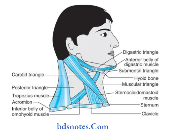
Answer. Each side of neck is the quadrilateral space which is subdivided by sternocleidomastoid into anterior triangle and posterior triangle.
These triangles are further subdivided into
- Anterior Triangle
- Posterior Triangle

Anterior Triangle
Boundaries
- Anterior: Anterior midline of the neck extending from symphysis menti above to the middle of suprasternal notch below.
- Posterior: Anterior border of sternocleidomastoid
- Base: Lower border ofthe body ofmandible and line joining the angle of mandible with the mastoid process
- Apex: Suprasternal notch, at the meeting point between anterior border of sternocleidomastoid and anterior midline.
Subdivisions of anterior triangle
The anterior triangle in subdivided by the diagastric muscle and superior belly of omohyoid into following:
- Submental triangle
- Digastric triangle
- Carotid triangle
- Muscular triangle.
Submental triangle
This triangle is complete only when the neck is seen from the front. Each half of the triangle is visible when viewed from side.
Boundaries
- On each side: Anterior belly of diagastric
- Base: Body of hyoid bone
- Apex: Chin or symphysis menti
- Floor: Oral diaphragm formed by the mylohyoid muscles.
Digastric Triangle
Boundaries
- Anteroinferior: Anterior belly of digastric
- Posteroinferior: Posterior belly of digastric
- Base: Base of the mandible and an imaginary line joining the angle of mandible to the mastoid process
- Apex: Intermediate tendon of diagastric muscle bound down to hyoid bone by a facial sling.
- Floor: Is formed by mylohyoid muscle (anteriorly), hyoglossus muscle and small partofmiddle constrictor(posteriorly).
- Roof: Is formed by the investing layer of deep cervical fascia which splits to enclose the submandibular salivary gland.
Carotid triangle
Boundaries
- Superior: Posterior belly of digastric and stylohyoid
- Anteroinferior: Superior belly of omohyoid
- Posterior: Anterior border of sternocleidomastoid
- Roof: Is formed by investing layer of deep cervical fascia
- Floor: Is formed by four muscles
1. Thyrohyoid
2. Hyoglossus
3. Middle constrictor of pharynx
4. Inferior constrictor of pharynx.
Muscular triangle
Boundaries
- Anterior: Anterior midline of the neck
- Antero-superior: Superior belly of the omohyoid
- Posteroinferior: Anterior border of sternocleidomastoid.
Posterior triangle
Boundaries
- Anterior: Posterior border of sternocleidomastoid
- Posterior: Anterior border of trapezius
- Base: Middle third of the clavicle
- Apex: Meeting point of sternocleidomastoid and trapezius on the superior nuchal line.
- Roof: Is formed by investing layer of deep cervical fascia stretching between sternomastoid and trapezius muscles
- Floor: Is muscular and is formed by following muscles
From above downwards
- Semispinalis capitis
- Splenius capitis
- Levator scapulae
- Scalenus posterior
- Scalenus medius
- Outer border of 1st rib.
Sub-divisions of Posterior triangle
- Occipital triangle
- Supraclavicular triangle
Occipital Triangle (From above downwards)
Occipital artery at apex
Spinal part of accessory nerve
Four cutaneous branches of cervical plexus of nerves
1. Lesser occipital
2. Great auricular
3. Transverse cervical
4. Supra clavicular.
Muscular branches of C3 and C4 nerves
Dorsal scapular nerve.
Supraclavicular triangle
Trunks of brachial plexus of nerves with their branches
- Dorsal scapular
- Long thoracic
- Nerve to subclavius.
Subclavian artery — 3rd part
Subclavian vein
External jugular vein
Supraclavicular lymph nodes.

Leave a Reply