Mycobacteria
Question 1. Describe morphology, staining characteristics, cultural characteristic,s and pathology produced by Mycobacterium tuberculosis.
Or
Describe the morphology and cultural characteristics
Answer:
Morphology
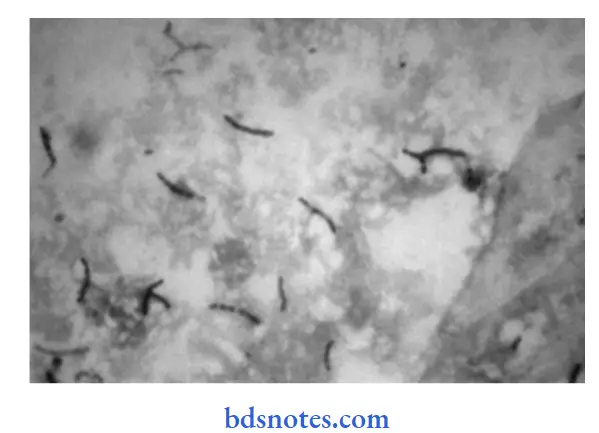
- It is slender, gram-positive, acid-fast bacilli.
- The size of acid-fast rods is 2 to 3µ × 0.4µ.
- It is non-sporing, non-capsulated, and non-motile.
- It is a straight or slightly curved rod with round ends.
- Beaded and the barred forms are seen in the sputum.
- It occurs singly, in pairs, in bundles/clumps.
Read And Learn More: Microbiology Question And Answers
Staining Characteristics
- Ziehl-Neelsen Staining: Bacilli appear red in blue background.
- Acid-fastness: Acid-fastness is due to the structural integrity of bacilli and the presence of mycolic acid in the cell wall.
- Fluorescent microscopy: Smear is stained with a florescent dye, i.e. auramine O or rhodamine illuminated by ultraviolet light, examine under florescent microscope bacteria appear yellow and florescent against the dark background.

Cultural Characteristics
- It is an obligate aerobes
- Optimum temperature: 35 to 37°C.
- Optimum pH: 6.8 to 7.4
- The rate of growth is very slow, i.e. it takes 2 to 8 weeks to
- Generation time is 14 to 15 hours.
- It grows well on enriched media which consists of serum, potato, blood, and egg.

- Solid media:
- Lowenstein-Jensen (LJ) media is most commonly used. It is a solid media. It consists of beaten eggs, asparagine, mineral salts, malachite green, and glycerol.
- The addition of glycerol improves the growth of a human type of M. tuberculosis.
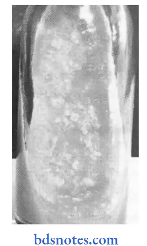
- The colonies of M. tuberculosis are dry, rough, raised, and irregular colony. It is a creamy white at first and becomes yellowish or buff colored later on.
- M. tuberculosis has eugenic growth on culture
- Liquid Media:
- Bacilli grow as surface pellicles due to the hydrophobic properties of their cell wall.
- Diffuse growth is obtained by the addition of detergent Tween 80 in
Dubo’s medium. - Tween 80 wets the surface and permits their growth diffusely. Virulent strains tend to grow as serpentine cords in liquid media, while avirulent strains grow in a more dispersed fashion.
Pathology Produced


Question 2. Classify the mycobacterium group of organisms. Describe The laboratory diagnosis of mycobacterium tuberculi.
Or
Laboratory diagnosis of Mycobacterium tuberculosis.
Answer:
Classification of Mycobacterium Group of Organisms
1. Cultivable
- Tubercle bacilli (mammalian).
- Human type M. tuberculosis
- Bovine type— M. bovis
- Vale type— M. microti
- African type— M. africanum.
- Atypical mycobacteria.
- Photochromogens
- Scotochromogens
- Non-photochromogens
- Rapid growers
- Mycobacteria Causing Skin Ulcers
- M. ulcerans
- M. balnei.
- Saprophytic Mycobacteria
- M. smegmatis
- M. phlei.
2. Non-cultivable
- M. leprae.
Laboratory Diagnosis
Specimen collection depends on the site of involvement. Tuberculosis may involve lungs (pulmonary) or sites other than the lungs (extrapulmonary). As a specimen, sputum is used for pulmonary tuberculosis, CSF for tubercular meningitis, 3 days morning sample of urine for renal tuberculosis, and aspirated fluid for bone and joint tuberculosis.
1. Microscopy:
The smear is made from the specimen and stained by the ZiehlNeelsen technique. It is examined under oil immersion lens. The acid-fast bacilli (AFB) appear as bright red bacilli against a blue background. To detect the bacilli microscopically, there should be at least 50,000 bacilli per mL of sputum.
2. Culture:
Culture is a very sensitive method for the detection of tubercle bacilli. The concentrated material is inoculated on two bottoms of the Lowenstein-Jensen medium. In the case of CSF, it is centrifuged, and the deposit is used for culture and smear examination. The culture media are incubated at 37 °C in the dark and in the light. Cultures are examined first after 4 days and thereafter weekly till 8 weeks. The tubercle bacilli usually grow in 2 to 8 weeks. In a positive culture, characteristic colonies appear on the culture medium.
3. Animal Inoculation:
0.5 mL of the concentrated specimen is inoculated intramuscularly into the thigh of two tuberculin negative healthy guinea pigs. The animals are weighed prior to inoculation and thereafter at a weekly interval. They are tuberculin tested after 3–4 weeks. There is the progressive loss of weight, and the tuberculin test becomes positive in animals that develop tuberculosis. The animal is killed after 6 weeks.
4. Serology:
Serology includes the detection of antimycobacterial antibodies in patient serum. Various methods such as ELISA, radioimmunoassay, latex agglutination assay have been employed. Several antigens like BCG, antigens 5 and 6, 64 kDa, antigens 60 and 32 kDa protein have been tried for the detection of antibodies against them.
5. Molecular Methods:
Polymerase chain reaction (PCR) is a rapid method in the diagnosis of tuberculosis. It is based on DNA amplification and has been
used to detect M. tuberculosis directly in clinical specimens.
Question 3. Write a conventional method of laboratory diagnosis of pulmonary tuberculosis.
Answer:
The conventional method for diagnosis of pulmonary tuberculosis is Ziehl-Nelsen staining.
Principle
Bacteria with lipid cell walls bind carbol fuschin tightly and resist destaining with strong decolorizing agents such as alcohol and acids. Acid-fast negative bacteria readily lose stains when treated with acid-alcohol solution. Following counter staining with methylene blue, the decolored acid-fast negative organism and other cells take blue color in contrast with red-colored acid-fast organisms.
Reagents
- Carbol-fuchsin solution
- 20% sulphuric acid
- Methylene blue or malachite green
Procedure
- Prepare smear from sputum specimen on glass slide and
heat fix it. - Place heat fied slide on staining reck and flood with carbol
fuchsin stain. - Heat gently on the burner until steam rises. Avoid boiling and continue heating for 5 minutes.
- The stain must not be allowed to evaporate or dry on the slide. If necessary, add more stain to the slide and reheat.
- Heating is necessary for the penetration of stains in the cell wall.
- Wash the slide underwater and remove all excess stains.
- Cover slide with 20% sulphuric acid for 20 seconds.
- The red color of the smear changes to yellowish brown.
- Wash with tap water
- Cover the slide with methylene blue or malachite green for 1 minute.
- Wash with tap water and allow the slide to dry in the air.
- Observe the stained slide in a microscope under 100X after putting a drop of cedar wood oil.
Results
- Acid-fast organisms: Bright red bacilli on blue background
- Other organisms: Dark blue.
Question 4. Write a short note on LJ media.
Answer:
LJ media is Lowenstein-Jensen media:
- LJ media is an enriched media that is commonly used for the growth of tubercle bacilli.
- LJ media consists of beaten eggs, asparagine, mineral
salts, malachite green, and glycerol or sodium pyruvate. - On heating, it solidifies.
- It is the media which is solid without the incorporation of agar.
- In LJ media, egg is the solidifying agent.
- Malachite green inhibits the growth of organisms other than mycobacteria and imparts color to the medium.
- Glycerol improves the growth of M. tuberculosis.
- Sodium pyruvate improves the growth of both M. tuberculosis and M. bovis.
- The colonies of M. tuberculosis are dry, rough, buff-colored, and raised with wrinkled surface.
- The colonies of M. bovis are flat, smooth, moist, and white and break up on touch.
- M. tuberculosis has eugonic growth, while M. bovis have dysgenic growth on LJ media.

Leave a Reply