Question 1. Describe the neuromuscular junction with the help of a diagram and explain the mechanism of transmission of nerve impulse across.
Answer:
Neuromuscular junction:
- Neuromuscular junction Definition:
- The junction between the terminal branch of the nerve fiber and muscle fiber is called neuromuscular junction.
- Neuromuscular junction Structure:
- Muscle fiber.
- It contains small thickened part in the midpoint called motor end plate.
- This shows a small depression called synaptic trough or synaptic gutter.
- Nerve fiber.
- Its each terminal branches called axon terminal.
- As it approaches close to muscle fiber it loses myelin sheath, expands and form synaptic knob.
- Muscle fiber.
Read And Learn More: BDS Previous Examination Question And Answers
- Neuromuscular junction Junction:
- Synaptic knob fits into the synaptic gutter.
- The membrane of the nerve ending is called the presynaptic membrane.
- the membrane of the muscle fiber is called postsynaptic membrane.
- Space present between these two is called synaptic cleft.
- Synaptic cleft contains basal lamina to which large quantity of acetylcholinesterase is attached.
- The postsynaptic membrane is thrown into numerous folds called subneural clefts.
- The axon terminal contains.
- Synaptic vesicles.
- It contains acetylecholine.
- Mitochondria.
- It contains ATP, a source of energy.
- Acetylcholine is synthesis by it.
- Synaptic vesicles.
- The postsynaptic membrane contains nicotinic acetylcholine receptors.
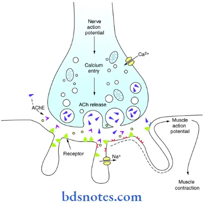
Mechanism of transmission of nerve impulse:
Transmission Of Nerve Impulse Series of events:
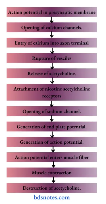
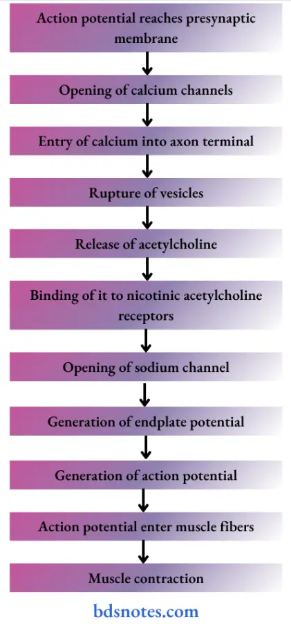
- Nerve impulse/action potential reaches the presynaptic nerve ending.
- This increases the permeability of presynaptic membrane for calcium ions by opening voltage gated calcium channels.
- Calcium enters axon terminal and causes rupture of vesicles.
- Thus acetylcholine is release from these vesicles by exocytosis and enters synaptic cleft.
- This acetylcholine now attached to the nicotinic acetylcholine receptors present in post synaptic membrane.
- It opens the ligand gated channels for sodium and thus sodium ions enters the neuromuscular junction.
- Increased permeability of Na+ causes depolarization of the post synaptic membrane and generates end plate potential.
- When more and more acetycholine is released continuously, the miniature end plate potentials are added together.
- When endplate potential reaches a threshold of 3040 mV, it depolarizes the surface membrane of the muscle and results in generation of action potential.
- Once this action potential reaches inside the muscle fiber then the muscle gives mechanical response by contraction.
- The acetylcholine that is released is destroyed very quickly by the enzyme acetylcholinesterase.
Question 1. Regulation of muscle tone.
Answer:
Muscle tone:
- Muscle tone Definition:
- The muscle fibers always maintain a state of slight contraction with certain degree of vigor and tension.
- This property of muscle is called tone.
- Muscle tone Regulation;
- In skeletal muscle.
- Regulation of muscle tone in skeletal muscle is neurogenic.
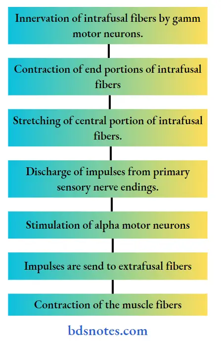
- It is due to continuous discharge of impulses from gamma motor neurons in anterior gray horn of spinal cord.
- The gamma motor neurons innervate the intrafusal fibers present in the musle spindle.
- Muscle spindle is the proprioreceptor situated in the muscle.
- The impulses from the gamma motor neuron causes contraction of end portions of intrafusal fibers.
- So, the central portion of the intrafusal fibers is stretched and activated.
- It leads to discharge of impulses from the primary sensory nerve endings.
- These impulses stimulates the alpha motor neurons of the spinal cord.
- Now, these neurons send impulses to extrafusal fibers.
- This causes contraction of the muscle fibers.
- The gamma motor neurons, muscle spindle and muscle tone are all controlled by basal ganglia.
- In cardiac muscle.
- Regulation of muscle tone in cardiac muscle is myogenic.
- The muscles themselves control the tone.
- The smooth muscle.
- In smooth muscle, tone is myogenic.
- It depends upon calcium level and number of cross bridges.
- In cardiac muscle.
- In skeletal muscle.
Question 2. Rigor mortis.
Answer:
- It refers to after death condition of the body which is characterized by stiffness of muscles and joints.
- It is derived from Latin word ‘rigor’ means ‘stiff’
Rigor mortis Mechanism:
- Soon after death, the cell membrane becomes highly permeable to calcium.
- As a result there is excessive influx of calcium into muscle which promotes the formation of actinomyosin complex.
- This causes contraction of muscles.
- Due to repeated contraction, the muscles become stiff after few hours of death.
Rigor mortis Cause:
- Normally, muscle contraction is compensated by relaxation.
- But for relaxation muscle needs ATP which causes exit of calcium.
- After death, due to repeated and excessive muscle contraction all ATP is used up and gets exhausted.
- Even new ATP cannot be produced due to lack of oxygen.
- As a result, the muscle remain in contracted state.
Rigor mortis Importance:
- Rigor mortis is important medico legally.
- It determines the time of death.
- Onset of stiffness start between 10 min and 3 hours after death.
- This depends on condition of the body and environmental temperature at the time of death.
- If body is active and temperature is high, stiffness occurs quickly.
- Stiffness occurs first in facial muscle and then spreads to other parts.
- Maximum stiffness occurs around 12-24 hours after death and remains for 1-3 days.
Question 3. Motor unit.
Answer:
Motor unit Definition:
- Each ventral horn cell along with its motor nerve is called motor neuron.
- The single motor neuron, its axon terminal and the muscle fibers innervated by it are together called motor unit.
Motor unit Properties:
- The number of muscle fibers in a motor unit varies inversely with the precision of the movement performed by the part.
- The number of muscle fibers is small in the motor units of the muscles controlling fine, graded and precise movements.
- The muscles concerned with coarse movements have motor units with large number of muscle fibers.
- All the muscle fibers in a motor unit are of same type.
- Depending on the type of muscle fibers, they innervate, motor units are of two types slow and fast.
- Recruitment of motor unit.
- With minimal voluntary activity of muscle a few motor units are discharge and with increasing voluntary efforts more motor units are put into action.
- This process is called recruitment of motor units.
- The graded response in the muscle is directly proportional to the number of motor units activated.
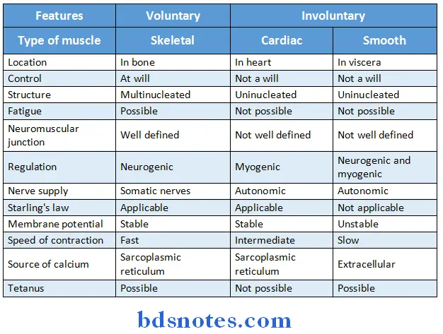
Question 4. State the difference between voluntary and involuntary muscles.
Answer:
Question 5. Describe with the help of diagram the lonic basis of action potential.
Answer:
Action potential:
- Resting membrane potential
- It occurs due to the movement of ions causing ionic imbalance across the cell
- This imbalance is brought by
- Sodium potassium pump
- It actively transports Nat and K+ ions in opposite direction
- By this pump, 3 Na+ ions are pumped out while 2 K+ ions are moved inside the cell using ATP
- This results in more positivity outside than inside the cell
- Selective permeability of membrane
- Transport channels are selective for movement of specific ions
- Channels for negatively charged substances remains closed as a result such ions remains inside the cell leading to more negativity inside than outside
- Action potential
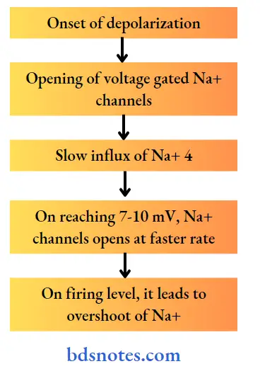
Similarly K+ channels opens and leads to efflux of K+ ions
Na+ channels are short lived while K+ channels remain open for longer duration
This results in efflux of more K+ ions producing more negativity inside the cell
Question 1. Types of muscles.
Answer:
1. Depending upon the presence or absence of striations.
- Straited muscle.
- These muscle contains cross striations.
- Example: skeletal muscle, cardiac muscle.
- Nonstraited muscle.
- They do not contains cross striation.
2. Depending upon action.
- Voluntary muscle.
- The activities of it can be controlled at will.
- Example: Skeletal muscle.
- Involuntary muscle.
- The activites of it cannot be controlled at will.
- Example: Cardiac smooth muscle.
3. Depending upon the location.
- Skeletal muscle.
- Found in association with bone.
- Cardiac muscle.
- Form musculature of heart.
- Smooth muscle.
- Present in hallow viscera.
Question 2. Sarcomere.
Answer:
- It is the structural and functional unit of the skeletal muscle.
- The cross striations which are characteristic feature of skeletal muscle are due to alternate dark and light cross band.
- The dark line is highly refractive and is called ‘A’ band.
- In the middle of `A’ band there is a light area called ‘H’ zone.
- In the middle of ‘H’ zone lies the middle part of myosin filament called ‘M’ line.
- The light band is low refractive and is called ‘I’ band.
- In the centre of each I band is found a narrow line of highly refractive material called ‘Z’ line.
- The substance included between two adjacent ‘Z’ line is called “Sarcomere”.
- The dark line is highly refractive and is called ‘A’ band.

Question 3. Neuromuscular transmission.
Answer:
- It includes following steps:
Question 4. Isotonic and isometric contraction:
Answer:
1. Isotonic contraction:
- In this type of muscular contraction, the length of the muscle changes while the tension remains constant.
- The muscle is allowed to shorten and lift a load.
- Example: Moving legs in walking.
2. Isometric contraction:
- In this type of muscular contraction, the length of the muscle remains same while the tension is increased.
- Muscle is not allowed to shorten.
- Example: Pulling of any heavy object.
Question 5. Frank starling’s law.
Answer:
- It states that the force of contraction of muscle is directly proportional to its initial length.
- Larger the initial length, greater will be the force of contraction.
Frank starling’s law Lamination:
- As the muscle is stretched the developed tension increases to a maximum and then declines as stretch becomes more extreme.
Frank starling’s law Significance:
- It is applicable to cardiac muscle.
- Thus, it helps to explain the blood ejected by each of the ventricle per heart beat.
Question 6. Membrane potential/resting membrane potential.
Answer:
Membrane potential Definition:
- The electrical potential difference across the cell membrane i.e., inside and outside of the cell under resting condition is called membrane potential or resting membrane potential.
- When two electrodes are placed on the cell surface and connected through a suitable amplifier to a cathode ray oscilloscope, no potential difference is observed.
- But, if one of the electrodes is inserted inside the cell, a constant potential difference is observed across the cell membrane.
- This is called resting membrane potential.
Membrane potential Genesis:
- There is negativity inside and positivity outside the cell membrane.
- It is due to distribution of ions across the cell membrane.
- Some K+ diffuses out of the cell while some anions like proteins stay inside the cell.
Question 7. Action potential.
Answer:
Action potential Definition:
The sequence of changes which occur in the membrane potential following excitation is called action potential.
Action potential Phases:
1. Depolarization:
- When the impulse reaches the muscle, the polarized condition is altered.
- The interior of the muscle becomes positive and outside become negative.
2. Repolarization:
- Within a short time, the potential reverses back to the resting membrane potential.
- Interior of the muscle become negative and outside becomes positive.
- This is called repolarization.
Action potential Properties:
- It is propagative.
- It obeys all or none law.
- It has refractory period.
- It is biphasic.
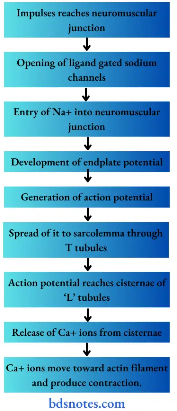
Question 8. excitation contraction coupling.
Answer:
Excitation Contraction Coupling Definition:
- excitation of muscle fiber causes development of action potential which through sequence of events produces muscular contraction.
- These both are coupled by Ca2+
- The process by which depolarization of the muscle fiber initiates contraction is called excitation contraction coupling.
Excitation Contraction Coupling Stages:
Question 9. Sliding theory/ratchet/walk along theory.
Answer:
The process by which the shortening of the contractile elements in the muscle is brought about is the sliding of actin filament over the myosin filament.
Events:
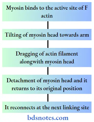
- Thus, myosin head move back and forth and bring all the actin filament towards the center of sarcomere.
- This forms actomyosin complex which cause muscular contraction.
Question 10. Myasthenia Gravis.
Answer:
Myasthenia Gravis Definition:
- It is a neuromuscular diseases characterized by grave weakness of the muscle.
Myasthenia Gravis Cause:
- It is autoimmune disease
- Caused due to formation of circulating antibodies against nicotinic acetycholine receptors.
Myasthenia Gravis Symptoms:
- Muscles mostly involved are: facial extraocular, swallowing and chewing muscles.
- Rapid onset of fatigue.
- Slow and weak muscular contraction.
- Marked generalised weakness of muscles.
- In severe condition.
- Paralysis of muscles.
- Death due to paralysis of respiratory muscles.
Question 11. Name the drug that stimulates neuromuscular junction.
Answer:
- Neostigmine.
- Physostigmine
- Di-isopropyl fluorophosphates.
Question 12. Properties of cardiac muscle.
Answer:
1. Electrical properties:
- excitability:
- Cardiac muscle forms a wave of depolarization in response to a stimulus.
- Autorhythmicity:
- The heart continues to beat for quite some time even after all nerves to it are cut due to pacemaker.
- Conductivity:
- Cardiac muscle spreads the impulses from SA nodes to all other parts of the heart.
2. Mechanical properties:
- Contractility:
- It is ability of the tissue to shorten in length after receiving a stimulus.
- All or none law:
- When a stimulus is applied, whatever may be the strength, the whole cardiac muscle responds to the maximum or it does not give response at all.
- Refractory period:
- It is the period in which the muscle does not show any response to a stimulus.
Question 13. What is clonus and tetanus?
Answer:
Clonus:
- It is a series of rapid and repeated involuntary jerky movements which occurs while eliciting deep reflexes
- Clonus occurs in individuals with upper motor neuron lesions due to hypertonicity of muscles and exaggeration of deep reflexes
Tetanus:
- It is sustained contraction of muscle due to repeated stimuli with high frequency
- The muscle relaxes only after the stoppage of stimuli or when the muscle is fatigued
- If frequency of stimuli is less, partial fusion of contraction occurs leading to incomplete tetanus
Question 14. Name the muscle proteins
Answer:
1. Myosin molecule
- Forms myosin filament
- It is globulin polypeptide containing 2 heavy chains and 4 light chains
- These chains twist around each other
2. Actin molecule
- Forms actin filament
- Its active site gives attachment to myosin molecule
3. Tropomyosin
- Present in actin filament
- In relaxed state, covers all active sites of actin filament
4. Troponin
- Present in actin filament
- Subunits:
- Troponin I-attached to F actin
- Troponin T-attached to tropomyosin
- Troponin C-attached to calcium ions
Question 15. Mention the neurotransmitter that is secreted at the neuromuscular junction and what is myasthenia gravis.
Answer:
- Acetylcholine is the neurotransmitter that is secreted at the neuromuscular junction.
Myasthenia gravis:
- It is a neuromuscular disease characterized by grave weakness of the muscle.
Myasthenia gravis Symptoms:
- Muscles mostly involved are facial, extra ocular, swallowing and masticatory muscles.
- Rapid onset of fatigue
- Slow and weak muscular contraction
- Marked generalized weakness of muscles
Question 16. What is chronaxie, rheobase and utilization time
Answer:
Rheobase:
- The weakest current strength which can excite a tissue if allowed to flow through it for an adequate time is called rheobase
Utilization time:
- The time for which current must be applied is called utilization time.
Chronaxie:
- The length of time for which a current of twice rheobase intensity must be applied to produce a response is called chronaxie.
Question 17. Stretch reflex.
Answer:
Stretch reflex is the reflex contraction of muscle when it is stretched.
Stimulus for the reflex is stretch to the muscle, response is contraction of the muscle and the neurotransmitter released is glutamic acid.
Stretch reflex is the basic reflex involved in maintenance of posture
It is also called myotatic reflex
It is monosynaptic reflex and quickest of all reflex.

Leave a Reply