Microscopy And Morphology Of Bacteria
Question 1. Write a short note on bacterial capsules.
Answer:
Bacterial Capsule Introduction::
A bacterial capsule is a thick gelatinous, circumscribed envelope situated just outside the cell wall of some bacteria formed by the active secretion of bacteria itself. When the capsule is loose and irregularly arranged, it is known as slime layer.
Bacterial Capsule Composition:
The main constituent in water is 98% and the rest is 2%, i.e. polysaccharides, polypeptides, lipoprotein, and hyaluronic acid. Bacteria form capsule which consists of either homopolysaccharides or heteropolysaccharides.
Types Of Bacterial Capsules:
It is of two types, i.e.
- Microcapsule: Capsule which has a width of more than 0.2 µ and is demonstrated by light microscope.
- Microcapsule: Capsule which has a width of less than 0.2 µ and is not demonstrated by light microscope.
Read And Learn More: Microbiology Question And Answers
Bacterial Capsule Functions:
- Protection: The capsule protects bacteria against antibacterial substances like bacteriophages, phagocytes, etc.
- Virulence: Capsule determines the virulence of bacteria. Certain bacteria are pathogens only in the capsulate state. Loss of capsule by mutation subculture renders virulent bacteria.
- Antigenicity: Capsular material is antigenic. Capsular antigens determine the antigenic specificity of bacteria. Capsule is site for specifi antigens and haptens. Capsular antigens may diffuse into body fluids and thus helps in serological diagnosis.
Demonstration of Capsule:
- India Ink staining: As ink cannot penetrate the capsule, it appears as a clear halo around. Bacterium as ink cannot penetrate the capsule.
- Serological methods: Since capsular material is antigenic so it can be demonstrated by mixing with specific anti-capsular serum.
- When the suspension of capsulated bacterium is mixed with specific anti capsular serum and is examined under the microscope, the capsule appears to be swollen.
- This is called as capsular swelling reaction or Quellung phenomenon. It is employed for typing of pneumococci.
- Special capsule staining: It employs copper salt as a mordant for staining of the capsule.
Question 2. Write a short note on flagella.
Answer:
Flagella Introduction: Flagella are filamentous, cytoplasmic appendages which are protruding through the cell wall and are found in all motile bacteria except spirochetes. They are contractile, extremely thin elongations of 5 to 20 µm in length and 0.01 to 0.02 µ in diameter.
Chemical Nature Of Flagella:
- Flagella is composed of a protein known as flagellin.
Parts of Flagella:
Flagella consists of three parts, i.e.
- Filament
- Hook
- Basal body
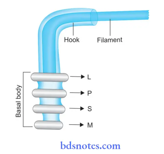
- Flagella Filament: It lies external to the cell and is connected to the hook at the cell surface. The filament is made of flagellin.
- Flagella Hook: This is a short curved structure that connects the filament and the basal body. The hook is broader than the filament. It is proteinaceous in nature and is embedded in the cell envelope.
- Flagella Basal Body:
- It is a circular structure that is embedded in the cell envelope and consists of a central rod that bears four rings, i.e. L, P, S, and M.
- All four rings are present in gram-negative bacteria, while in gram-positive bacteria S and M rings are present.
- S and M rings are needed for motility, while L and P rings are needed for support.
- In Gram-negative bacteria:
- L is located in LPS
- P is located in the inner peptidoglycan
- S is located just above the cell membrane
- M is embedded in the cell membrane.
Types of Flagella:
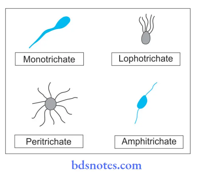
Depending on position and arrangement of flagella/polar: At one/both ends
- Monotrichous: Single flgella at one pole (Vibrio)
- Amphitrichous: At both poles (Listeria monocytes)
- Lophotrichous: Tuft of flgella at one/both poles (e.g. spirilla, Pseudomonas)
- Peripheral/Peritrichous: Flagella is present all around the periphery, for example. Salmonella proteins, E. coli.
Depending On The Configuration Of The Filament:
- Monomorphic/conventional: All flagella are of the same length.
- Dimorphic: Two flagella having different wavelengths.
Demonstration Of Flagella:
The following methods are used for demonstrating the flagella:
- Electron microscopy
- Dark ground illumination
- Special staining method—by increasing the thickness
- Indirect methods:
- Hanging drop preparation
- Spreading growth on semisolid agar
- By Craigie’s or U-tube technique
Functions Of Flagella:
- It helps the bacteria in motility and pathogenicity.
- Flagella contain specific antigens which induce specific antibodies which help in serodiagnosis.
Question 3. Write a brief on spores of bacteria.
Or
Write a short note on bacterial spores.
Answer:
Bacterial Spores:
- Spores are highly resistant resting phase of bacterium formed in unfavorable environmental conditions like starvation and extremes of temperature.
- All bacterial spores are formed in the parent cell which is known as an endospore.
- Each of the bacteria forms one spore which on germination forms form a single vegetative cell, so it is not the method of reproduction.
Bacterial Sporulation:
- Bacterial cells undergo spore formation when nutritional conditions are unfavorable. This is known as sporulation.Spore develops from a portion of the protoplasm near one end of the cell.
- The remaining part of the cell is known as sporangium.Bacterial DNA replicates and divide into two DNA molecules. Out of these one molecule is incorporated in forespore and another into sporangium.
- A transverse septum grows across the cell from the cell membrane which divides the forespore and sporangium. Forespore is encircled by the septum as double-layered membrane.
- Inner layer become the spore membrane and the outer layer become thickened spore coat. Between two layers lies the spore cortex.
Parts Of Bacterial Endospore:
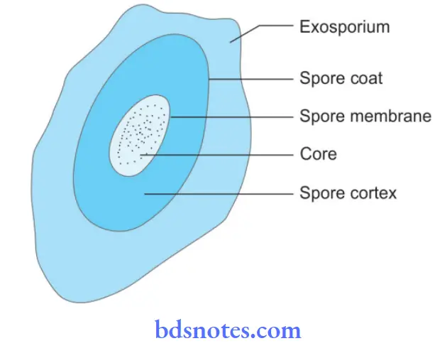
- Core: It is a spore protoplast that consists of a nucleus and some of the unique enzymes, i.e. dipicolinic acid synthetase which synthesize dipicolinic acid which makes spore resistant to heat and other components.
- Spore wall: It is the innermost layer that surrounds the inner spore membrane which consists of peptidoglycan and becomes the future cell wall.
- Cortex: It is the thickest layer surrounding the spore wall which contains unusual type of peptidoglycan with fewer cross–links, extremely sensitive to lysozyme.
- Coat: Composed of keratin-like protein with many disulphide bonds. It is impermeable which makes the spore resistant to antibacterial chemical agents.
- Exosporium: Lipoprotein membrane containing some carbohydrates.
Morphologic Types Of Bacterial Spores:
- According to spore diameter:
- Spores with bulging of the cell wall.
- Spores without bulging of the cell wall.
- According to spore position:
- Central/equatorial, for example, B. Anthracis.
- Subterminal, for example, C. Tetani.
- Terminal/drumstick: C. Tetani.
- According to spore shape:
- Round to oval.
- According to maturity:
- Immature.
- Mature.
Demonstration of Bacterial Spores:
- Gram staining: Spores appear as an unstained refractile body within the cell.
- Modified Ziehl–Neelsen stain: Spores appear as acid-fast. Ziehl–Neelsen stain with 0.25 to 0.5% sulphuric acid as decoloring agent is used for spore staining.
Uses Of Bacterial Spores:
- Spores make the survival of certain organisms possible under unfavorable conditions like dry state.
- Spores of some bacteria act as an indicator for proper sterilization.
Germination Of Bacterial Spores:
Germination of the spore occurs when conditions become favorable forming one vegetative cell.
The following are the stages involved in germination:
- Bacterial Spores Activation: It occurs in nutritionally rich medium. Damage to the spore coat is produced by heat, abrasion, acidity, etc.
- Bacterial Spores Initiation: During this stage cortex peptidoglycan and a variety of other components are degraded, water is taken up and calcium dipicolinic acid is released.
- Bacterial Spores Outgrowth: A new vegetative cell with a spore protoplast emerges out. A period of active synthesis occurs that terminates in cell division.
Question 4. Write a short note on Gram’s stain.
Or
Write in brief Gram’s staining.
Answer:
Gram’s Stain Introduction: Gram’s stain is the most commonly used staining method discovered by histologist Christian Gram in 1884. Gram’s staining is the diffrential staining method which diffrentiates organisms into gram-positive and gram-negative as per the Gram’s reaction.
Gram’s Staining Of Principle:
- The air-dried fied smear of bacteria, picks up the stain and looks purple when treated with the crystal violet stain (Gram stain) and iodine.
- Iodine enhances the staining reaction between dye, i.e. crystal violet, and internal cellular contents of bacteria.
- The alcohol (or acetone) treatment decolorizes gram-negative bacteria while gram-positive bacteria retain the color (purple).
- Counterstaining with safranine (or basic fuchsin) stains the gram-negative bacteria red.
Gram’s Staining Of Reagents:
- Crystal violet or methyl violet or gentian violet stain
- Gram’s iodine solution
- Alcohol or acetone
- Safranine or carbol fuchsin or neutral red.
Gram’s Staining Of Procedure:
1. Smear Preparation :
- Take a grease-free dry slide and make an oval-shaped mark at the center by using a glass marker.
- Sterilize the inoculating (nichrome) loop on the flame of a Bunsen burner.
- Transfer a loopful of culture by the sterile nichrome loop and make a smear in the premarked area of the slide.
- Allow the smear to dry in the air.
- Fix the dry smear by passing the slide 3 to 4 times through the flame quickly with the smear side facing up.
2. Gram Staining:
- Place the slide on the staining glass rods.
- Cover the smear with crystal violet, methyl violet, or gentian violet stain and leave for l minute.
- Wash carefully under running water.
- Flood the Smear with Gram’s iodine and wait for 1 minute
- Drain of the iodine with water
- Decolorize the smear with alcohol or ketone for 20-30 seconds.
- Gently wash the slide under running tap water.
- Counterstain smear with safranin or carbol fuchsin or neutral red for 10 seconds
- Drain excess stain and allow the stained smear to dry in the air.
- As staining is completed watch slide in microscope under 100X after putting drop of cedar wood oil.
Gram’s Staining Of Observations:
- Gram-positive bacteria: These bacteria resist decolorization and retain the color of the primary stain. These bacteria appear violet. Examples: Pneumococci, streptococci, and staphylococci are gram-positive cocci Clostridia, Corynebacterium, and Bacillus spp. are grampositive bacilli.
- Gram-negative bacteria: They are decolorized by an alcohol, so they take counterstain and appear red. Examples: Gonococci and meningococci are gramnegative cocci. E. coli, Salmonella, Shigella, V. cholerae are gram-negative bacilli
- Background or pus cells appear red in color
- Yeast cells appear dark purple in color.
Question 5. Write a short note on ZN staining.
Answer:
ZN Staining Introduction:
Organisms that are not easily stained by ordinary staining methods, once stained resist decolorization by acids are known as acid-fast organisms and method used for staining the organism is known as acid-fast stain.
- As acid-fast staining was devised by Ehrlich in 1883 and modified by Ziehl–Neelsen in 1885. So it is known as the Ziehl Neelsen staining method.
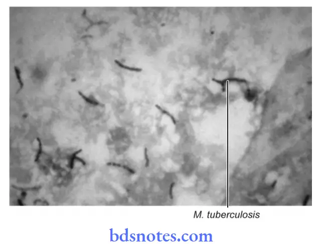
- This method is a differential staining method that differentiates acid-fast organisms, i.e. mycobacteria from other organisms, i.e. nonacid-fast.
Principle Of ZN Staining:
- Bacteria with lipid cell walls bind carbol fuschin tightly and resist destaining with strong decolorizing agents such as alcohol and acids.
- Acid-fast negative bacteria readily loose stain when treated with an acid-alcohol solution.
- Following counterstaining with methylene blue the decolored acid-fast negative organism and other cells take blue color in contrast with red colored acid-fast organisms.
Reagents Of ZN Staining:
- Carbol-fuchsin solution
- 20% sulphuric acid
- Methylene blue or malachite green.
Procedure Of ZN Staining:
- Prepare a smear from the sputum specimen on a glass slide and heat fi it.
- Place heat fied slide on staining reck and flood with carbol fuchsin stain.
- Heat gently on the burner until steam rises. Avoid boiling and continue heating for 5 minutes. The stain must not be allowed to evaporate or dry on the slide.
- If necessary, add more stain to the slide and reheat.
- Heating is necessary for the penetration of stain in the cell wall.
- Wash the slide under water and remove all excess stains.
- Cover the slide with 20% sulphuric acid for 20 seconds.
- The red color of the smear changes to yellowish brown.
- Wash with tap water.
- Cover slide with methylene blue or malachite green for 1 minute.
- Wash with tap water and allow slide to dry in air.
- Observe stained slide in a microscope under 100X after putting a drop of cedar wood oil.
Results Of ZN Staining:
- Acid-fast organisms: Bright red bacilli on blue background
- Pus cells, epithelial cells, and other organisms: Dark blue or green.
Question 6. Write a short note on acid-fast bacilli.
Or
Write briefly on acid-fast bacilli.
Answer:
Acid-Fast Bacilli:
- Acid-fast bacteria are those which retain a concentrated dye (Carbol fuchsin) and resist decol-orization by strong 20% sulphuric acid, thus being stained red.
- Acid-fastness is due to the presence of high lipid content, fatty acid, and higher alcohols found in the cell wall of Mycobacterium.
- Mycolic acid, a waxy substance is present in the cell wall which do not allow the stain to penetrate easily inside these organisms.
- Acid fastness mainly depends on the integrity of the cell wall.
- For example: Mycobacteria has lipid-rich cell wall that prevent staining by ordinary dyes but on forced staining with concentrated hot dyes like Carbol fuchsin, they retain the dye and resist decolorization with strong acid.
- Such bacteria are acid-fast.
- Acid-fast organism is usually alcohol-fast.
- Ziehl–Neelsen is the acid-fast stain which is used to stain acid-fast bacilli.
- In Ziehl-Neelsen staining carbol fuchsin which is the phenolic solution of basic fuchsin is applied with heat.
- Heat and phenol facilitate the penetration of dye.
- Subsequently, when decolorization by acid is done the dye does not comes out as it is soluble in phenol and phenol is more soluble in lipid.
- Substances hence there is no decolorization and they retain the color of basic fuchsin.
Question 7. Write a short note on bacterial motility.
Answer:
Bacterial Motility:
- Flagella are the organ of bacterial motility.
- All motile bacteria, except spirochaetes possess one or more flagella.
Parts of Flagella:
Flagella consists of three parts, i.e.
- Filament
- Hook
- Basal body.
 Flagella Filament: It lies external to the cell and is connected to the hook at the cell surface. The filament is made of flagellin.
- Flagella Hook: This is a short curved structure which connects the filament and the basal body. Hook is broader than the filament. It is proteinaceous in nature and is embedded in the cell envelope.
- Flagella Basal Body:
- It is a circular structure that is embedded in the cell envelope and consists of a central rod that bears four rings, i.e. L,P, S, and M.
- All four rings are present in Gram-negative bacteria while in Gram-positive bacteria S and M rings are present.
- S and M rings are needed for motility while L and P rings are needed for support.
- In Gram-negative bacteria:
- L is located in LPS
- P is located in the inner peptidoglycan
- S is located just above the cell membrane
- M is embedded in the cell membrane.
Types of Flagella:
1. Depending on the Position and Arrangement of Flagella/ Polar: At One/Both Ends:
- Monotrichous: Single flgella at one pole (Vibrio)
- Amphitrichous: At both poles (Listeria monocytogens)
- Lophotrichous: Tuft of flagella at one/both poles (example spirilla, Pseudomonas)
- Peripheral/Peritrichous: Flagella is present all around the periphery, an example of Salmonella proteins, E. coli.
2. Depending on the Configuration of Filament:
- Monomorphic/conventional: All flagella are of same length.
- Dimorphic: Two flagella having different wavelengths.
3. Functions of Flagella:
- It helps the bacteria in motility and pathogenicity.
- Flagella contain specific antigens which induce specific antibodies which help in serodiagnosis.
Question 8. Differentiation between differential stain vs. negative stain.
Answer:
Differentiation between differential stain vs. negative stain: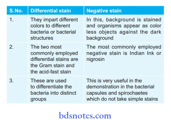
Question 9. Write a short note on Koch’s phenomenon.
Answer:
Koch’s Phenomenon
The response of a tuberculous animal to reinfection was best explained by Robert Koch.
- When a healthy guinea pig is inoculated subcutaneously with virulent tubercle bacilli, the puncture site heals quickly and there is no immediate visible reaction.
- After 10-14 days, a nodule appears at the site of injection which ulcerates and ulcer persists till the animal dies of progressive tuberculosis.
- The regional lymph nodes are enlarged and caseous.
- 4 On the other hand, virulent tubercle bacilli are injected in a guinea pig that had received a prior injection of tubercle bacilli 4-6 weeks earlier, an indurated lesion appears at the site of injection in a day or two which undergoes necrosis in another day or so to form a shallow ulcer.
- This ulcer heals rapidly without the involvement of the regional lymph nodes or tissues.
- This is called Koch phenomenon. This phenomenon is a combination of hypersensitivity and immunity.
Question 10. Write a short note on bacterial spores, sporulation, and germination.
Answer:
Bacterial Spores Introduction:
Some bacteria particularly members of the genera Bacillus and Clostridium have the ability to form highly resistant resting stages called spores. Each bacterium forms one spore which on germination forms a single vegetative cell. All bacterial spores are formed in the parent cell which is known as endospore. Each of the bacteria forms one spore which on germination forms form a single vegetative cell, so it is not the method of reproduction.
Bacterial Sporulation:
- Bacterial cells undergo spore formation when nutritional conditions are unfavorable. This is known as sporulation.
- Spore develops from portion of the protoplasm near one end of the cell. The remaining part of the cell is known as sporangium.
- Bacterial DNA replicates and divides into two DNA molecules.
- Out of these one molecule is incorporated in the forespore and another into sporangium.
- A transverse septum grows across the cell from cell membrane which divides the forespore and sporangium.
- Forespore is encircled by the septum as a double-layered membrane.
- Inner layer become the spore membrane and the outer layer becomes thickened spore coat.
- Between two layers lies the spore cortex.
Bacterial Germination:
Germination of the spore occurs when conditions become favorable forming one vegetative cell.
The following are the stages involved in germination:
- Activation: It occurs in nutritionally rich medium. Damage to the spore coat is produced by heat, abrasion, acidity, etc.
- Initiation: During this stage cortex peptidoglycan and a variety of other components are degraded, water is taken up and calcium dipicolinic acid is released.
- Outgrowth: A new vegetative cell with a spore protoplast emerges. A period of active synthesis occurs that terminates in cell division.
Question 11. Write a short note on Koch’s postulates.
Answer:
Robert Koch’s:
Robert Koch postulated the criteria for proving that a microorganism isolated from the disease was indeed casually related to it. As per Koch’s postulates, a microorganism is accepted as the causative agent of particular infectious disease.
When the following conditions are fulfilled:
- Organisms should be constantly associated with lesions of the disease.
- It should be possible to isolate organisms in pure culture from the lesions of the disease.
- Isolated organisms when inoculated in suitable laboratory animals should produce a similar disease.
- It should be possible to reisolate the organism in pure culture from the lesions produced in experimental lesions.
- Specific antibodies to organisms are demonstrable in the serum of patients.
- Koch’s postulates have proved to be useful in confirming doubt claims made regarding causative agents of infectious diseases.
Question 12. Write a short note on the bacterial growth curve.
Answer:
Bacterial Growth Curve:
When a bacteria is inoculated into a suitable culture medium and incubated, its growth occurs in definite course.
When the bacterial count of such culture is observed at different intervals and is plotted in relation to time, a growth curve is formed which is known as a bacterial growth curve.

The bacterial growth curve has the following four phases:
1. Lag phase: As inoculation of the culture medium is completed, multiplication does not begin immediately.Period between inoculation and the beginning of multiplication is known as the lag phase.
During this phase, organisms adapt to the new environment during which necessary enzymes and intermediate metabolites are released by bacteria in adequate quantities for multiplication to occur.
There is an increase in the size of the cells but there is no increase in numbers. The duration of the lag phase varies with the species, nature of the culture medium and temperature, etc.
2. Log (Logarithmic) or exponential phase:
1As cell division begins and their numbers increase exponentially or by geometric progression with time. If the logarithm of the viable count is plotted against time, a straight line is formed.
3. Stationary phase:
As the long phase is over, bacterial growth ceases completely due to the exhaustion of nutrients and the accumulation of toxic products. The number of progeny cells formed are enough to replace the number of cells that die. The number of viable cells remains stationary as there is almost a balance between the dying cells and the newly formed cells.
4. Phase of decline:
After the stationary phase, the bacterial population decreases due to the death of cells. The decline phase starts due to exhaustion of nutrients, accumulation of toxic products, and autolytic enzymes. There is a decline in the viable count and not in the total count. With autolytic bacteria, even the total count shows a phase of decline.
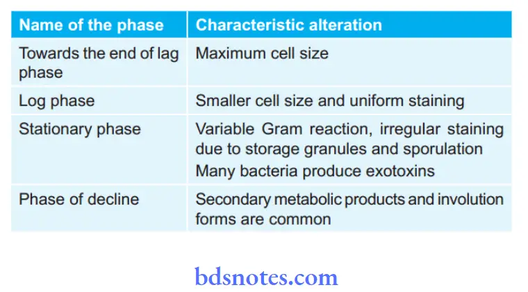
Morphological and Physiological Alterations of Bacteria during Different Phases of Growth:
Question 13. Describe Gram’s stain and ZN stain.
Answer:
Gram’s Stain Introduction:
Gram’s stain is the most commonly used staining method discovered by histologist Christian Gram in 1884. Gram’s staining is the diffrential staining method which diffrentiates organisms into gram-positive and gram-negative as per the Gram’s reaction.
Gram’s Staining Of Principle:
- The air-dried fied smear of bacteria, picks up the stain and looks purple when treated with the crystal violet stain (Gram stain) and iodine.
- Iodine enhances the staining reaction between dye, i.e. crystal violet, and internal cellular contents of bacteria.
- The alcohol (or acetone) treatment decolorizes gram-negative bacteria while gram-positive bacteria retain the color (purple).
- Counterstaining with safranine (or basic fuchsin) stains the gram-negative bacteria red.
Gram’s Staining Of Reagents:
- Crystal violet or methyl violet or gentian violet stain
- Gram’s iodine solution
- Alcohol or acetone
- Safranine or carbol fuchsin or neutral red.
Gram’s Staining Of Procedure:
1. Smear Preparation :
- Take a grease-free dry slide and make an oval-shaped mark at the center by using a glass marker.
- Sterilize the inoculating (nichrome) loop on the flame of a Bunsen burner.
- Transfer a loopful of culture by the sterile nichrome loop and make a smear in the premarked area of the slide.
- Allow the smear to dry in the air.
- Fix the dry smear by passing the slide 3 to 4 times through the flame quickly with the smear side facing up.
2. Gram Staining:
- Place the slide on the staining glass rods.
- Cover the smear with crystal violet, methyl violet, or gentian violet stain and leave for l minute.
- Wash carefully under running water.
- Flood the Smear with Gram’s iodine and wait for 1 minute
- Drain of the iodine with water
- Decolorize the smear with alcohol or ketone for 20-30 seconds.
- Gently wash the slide under running tap water.
- Counterstain smear with safranin or carbol fuchsin or neutral red for 10 seconds
- Drain excess stain and allow the stained smear to dry in the air.
- As staining is completed watch slide in microscope under 100X after putting drop of cedar wood oil.
Gram’s Staining Of Observations:
- Gram-positive bacteria: These bacteria resist decolorization and retain the color of the primary stain. These bacteria appear violet. Examples: Pneumococci, streptococci, and staphylococci are gram-positive cocci Clostridia, Corynebacterium, and Bacillus spp. are grampositive bacilli.
- Gram-negative bacteria: They are decolorized by an alcohol, so they take counterstain and appear red. Examples: Gonococci and meningococci are gramnegative cocci. E. coli, Salmonella, Shigella, V. cholerae are gram-negative bacilli
- Background or pus cells appear red in color
- Yeast cells appear dark purple in color.
ZN Staining Introduction:
Organisms that are not easily stained by ordinary staining methods, once stained resist decolorization by acids are known as acid-fast organisms and method used for staining the organism is known as acid-fast stain.
- As acid-fast staining was devised by Ehrlich in 1883 and modified by Ziehl–Neelsen in 1885. So it is known as the Ziehl Neelsen staining method.
- This method is a differential staining method that differentiates acid-fast organisms, i.e. mycobacteria from other organisms, i.e. nonacid-fast.

Principle Of ZN Staining:
- Bacteria with lipid cell walls bind carbol fuschin tightly and resist destaining with strong decolorizing agents such as alcohol and acids.
- Acid-fast negative bacteria readily loose stain when treated with an acid-alcohol solution.
- Following counterstaining with methylene blue the decolored acid-fast negative organism and other cells take blue color in contrast with red colored acid-fast organisms.
Reagents Of ZN Staining:
- Carbol-fuchsin solution
- 20% sulphuric acid
- Methylene blue or malachite green.
Procedure Of ZN Staining:
- Prepare a smear from the sputum specimen on a glass slide and heat fi it.
- Place heat fied slide on staining reck and flood with carbol fuchsin stain.
- Heat gently on the burner until steam rises. Avoid boiling and continue heating for 5 minutes. The stain must not be allowed to evaporate or dry on the slide.
- If necessary, add more stain to the slide and reheat.
- Heating is necessary for the penetration of stain in the cell wall.
- Wash the slide under water and remove all excess stains.
- Cover the slide with 20% sulphuric acid for 20 seconds.
- The red color of the smear changes to yellowish brown.
- Wash with tap water.
- Cover slide with methylene blue or malachite green for 1 minute.
- Wash with tap water and allow slide to dry in air.
- Observe stained slide in a microscope under 100X after putting a drop of cedar wood oil.
Results Of ZN Staining:
- Acid-fast organisms: Bright red bacilli on blue background
- Pus cells, epithelial cells, and other organisms: Dark blue or green.
Question 14. Write a short note on capsule of bacteria and fungus.
Answer:
Bacterial Capsule Introduction::
A bacterial capsule is a thick gelatinous, circumscribed envelope situated just outside the cell wall of some bacteria formed by the active secretion of bacteria itself. When the capsule is loose and irregularly arranged, it is known as slime layer.
Bacterial Capsule Composition:
The main constituent in water is 98% and the rest is 2%, i.e. polysaccharides, polypeptides, lipoprotein, and hyaluronic acid. Bacteria form capsule which consists of either homopolysaccharides or heteropolysaccharides.
Types Of Bacterial Capsules:
It is of two types, i.e.
- Microcapsule: Capsule which has a width of more than 0.2 µ and is demonstrated by light microscope.
- Microcapsule: Capsule which has a width of less than 0.2 µ and is not demonstrated by light microscope.
Bacterial Capsule Functions:
- Protection: The capsule protects bacteria against antibacterial substances like bacteriophages, phagocytes, etc.
- Virulence: Capsule determines the virulence of bacteria. Certain bacteria are pathogens only in the capsulate state. Loss of capsule by mutation subculture renders virulent bacteria.
- Antigenicity: Capsular material is antigenic. Capsular antigens determine the antigenic specificity of bacteria. Capsule is site for specifi antigens and haptens. Capsular antigens may diffuse into body fluids and thus helps in serological diagnosis.
Demonstration of Capsule:
- India Ink staining: As ink cannot penetrate the capsule, it appears as a clear halo around. Bacterium as ink cannot penetrate the capsule.
- Serological methods: Since capsular material is antigenic so it can be demonstrated by mixing with specific anti-capsular serum.
- When the suspension of capsulated bacterium is mixed with specific anti capsular serum and is examined under the microscope, the capsule appears to be swollen.
- This is called as capsular swelling reaction or Quellung phenomenon. It is employed for typing of pneumococci.
- Special capsule staining: It employs copper salt as a mordant for staining of the capsule.
The Capsule of Bacteria Fungus:
There are a few encapsulated fungal species. The best-characterized fungal capsule belongs to Cryptococcus neoformans. The capsule of this microorganism has been extensively studied because it is the main virulence factor of this pathogenic organism.
Bacteria Components And Structure
- The most characteristic feature of C. neoformans is a polysaccharide capsule that surrounds the cell body.
- The polysaccharide capsule confers a strong negative charge by virtue of the glucuronic acid residues on its main polysaccharide component.
- The polysaccharides that constitute the capsule are found in two different locations. The first location is attached to the cell wall forming the physical structure defined as the capsule.
- These polysaccharides are also constitutively released by the cell into the surrounding medium and environment, and they can be isolated as exopolysaccharides after certain purification protocols.
- It is not known whether the capsule’s release into the medium is an active phenomenon regulated by the cell, or if it is just an unspecific capsule shedding.
- An intriguing finding is that capsule structure is very variable depending on the strain (and on its the environment). It also changes during the course of an infection.
Demonstration Of Fungal Capsule:
- The capsule is not visible by regular microscopy because it is highly hydrophilic, and due to its high water content it has the same refraction index as the medium.
- However, it can be easily made visible by several techniques. The classic image of the capsule is that of a halo surrounding the cell made visible by suspending the yeast in Indian ink preparations.
- It can also be nicely observed by other microscopic techniques such as scanning electron microscopy and florescence.
Functions of Bacteria Of Fungal Capsule:
- Capsules act as a physicochemical barrier and prevent phagocytosis. This helps its spread in body.
- It acts as barrier against the action of antibiotics and increases invasive properties.
- It also helps to resist the action of lysosomal enzymes.
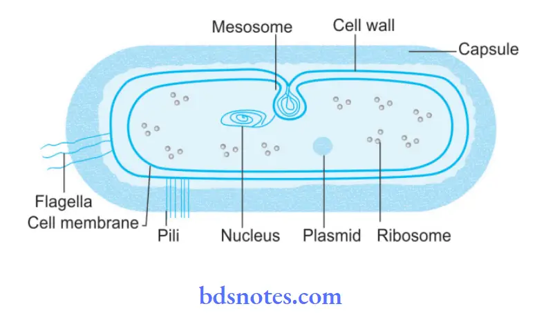
Question 15. Draw a neat diagram of the bacterial cell. Add a note on the bacterial cell wall.
Answer:
Diagram of Bacterial Cell:
Note on Bacterial Cell Wall:
- The cell wall is a tough and rigid structure that surrounds the bacterium like a shell.
- The cell wall is 10 to 25 nm in thickness and weighs about 20 to 25% of the dry weight of cell.
- The cell wall is absent in the mycoplasma group of organisms.
Chemical Nature Of Bacterial Cell:
The chemical composition of cell wall differs in gram-positive and gram-negative bacteria. Both gram-positive and gram-negative bacteria have a common component, i.e. mucopeptide or peptidoglycan which is primarily responsible for mechanical strength.
This layer is thicker than gram-positive bacteria as compared to gram-negative bacteria. In gram-positive bacteria cell wall is 16 to 18 nm thick and is monolayered. Peptidoglycan is the major component.
The gram-positive cell wall also consists of teichoic acids, i.e. glycerol teichoic acid or ribitol teichoic acid. Teichoic acid constitute a major surface antigen in gram-positive bacteria. In gram-negative bacteria cell wall is thin and is multilayered structure.
The following are the layers:
- Lipoprotein layer: It consists of the inner layer of peptidoglycan to the outer membrane.
- Outer membrane: It has proteins named as outer membrane proteins which are the target sites for phages, antibiotics, and bacteriocins.
- Lipopolysaccharide: The lipopolysaccharide consists of lipid A to which a polysaccharide is attached. Lipopolysaccharide constitutes endotoxin of gram-negative bacteria. The polysaccharide determines the major surface antigen, i.e. O antigen. The toxicity of bacteria is associated with lipid A.
- Periplasmic space: It lies between the inner and outer membranes and consists of various binding proteins for specific substrates.
Bacterial Cell Of Functions:
- It provides shape as well as rigidity to the cell.
- It provides mechanical support to the cell membrane.
- It takes part in cell division
- It plays a role in virulence and immunity.
- It maintains the osmotic pressure and protects the cell against osmotic damage.
- The cell wall provides the site for phage absorption.
Demonstration Of Bacterial Cell Wall:
The cell wall cannot be seen by a light microscope. Its demonstration is done by the following methods:
Plasmolysis of Bacterial Cell Wall:
As bacteria are placed in the hypotonic saline, there occurs the shrinkage of cytoplasm, while the cell wall retains its original shape and size.
- Microdissection
- Differential staining
- Reaction with specific antibody
- Electron microscopy

Leave a Reply