Muscle Tissue
Question 1. Briefly discuss the muscle tissue and classify it.
Answer.
The muscle tissue (also called muscle) is made up of muscle cells surrounded by connective tissue. The muscle cells are elongated hence, called muscle fibres. The muscle fibres contain contractile proteins, mainly actin and myosin.
The muscle fibres are specialized to shorten in length by contraction. The muscle tissue is responsible for the movements of the various parts of the body.
Muscle Tissue Classification Histologically, the muscle tissue is classified into three types:
- Skeletal muscle
- Cardiac muscle
- Smooth muscle
Question 2. Write a short note on the skeletal muscles.
Answer.
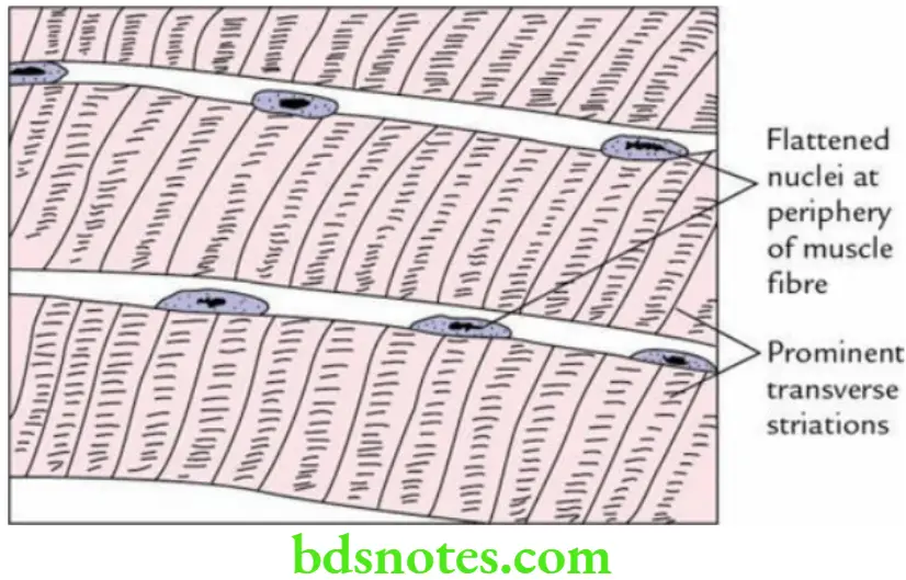
The skeletal muscles are striated and attached to the bones of the skeleton. They are found mainly in the limbs and trunk of the body.
Skeletal Muscles Histological features
- Muscle fibres are elongated, cylindrical and multinucleated.
- Muscle fibres present prominent cross-striations with alternating dark ‘A’ and light ‘I’ bands.
- Nuclei are flat and located at the periphery.
- Muscle fibres do not show branching.

Question 3. Write a short note on the cardiac muscle.
Answer.
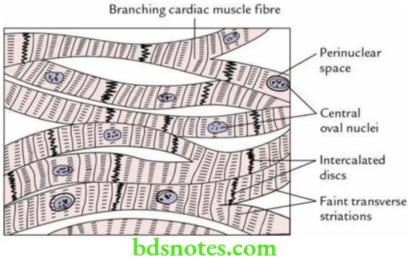
The cardiac muscle is striated, involuntary and located exclusively in the heart.
Cardiac Muscle Histological features
- Cardiac muscle fibres are short and thick. They branch and anastomose to form syncytium.
- Cardiac muscle fibres are joined end-to-end at junctional specializations called intercalated discs.
- Each cardiac muscle fibre has a centrally located single oval nucleus.
- Cardiac muscle fibres present faint cross-striations with alternating dark ‘A’ and light ‘I’ bands, i.e. they are not as conspicuous as in skeletal muscle.

Question 4. Write a short note on the smooth muscles.
Answer.
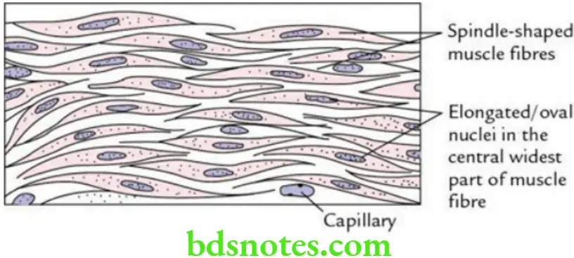
The smooth muscles are nonstriated, and involuntary and are located in the walls of hollow viscera, like the stomach and intestine, and in the walls of blood vessels.
Smooth Muscles Histological features
- Smooth muscle fibres are spindle-shaped.
- Smooth muscle fibres do not present cross-striations.
Read And Learn More: Selective Anatomy Notes And Question And Answers
- Each smooth muscle fibre has a centrally located elongated nucleus.

Compare the histological features of three types of muscle tissues (i.e. skeletal, cardiac and smooth muscles).
The comparison of three types of muscle tissues is given in Table.
Comparison Between Skeletal, Cardiac and Smooth Muscles

Blood Vessels
Question 1. Enumerate three layers/coats in the wall of the artery.
Answer.
The three layers/coats in the wall of an artery from inside to outside are as follows:
- Tunica intima
- Tunica media
- Tunica adventitia
The features of these coats are given in Table.
Features of Three Coats in the Wall of Artery

Question 2. Describe the histological features of an elastic artery (large artery) in brief.
Answer.
Elastic Artery Histological features
The characteristic histological features of an elastic artery (syn, large-sized artery) are as follows:
Tunica intima
- The subendothelial layer is prominent.
- Internal elastic lamina is not clearly visible.*
Tunica media
- The presence of a large number of elastic fibres arranged in the form of concentric and fenestrated laminae or sheets.
- The concentric layers of smooth muscle fibres in between the elastic lamellae.
Tunica adventitia
- Thin and made up of connective tissue. It contains longitudinally running collagen fibres, which merge with external elastic lamina.
- Contains vasa vasorum.
Elastic Artery Functions
- Conduct blood from the heart to medium-sized (muscular) arteries.
- Elastic recoil of the vessel wall ensures continuous flow of the blood through medium-sized arteries.
Elastic Artery Examples
- Aorta
- Pulmonary trunk
- Main branches of the aorta
- Brachiocephalic trunk
- Common carotid artery
- Left subclavian artery
Question 3. Describe the histological features of a muscular (medium-sized) artery in brief.
Answer.
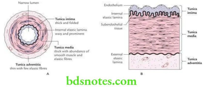
Muscular Artery Histological features
Tunica intima
- It presents a folded appearance.
- Internal elastic lamina is prominent and wavy (i.e. thrown into wavy folds).
- Subendothelial tissue is not prominent.
Tunica media
- The presence of a large/huge number of smooth muscle fibres arranged concentrically.
- About 75% mass of tunica media is formed by the smooth muscle fibres.
Tunica adventitia
- It is histologically similar to that of elastic artery but thicker than that.

Muscular Artery Functions Regulate the flow of blood according to need by altering the size of its lumen by contraction and relaxation of a huge number of smooth muscle fibres in its wall.
Muscular Artery Examples
- Brachial artery
- Radial artery
- Popliteal artery
Question 4. List the histological features of an arteriole.
Answer.
The arterioles are small arteries with diameter <0.5 mm.
Arteriole Histological features
- Tunica intima Consists of the only endothelial lining (the subendothelial layer and internal elastic laminae are absent).
- Tunica media is Made up of one to five layers of smooth muscle fibres.
- Tunica adventitia is Thin and poorly developed.
Lymphoid Tissue
Question 1. What is lymphoid tissue? List its main functions.
Answer.
The lymphoid tissue is a kind of specialized connective tissue. It is made up of a meshwork of reticular cells and reticular fibres (supporting framework) and a large number of lymphocytes occupying the spaces within the meshwork. The other cells present in the lymphatic tissue are plasma cells and macrophages.
Lymphoid Tissue Function Defence of the body
Note: The lymphoid tissue mainly consists of lymphocytes and macrophages, which protect the body against the invasion of microorganisms, for example, bacteria and viruses by producing specific immune responses.
Classify lymphoid organs. The lymphoid organs are classified into the following two types:
- Primary lymphoid organs, e.g. bone marrow and thymus
- Secondary lymphoid organs, e.g. lymph nodes and spleen
Question 2. Write a short note on ‘MALT’.
Answer.
The term MALT stands for mucosa-associated lymphoid tissue. It consists of noncapsulated, dense lymphatic nodules, or follicles formed by the aggregation of lymphocytes in the submucosa of gastrointestinal (GALT) and respiratory tracts (BALT).
MALT Function
The MALT provides immunological protection against invasion of the body by microorganisms, e.g. bacteria and viruses via vulnerably exposed absorptive surfaces of the gastrointestinal and respiratory tracts.
Question 3. Enumerate MALT associated with the gut (alimentary canal).
Answer.
The MALT associated with the gut includes:
- Tonsils
- Aggregated lymphoid nodules (Peyer’s patches)
- Aggregations lymphoid follicles in vermiform appendix
- Solitary nodules in the oesophagus, small intestine and large intestine

Leave a Reply