Larynx
Question 1. Define larynx and list its functions.
Answer.
The larynx is the first part of the lower respiratory tract (LRT). It is located on the front of neck opposite C3 to C6 vertebrae.
Larynx Functions
- Phonation
- Respiration
- Protection
- Deglutition
Question 2. Enumerate the cartilages forming the skeleton of larynx. List their types.
Answer.
The skeleton of larynx is formed by nine cartilages (three unpaired and three paired:
- Unpaired cartilages
- Epiglottis
- Thyroid
- Cricoid
- Paired cartilages
- Arytenoid
- Corniculate
- Cuneiform
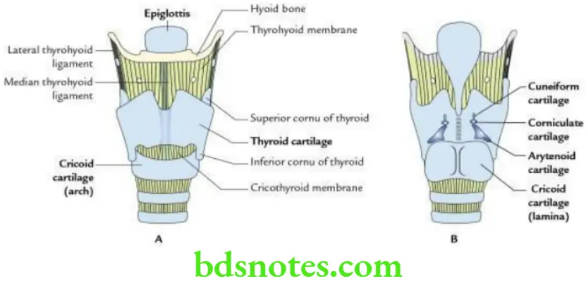
Cartilages Forming the Skeleton of Larynx Types
- The thyroid, cricoid and most of arytenoid are made up of hyaline cartilage.
- The epiglottis, corniculate, cuneiform and apices of arytenoids are made of yellow elastic cartilage.
Question 3. Name the safety muscle of larynx and give the reason why it is so named.
Answer.
The posterior cricoarytenoid is a safety muscle of larynx.
It is so named because it is the only intrinsic muscle of larynx, which abducts the vocal cords to allow the entry of air into the LRT. All the other intrinsic muscles of larynx adduct the vocal cords and restrict the entry of air into the LRT.
Question 4. What are the boundaries of laryngeal inlet?
Answer.
- Anterior: Epiglottis
- On each side: Aryepiglottic fold
- Posterior: Interarytenoid fold
Question 5. List the subdivisions of laryngeal cavity. Mention the narrowest part of the laryngeal cavity.
Answer.
The laryngeal cavity is divided into three parts:
- Vestibule: Between laryngeal inlet and vestibular folds.
- Ventricle (sinus): Between vestibular folds above and vocal folds below.
- Infraglottic part: Below the vocal folds, and up to the lower border of cricoid cartilage.
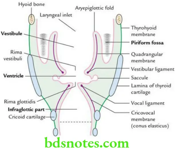
The glottis (i.e. space between the two vocal folds) is the narrowest part of the laryngeal cavity.
Question 6. Enumerate the intrinsic muscles of larynx.
Answer.
The intrinsic muscles of larynx are:
- Cricoarytenoid
- Posterior cricoarytenoid
- Lateral cricoarytenoid
- Transverse arytenoid
Read And Learn More: Selective Anatomy Notes And Question And Answers
- Oblique arytenoid
- Aryepiglotticus
- Thyroarytenoid
- Thyroepiglotticus
Question 7. List the origin, insertion, nerve supply, actions and applied anatomy of cricothyroid muscle.
Answer.
Cricothyroid Muscle Origin
Anterolateral part of the arch of cricoid cartilage.
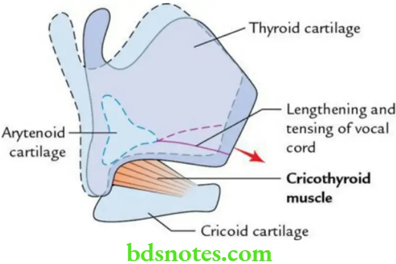
Cricothyroid Muscle Insertion
Inferior cornu and adjoining part of the lower border of thyroid cartilage.
Cricothyroid Muscle Nerve supply
External laryngeal nerve.
Cricothyroid Muscle Actions
It lengthens and tenses vocal cords by tilting the thyroid cartilage forwards. It also causes adduction of vocal cords.
Cricothyroid Muscle Applied anatomy
The cricothyroid muscle is an important muscle for pitch and tone of voice. Hence, its paralysis may alter the voice significantly, which is noticeable especially in singers.
Question 8. What is rima glottidis? Mention its boundaries.
Answer.
The rima glottidis is the narrowest part of the laryngeal cavity. It is an anteroposterior cleft, bounded in front by angle of thyroid cartilage, behind by interarytenoid fold and on each side by vocal fold (in anterior three-fifth) and by vocal process of arytenoid cartilage (in posterior two-fifth).
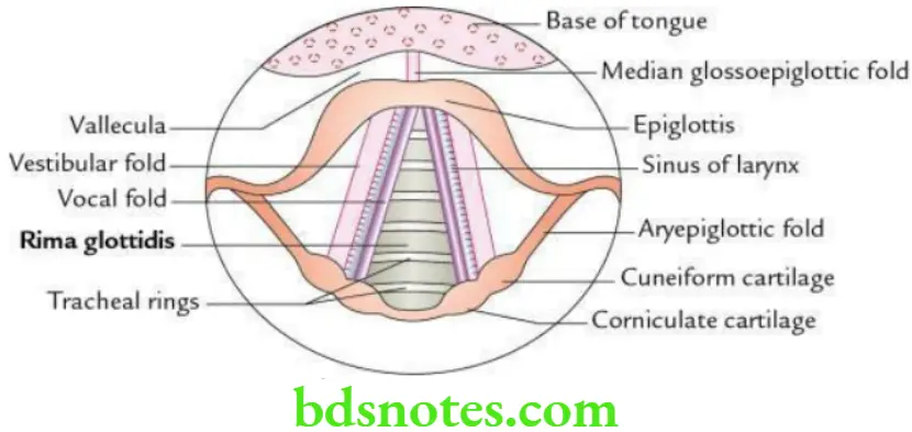
Question 9. Draw a labelled diagram to show structures seen in the laryngeal cavity during laryngoscopy.
Answer.
The structures seen in the laryngeal cavity during laryngoscopy.
Question 10. Write a short note on vocal cords.
Answer.
They are a pair of folds extending anteroposteriorly within the laryngeal cavity. The gap between the right and left vocal folds is called rima glottidis. Each vocal cord is made up of vocal ligament (medially) and vocalis muscle (laterally).
The vocal ligament extends from the tip of vocal process of arytenoid cartilage posteriorly to the inner aspect of thyroid cartilage anteriorly. The vocalis muscle (the detached medial part of thyroarytenoid muscle) also extends from inner aspect of thyroid cartilage (anteriorly) to the vocal process of the arytenoid cartilage (posteriorly). The posterior part of vocal folds is formed by arytenoid cartilages. Each vocal cord is covered by mucous membrane.
Question 11. Write a note on changes in size and shape of rima glottidis.
Answer.
Functionally rima glottidis consists of two parts:
- Intramembranous part (anteriorly three-fifth) between vocal cords
- Intracartilaginous part (posteriorly one-fifth) between arytenoid cartilage
The changes in size and shape of rima glottidis takes place during respiration and speech. These are:
- During quiet respiration, vocal cords lie only, at some distance.
- During full respiration, vocal cords move apart.
- During whispering, only intracartilaginous part widens.
- During high-pitched voice, both intramembranous and intracartilaginous parts adduct and rima glottidis is reduced to a linear chink.
Question 12. Why vocal cords do not swell much in laryngitis? Explain its anatomical basis.
Answer.
The laryngitis is the inflammation of the larynx. The vocal cords do not swell much in laryngitis due to the following reasons:
- They are lined by stratified squamous epithelium (cf. rest of the laryngeal cavity is lined by pseudostratified ciliated columnar epithelium).
- The mucous membrane of the vocal cords is firmly attached to the vocal ligaments.
- There is no submucous tissue and glands in the vocal cords.
Question 13. What are vocal nodules/singer’s nodules?
Answer.
The vocal nodules are bilateral swellings in the vocal cords at the junction of their anterior one-third and posterior two-third.
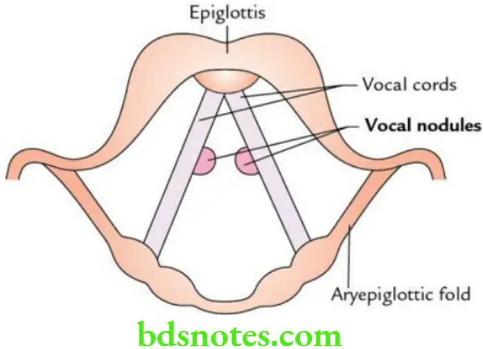
When the vocal cords vibrate during phonation, they come in maximum contact with each other at the junction of their anterior one-third and posterior two-third. The inflammatory swellings (inflammation due to friction) which appear at these sites are called vocal nodules.
The size of vocal nodules varies from pin-head to split-pea and their colour varies from reddish (in early stage) to whitish (in later stage).
Question 14. Enumerate the effects of lesions of laryngeal nerves.
Answer.
The effects of lesions of laryngeal nerve.
Effects of Lesions of Laryngeal Nerves

Question 15. Describe piriform fossa in brief and discuss its clinical importance.
Answer.
The piriform fossa is a deep recess in the lateral wall of the laryngopharynx, one on each side of laryngeal inlet.
Piriform Fossa Boundaries
Medially: Aryepiglottic fold
Laterally: Thyrohyoid membrane and thyroid cartilage
Piriform Fossa Clinical importance
The foreign bodies like fish bones and safety pins may be lodged in piriform fossa. If care is not taken during the removal of these foreign bodies, the instrument used for the removal of foreign bodies may pierce the mucous membrane lining, the floor of fossa and damage the internal laryngeal nerve and vessels, which lies just beneath it.

Leave a Reply