Question 1. Describe anatomy of tongue. Add a note on its development
(or)
Describe surface features of dorsum of the tongue. How do you connect its epithelial innervations to its development?
(or)
Development of tongue
Answer:
Development of Tongue:
- It is a muscular organ situated in the floor of the mouth
- The tongue has
- A root
- A tip
- A bodywhich has
- A curved upper surface or dorsum
- An inferior surface
Read And Learn More: BDS Previous Examination Question And Answers
Dorsum of tongue:
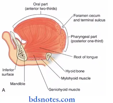
- It is divided into 2 parts by a faint Vshaped groove, the sulcus terminalis.
- They are
1. Oral part or anterior twothird
- Site: It is placed on the floor of the mouth
- Surface & Margins:
- Its margins are free & in contact with the gums & teeth
- Its superior surface shows a median furrow & is covered with rough papillae
2. Pharyngeal part or posterior onethird
Development of Tongue Site:
- It lies behind the palatoglossal arches & the sulcus terminalis
Development of Tongue Surface:
- Posterior surfaceforms the anterior wall of the oropharynx
- The mucous membrane is devoid of papillae
- It consists of lingual tonsil & mucous glands
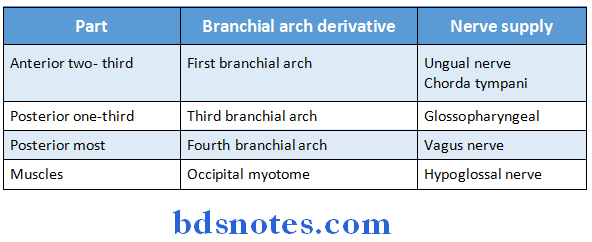
Development of tongue & nerve supply:
Epithelium & muscular development:
Question 3. Give an account of musculature of tongue & its development. Write briefly about lymphatic drainage of tongue (or) Lymphatic drainage of tongue (or) Lymphatic drainage of tongue
Answer:
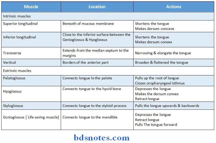
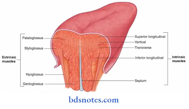
- Tongue is a muscular organ
- It consists of 4 intrinsic & 4 extrinsic muscles
Lymphatic Drainage:
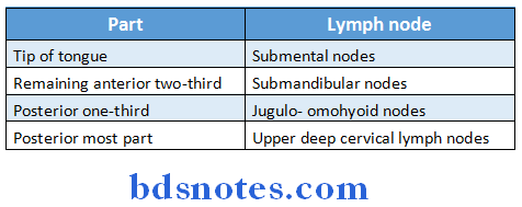
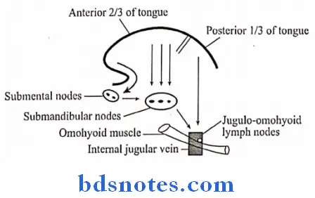
Question 4. Describe in detail about tongue including its blood supply & innervations
Answer:
Tongue Blood Supply:
- It is muscular organ
Tongue Blood Supply Location:
- It is situated in the floor of mouth
Tongue Blood Supply Parts:
1. Oral part
- It lies in the mouth
2. Pharyngeal part
- It lies in the pharynx
3. Sulcus terminalis
- V-shaped sulcus, separating oral & pharyngeal part
Tongue Blood Supply Functions:
- Taste
- Speech
- Deglutition
- Mastication
Tongue Blood Supply External features:
- The tongue has
-
- Root:
- Attachments
- Abovestyloid process & soft palate
- Belowhyoid bone
- Between themgeniohyoid & mylohyoid muscles
- Attachments
- TIP:
- It forms anterior free end
- Location: It lies behind upper incisor teeth
- Body:
- It has
- A curved upper surface/dorsum It is divided into
- Oral/anterior 2/3rd
- Placed on the floor of mouth
- It shows foliate papillae just in front of the palatoglossal arch
- Pharyngeal part/posterior 1/3rd
- It lies behind paltoglossal arches & the sulcus terminalis
- It constitute lingual tonsil
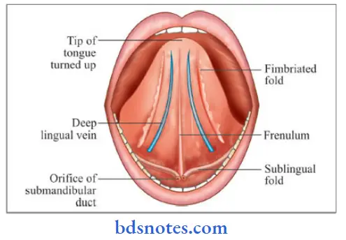
- An inferior surface
- It shows median frenulum linguae
- On either side, there is presence of deep lingual veins
- Oral/anterior 2/3rd
- A curved upper surface/dorsum It is divided into
- It has
- Root:
Posteriormost part of tongue:
- It is connected to the epiglottis by
- Median glossoepiglotic fold
- Right & left lateral glossoepiglotic fold
- It separates vallecula which is a depression, from the piriform fossa
Papillae of the tongue:
1. Vallate or circumvallate papillae
- Situated immediately in front of the sulcus terminalis
2. Fungiform papillae
- Present near the tip & margins of the tongue & scattered over the dorsum
3. Filiform papillae
- Covers the presulcal area
4. Foliate papillae
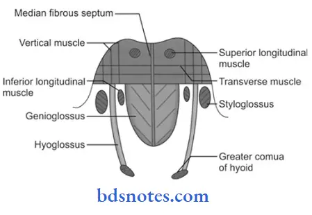
Muscles of tongue:
1. Intrinsic Muscles:
- Superior longitudinal
- Inferior longitudinal
- Transverse
- Vertical
2. Extrinsic muscles:
- Genioglossus
- Hyoglossus
- Styloglossus
- Palatoglossus
Muscles of tongue Blood Supply:
- Arterial Supply:
- Lingual arterybranch of external carotid
- Tonsillarbranch of facial artery
- Ascending pharyngealbranch of external carotid
- Venous Drainage:
- Two venae comitantes
- Accompany the lingual artery
- One vena comitantes
- Accompany hypoglossal nerve
- Deep lingual veinprincipal vein
- These veins unite at the posterior border of the hyoglossus
- It forms lingual vein
- It ends in internal jugular vein
- Two venae comitantes
Question 5. Describe tongue under the following headings
- Parts & gross external features
- Nerve supply
- Development including anomalies
Answer:
Parts & gross external features:
Anomalies:
- Glossia
- It is complete absence of tongue
- Hemiglossia
- Onehalf of anterior twothird is absent
- Lingual thyroid
- Thyroglossal duct is absent
- Persistence of Thyroglossal duct on tongue
- Ankyloglossia
- Short frenulum linguae
- Bifid tongue
Question 5. Describe tongue under following headings
- Development
- Microscopic anatomy
- Applied importance
Answer:
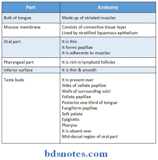
Tongue Microscopic anatomy:
Tongue Applied importance:
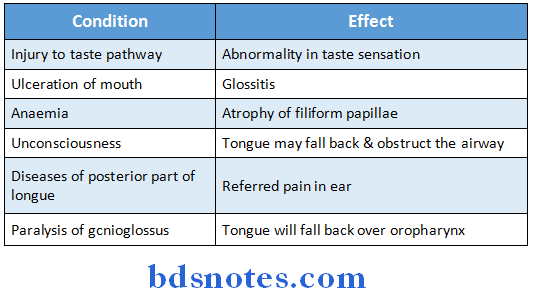
Tongue Other importance:
- The presence of rich network of lymphatics is responsible for acute glossitis
- The undersurface of tongue is examined for jaundice
- Lingual tonsil is a part of Waldeyer’s ring
- In case the tongue falls back, it is pulled mechanically
- Sublingual route of sorbitrate is effective for treatment of angina
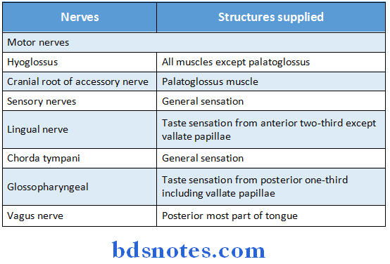
Question 6. Nerve supply of tongue
Answer:
Question 7. Hyoglossus muscle
Answer:
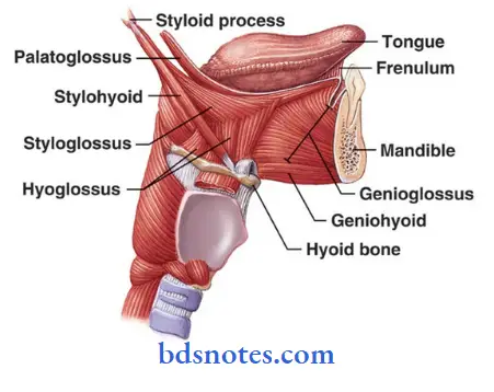
- It is an extrinsic muscle of tongue
- It connects the tongue to hyoid bone
Hyoglossus muscle Origin:
- Whole length of greater cornua
- Lateral part of hyoid bone
Hyoglossus muscle Insertion:
- Side of tongue between styloglossus & inferior longitudinal muscle of tongue
Hyoglossus muscle Actions:
- Depresses tongue
- Makes dorsum convex
- Retracts protruded tongue
Hyoglossus muscle Relations:
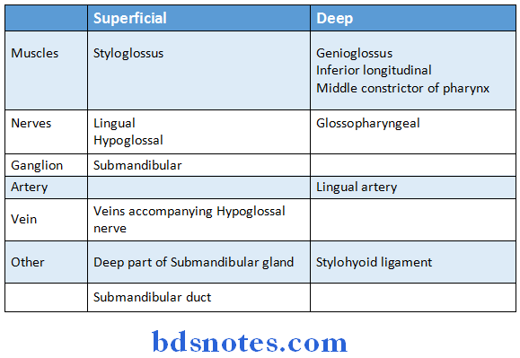
Question 8. Histology of tongue
Answer:
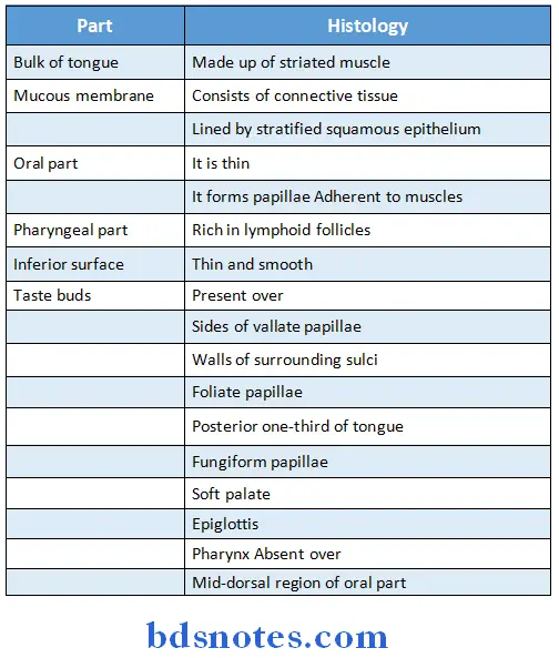
Question 9. Circumvallate papillae
Answer:
- It is large in size
- Size: 12 mm in diamter
- Number – 8 – 12
- Situation
- Immediately in front of sulcus terminalis
- Each papilla is cylindrical projection surrounded by circular sulcus
- The walls of papilla have taste buds
Question 10. Name extrinsic muscles of tongue
Answer:
Extrinsic Muscles of Tongue:
- Palatoglossus connects tongue to palate
- Hyoglossus connects tongue to hyoid bone
- Styloglossus connects tongue to styloid process
- Genioglossus connects tongue to mandible.
Question 11. Bifid tongue
Answer:
- Bifid tongue is a congenital structural defect of the tongue in which its anterior part is divided longitudinally for a greater or lesser distance
- Bifid tongue is a rare congenital anomaly usually associated with syndromes and infrequently associated with nonsyndromic cases.
- The syndrome most commonly seen with associated finding of bifid tongue is the orofacialdigital syndrome.
- Various other syndromes and nonsyndromic cases also have been found to have an associated finding of bifid tongue as an irregular feature.
Question 12. Types of papillae over the tongue
Answer:
Papillae over the tongue are:
1. Fungiform papillae
- These are slightly mushroomshaped if looked at in longitudinal section.
- These are present mostly at the dorsal surface of the tongue, as well as at the sides.
- Innervated by facial nerve.
2. Foliate papillae
- These are ridges and grooves towards the posterior part of the tongue found at the lateral borders.
- Innervated by facial nerve (anterior papillae) and glossopharyngeal nerve (posterior papillae).
3. Circumvallate papillae
- There are only about 10 to 14 of these papillae on most people, and they are present at the back of the oral part of the tongue.
- They are arranged in a circularshaped row just in front of the sulcus terminalis of the tongue.
- They are associated with ducts of Von Ebner’s glands, and are innervated by the glossopharyngeal
nerve.
4. Filiform papillae
- They are the most numerous but do not contain taste buds.
- They are characterized by increased keratinisation
Question 13. Foliate papillae
Answer:
- Foliate papillae are short vertical folds and are present on each side of the tongue.
- They are located on the sides at the back of the tongue, just in front of the palatoglossal arch of the fauces.
- There are four or five vertical folds and their size and shape is variable.
- The foliate papillae appear as a series of red colored, leaflike ridges of mucosa.
- They are covered with epithelium, lack keratin and so are softer, and bear many taste buds.
- They are usually bilaterally symmetrical.
- Sometimes they appear small and inconspicuous, and at other times they are prominent.
- Because their location is a high risk site for oral cancer, and their tendency to occasionally swell, they may
- be mistaken as tumors or inflammatory disease.
- Taste buds are scattered over the mucous membrane of their surface.
- Serous glands drain into the folds and clean the taste buds.

Leave a Reply