Skeletal System
The skeletal system is made up of bones and cartilage. It provides a strong and flexible framework for the body.
Bones
Question 1. What is bone?
Answer.
The bone is a specialized connective tissue with a mineralized matrix of calcium salts, for example, calcium hydroxyapatite crystals. It provides hardness to the human skeleton.
Classify bones according to their shape.
Classification of Bones
- Long bones: They consist of a shaft and two ends. The elongated tubular shaft or body is called diaphysis. It contains a medullary cavity within it. The expanded ends are called epiphyses. Examples: Long bones of limbs such as femur, tibia and fibula in the lower limb and humerus, radius and ulna in the upper limb.
- Short long bones (also called miniature long bones): They have a shaft and only one epiphysis. Examples: Metatarsals, metacarpals and phalanges.
- Short bones: They are smaller in size and usually cube-shaped. Examples: Carpal and tarsal bones.
- Flat bones: They are flat (plate-like) and consist of two layers (plates) of compact bone with spongy bone, filled with bone marrow between them. Examples: Bones forming the skull cap such as frontal, parietal and occipital. The ribs, sternum and scapula are also classified as flat bones.
- Irregular bones: They have an irregular shape. Examples: Bones of the face and base of the skull; vertebrae and hip bones.
- Pneumatic bones: They contain cavities within them, which are filled with air. These bones are confined to the skull. Examples: Paranasal bones such as maxilla, ethmoid, sphenoid and frontal bones.
- Accessory bones: They are sometimes present in the limbs and skull. Examples: Os trigonum (os vesalianum), os cubiti in the limbs and wormian bones in the skull.
- Sesamoid bones: They develop in the tendons of muscles and are devoid of periosteum. Examples: Patella and fabella.
Question 2. What are the functions of the bones?
Answer.
Functions of the bones:
- Form the skeletal framework of the body, thus providing shape to the body.
- Protect vital organs such as the brain, spinal cord, heart and lungs.
- Provide a surface for attachments to the skeletal muscles.
- Act as a storehouse for mineral salts, for example, calcium and phosphorus.
- Act as levers for the body movements by the muscles.
- Produce blood cells.
Question 3. Enumerate the structural components of a long bone.
Answer.
Structural components of a long bone
- Mineralized matrix
- Specialized cells, for example, osteoblasts, osteocytes and osteoclasts
- Periosteum
- Endosteum
- Medullary cavity
- Bone marrow
Question 4. What are the sites where red bone marrow is present in adults?
Answer.
The sites where red bone marrow is present in adults:
- Proximal ends of femur and humerus
- Ribs
- Sternum
- Skull
- Vertebrae
- Hip bone
Question 5. What are the parts of a long bone?
Answer.
The long bone consists of the following parts:
- Epiphyses: Ends of a long bone that ossifies from secondary centres.
- Diaphysis: Shaft of a long bone that ossifies from a primary centre. It consists of an outer cortex of compact bone and an inner medullary cavity filled with bone marrow.
Question 6. Write a short note on the periosteum.
Answer.
The periosteum forms the outer fibrous covering of the bone. It covers the whole of the bone surface, except where the bone is covered by articular cartilage. The periosteum is attached to the bone tissue by Sharpey’s fibres. It consists of two layers:
- An outer fibrous layer, which becomes continuous at the ends of the bone with the fibrous capsule of a joint. It protects the bone.
- An inner cellular layer containing osteoprogenitor cells (osteogenic cellular layer), ends at the epiphyseal line. This layer is responsible for the deposition of bone on the surface of the shaft and thus adds to the growth of bone in girth. It is also essential for fracture repair.
Functions
- Protects the bone, as it covers the outer surface of the bone.
- Helps in bone formation, helping bone growth at a young age and repairing fractures in adults, as it contains bone-forming cells.
Read And Learn More: Selective Anatomy Notes And Question And Answers
- Helps to provide nutrition to the bone, as it is richly supplied with blood vessels.
- Makes bone sensitive to pain as it is supplied with sensory nerves.
- Provides a medium for the attachment of ligaments, tendons and muscles to the bone.
Question 7. What are Sharpey’s fibres?
Answer.
- The periosteum is anchored to the outer part of bone tissue by Sharpey’s fibres.
- From the inner layer of the periosteum, coarse collagen fibres extend inwards to enter the bone matrix. These are called Sharpey’s fibres (perforating fibres). The Sharpey’s fibres enter the bone matrix like spikes of a shoe thus anchoring the periosteum to the bone.
Question 8. Classify bones according to their structure.
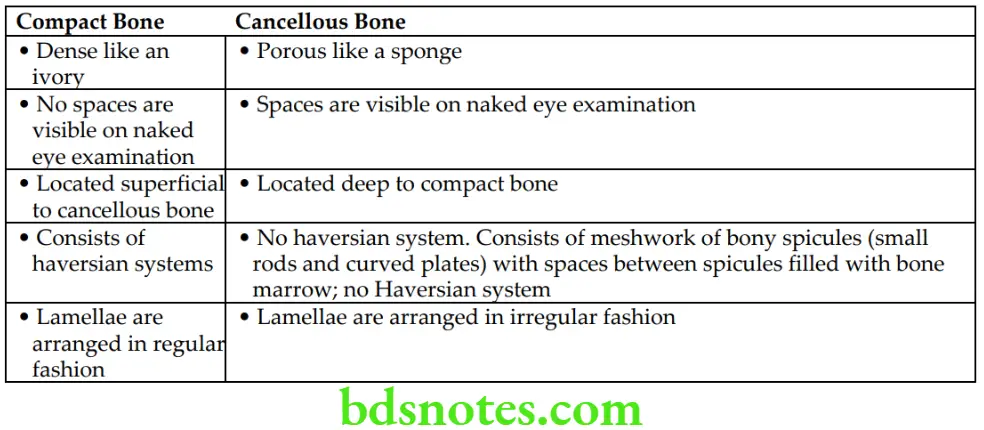
Answer: Structurally, bones are classified into two types: compact bone and cancellous bone. The differences between these two types are given in Table.
Differences Between Compact and Cancellous Bones

Question 9. Enumerate the parts of a growing/developing long bone.
Answer.
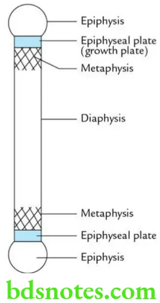
The parts of growing long bone:
- Epiphysis: It is the end of long bones that ossifies from the secondary centre.
- Diaphysis: It is the shaft/body of long bone that ossifies from the secondary centre.
- Metaphysis: It is part of the diaphysis near the epiphysis. It is a zone of active bone growth.
- Epiphyseal plate: It is a plate of hyaline cartilage between epiphysis and diaphysis. The epiphysial cartilage is responsible for the growth of bone in length. Hence, it is also called a growth plate.

Question 10. Write a short note on the arterial supply of a growing long bone.
Answer.
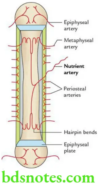
The growing long bone is supplied by the following arteries:
- Nutrient artery: It is tortuous and enters the middle of the shaft through the nutrient foramen. It runs obliquely through the cortex into the medullary cavity, where it divides into ascending and descending branches.
- Each branch in turn divides and redivides into parallel vessels, which run in metaphysis, where they terminate by forming hairpin bends. The ascending and descending branches also ramify in the endosteum and give twigs to adjoining canals.
- It supplies the medullary cavity and the inner two-thirds of the cortex. The nutrient foramen is directed opposite to the growing end of a long bone.
It anastomoses with periosteal and metaphyseal arteries.
- Juxta-epiphysial (metaphyseal) arteries: These are derived from arterial anastomosis around the joint. They pierce the metaphysis along the line of attachment of the joint capsule.
- Epiphysial arteries: These are derived from periarticular vascular arcades, found on the nonarticular bony surface and enter the bone distal to epiphyseal cartilage.
- Periosteal arteries: These ramify beneath the periosteum and supply the outer two-thirds of the cortex. The removal of periosteum may cause necrosis of underlying bone.

Metaphysics is the common site for osteomyelitis in children – mention its anatomical basis.
This is because the metaphysis is a zone of active growth. It is profusely supplied with blood by end arteries, which form ‘hairpin bends’. The bacteria and emboli are easily trapped in these hairpin bends leading to infarction and subsequently to osteomyelitis.
Question 11. Describe sesamoid bones briefly.
Answer.
The sesamoid bones are seed-like bony nodules found embedded in the tendons of muscles (in Arabic: Sesame = seed).
Characteristic features
- Develop in tendons of the muscles after birth.
- Are devoid of periosteum.
- Are devoid of Haversian systems.
- Ossify by multiple secondary centres.
Functions of bones
- Alter the direction of the pull of the muscle tendon.
- Minimize the friction of the tendon against the bone.
- Modify and sustain pressure.
- Provide an additional articular surface to a joint.
- Protect the tendons from wear and tear.
- Act as pulleys for muscular contraction.
Sites of bones
- Tendon of quadriceps femoris (patella)
- Lateral head of gastrocnemius muscle (fabella)
- Tendon of flexor carpi ulnaris (pisiform)
- Two bony nodules (sesamoid bones) beneath the head of 1st metatarsal in the tendon of flexor pollicis brevis
- Bony nodule in the tendon of adductor hallucis
- Bony nodule in the tendon of peroneus longus where it winds around the cuboid bone
Question 12. Define ossification and ossification centres.
Answer.
Ossification It is the process of bone formation.
Ossification centre The ossification centres are sites where bone formation begins.
There are two types of ossification centres:
- Primary and
- Secondary.
- Primary centre: It appears before birth in the centre of the shaft or body of bone which it forms.
- Secondary centre: It appears later, usually after birth at each end of the bone which it forms.
Question 13. Write a short note on endochondral/cartilaginous ossification.
Answer.
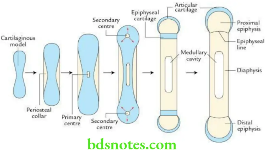
It is the process of formation of bone from the preformed cartilaginous model (premature long bone).
- The process of bone formation begins in the centre of the shaft of a long bone. This site where bone formation begins is called the primary ossification centre. This centre forms the diaphysis.
- Later, centres of ossification appear at different intervals at each end of the cartilaginous model. These are called secondary ossification centres. These centres form the epiphyses.
- The plate of hyaline cartilage separating the epiphysis and diaphysis is called epiphyseal plate/growth plate. It is essential for the growth of bone in length.
- When the epiphysis unites with the diaphysis, the epiphyseal plate is replaced by a linear scar called the epiphyseal line.

Question 14. What are the different types of epiphyses?
Answer.
There are four types of epiphyses:
- Pressure epiphyses: They are present at the ends of long bones. They are, therefore, articular in nature and take part in the transmission of weight, for example, the head of the femur, lower end of the radius and medial end of the clavicle.
- Traction epiphyses: They are nonarticular in nature and are not involved in the transmission of weight. They provide attachment to one or more muscle tendons, which exert traction on it, for example, greater and lesser trochanters of the femur, and greater and lesser tubercles of the humerus.
- Atavistic epiphyses: Phylogenetically, they represent a separate bone, which in man has become fused secondarily to another bone, for example, the coracoid process of the scapula, posterior tubercle of the talus (os trigonum).
- Aberrant epiphyses: They are not always present but appear sometimes, for example, epiphysis at the head of the 1st metacarpal.
Cartilage
Question 1. What is cartilage?
Answer.
The cartilage is a specialized connective tissue, with a rubbery matrix (gel-like matrix) due to the deposition of proteoglycans which provides firmness along with elasticity to the skeletal framework of the body. Phylogenetically, it is older than the bone tissue.
- It is made up of a dense network of collagen or elastic fibres, which provide tensile strength to it.
- Its fibres are embedded in a firm, jelly-like amorphous substance made up of mucopolysaccharides, which allows the cartilage to bear weight without bending.
- It is firm in consistency and has elasticity.
- It is an avascular tissue. The invasion of cartilage by blood vessels results in its calcification and death.
- It has no lymphatics.
- It is well adapted to coat the articular ends of the bone.

Leave a Reply