Special Connective Tissues
The special connective tissues include:
- Cartilage
- Bone
- Haemopoietic tissue
- Blood
- Lymph
Cartilage
Question 1. What is cartilage? List its properties.
Answer.
Cartilage is a modified connective tissue. It consists of all the three components of connective tissue, but differs from connective tissue proper in the sense that its ground substance is made up of gel-like material (containing chondromucoprotein), which provides it firmness and elasticity.
Properties Of Cartilage
- It is firm in consistency.
- It is relatively avascular.
- It derives its nutrition from adjacent tissues by diffusion or through small vessels passing through cartilaginous canals.
- It has limited regeneration capacity.
- It is prone to undergo calcification with advancing age.
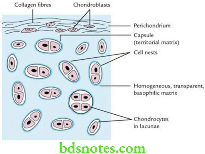
- It is usually surrounded by a fibrovascular membrane called perichondrium.
- It forms part of body skeleton.
- It has no lymphatics.
- It has no nerves; hence, it is insensitive to pain.
- Its growth occurs by appositional growth as well as interstitial growth.
Question 2. Write a short note on perichondrium.
Answer.
- The perichondrium is a vascular membrane which forms the covering of a cartilage.
- It is made up of two layers:
- Outer fibrous layer made up of collagen bundles. It has rich blood supply.
- Inner chondrogenic layer made up of chondroblasts. The chondroblast can change into chondrocytes. The chondroblasts can divide and secrete matrix, and it assists in the peripheral growth of the cartilage.
Classify The Various Types Of Cartilages.
The cartilages are classified into three types:
- Hyaline cartilage
- Yellow elastic cartilage
- Fibrocartilage
Question 3. Write a short note on the hyaline cartilage.
Answer.
It is the most widely distributed cartilage in the body.
Histological features
- It is transparent (Greek, hylos = transparent stone).
- It has homogeneous, basophilic intercellular substance.
- It stains blue (i.e. basophilic) with haematoxylin and eosin stain due to the presence of chondroitin and keratin sulphate in its ground substance.
Read And Learn More: Selective Anatomy Notes And Question And Answers
- The chondrocytes are present in groups of two to four cells within lacunae to form cell nests.
- The matrix around the cell nests is stained deeper to form lacunar capsule. Groups of 2 or more chondrocytes are known as cell nests.
- The fibres are not seen as a distinct entity.
- It is covered by perichondrium.

Function
Resists compressive and tensile forces.
Sites
Laryngeal cartilages (e.g. thyroid, cricoid), tracheal ring, costal cartilage, septal cartilage of nose, most of the fetal skeleton.
Question 4. Write a short note on the elastic cartilage.
Answer.
The elastic cartilage is also called yellow elastic cartilage.
Histological features
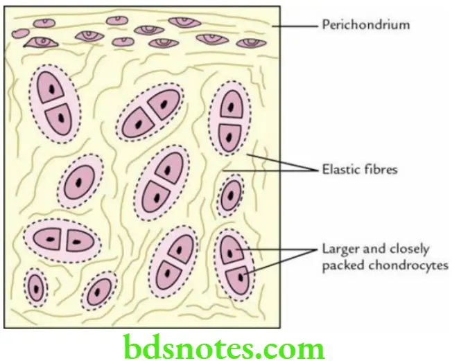
- It appears yellowish in fresh sections due to the presence of yellow elastic fibres.
- It contains large number of branching and anastomosing yellow elastic fibres in its ground substance. They are continuous with those of perichondrium.
- Its chondrocytes are larger, more numerous and closely packed than those of hyaline cartilage.
- The chondrocytes are seen in lacunae simply or in groups of two.
- It is covered by perichondrium.

Function
Provides not only shape and support to the organ but also elasticity/pliability.
Sites of distribution
Pinna of external ear, epiglottis, eustachian tube, etc.
Question 5. Write a short note on the fibrocartilage.
Answer.
Histological features
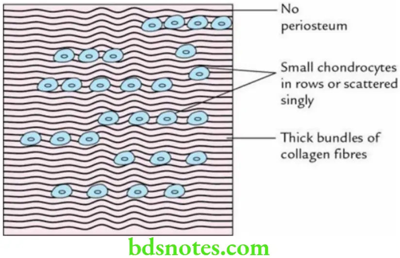
- Macroscopically, it looks like a dense connective tissue.
- It consists of large bundle of collagen (type I) fibres.
- Its chondrocytes are small and of the same size.
- The chondrocytes are frequently arranged in rows between the collagen bundles.
- It is not covered by perichondrium.

Function
Resists compression and shear forces.
Sites of distribution
Intervertebral discs, menisci of the knee joint, intra-articular discs of the synovial joints, glenoid labrum and acetabular labrum.
Question 6. List the differences among hyaline, elastic and fibrocartilages.
Answer.
The differences are given in the Table.
Differences Between Hyaline, Elastic and Fibrocartilage

Bone
Question 1. Define bone and list its functions.
Answer.
The bone is a specialized connective tissue in which the matrix is mineralized with calcium salts, making it hard and rigid. The calcium salts in the matrix also provide whitish look to the bones.
Functions Of Bones
The functions of bones are:
- To form rigid framework of the body.
- To provide surface for attachment of the muscles.
- To serve as storehouse of calcium and phosphorus.
- To form cavities to enclose and protect the various viscera.
- To manufacture blood cells, e.g. RBCs, WBCs and platelets.
- To act as levers for various movements of the body.
Question 2. Enumerate the various types of cells in the bone tissue.
Answer.
There are four types of cells in the bone tissue:
- Osteogenic cells: They are precursors of other cell types in the bone.
- Osteoblasts: They lay down the matrix of bone tissue.
- Osteocytes: They maintain bone matrix and are the main cells of bone tissue.
- Osteoclasts: They are involved in bone resorption.
Question 3. Define compact bone and list its histological features.
Answer.
The compact bone is dense and hard like an ivory, with no visible spaces on naked-eye examination.
Histological features
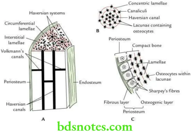
- It consists of Haversian systems. Each Haversian system (osteon) consists of a Haversian canal surrounded by 6–12 concentric bony lamellae.
- Haversian canals are connected with one another and communicate with marrow cavity through the bony canals called Volkmann’s canals.
- It has three types of bony lamellae:
- Concentric lamellae, around the haversian canals.
- Interstitial lamellae, between the haversian systems.
- Circumferential lamellae, subjacent to the periosteum and adjacent to the endosteum.
- It is covered by periosteum.

Sites
- Shaft of long bones.
- Outer layer of all bones.
Question 4. Write a short note on the spongy bone.
Answer.
The cancellous bone is porous and relatively less hard as compared to compact bone.
Histological features Of Bone
- It does not have haversian systems.
- The bone tissue is arranged in the form of interconnecting rods and thin plates called trabeculae.
- It contains large irregular communicating spaces between the adjoining trabeculae called marrow spaces filled with red bone marrow.
- The osteocytes are embedded in lacunae within the trabeculae, while osteoblasts and osteoclasts are present at the surface of trabeculae.
- It is covered by periosteum.
Sites
- Epiphyses of long bones.
- All short, flat and irregular bones.
Question 5. List Differences Between The Compact And Spongy Bones.
Answer.
They are given in Table.
Differences Between the Compact and Spongy Bones


Leave a Reply