Histochemistry Of Oral Tissues
Question 1. Describe the stages of tissue processing and write about fixation.
Answer:
Tissue processing:
- It is a long procedure and requires 24 hours.
- Stages:
1. Fixation:
- It is an important step in the preparation of tissue for microscopic examination.
- Goals:
- Preserves the structure.
- It terminates any ongoing biochemical reaction.
- It increases the strength and stability of treated tissue.
Read And Learn More: BDS Previous Examination Question And Answers
- Purposes:
- Coagulate proteins.
- Prevents autolysis of cell
- Fix cell constituents and tissue that withstand subsequent treatment with various reagents with minimum loss of anatomy.
- Steps:
- Killing tissue.
- Preservation of tissue.
- It is done by fixatives, commonly used – 10% formalin.
- Temperature:
- Done at room temperature.
- Time:
- It takes several hours to several days.
2. Dehydration:
- After fixing the tissue, it is washed with running water.
- Water cannot mix with paraffin.
- Hence, the water content is to be removed.
- It is done by passing the specimen by increasing the percentage of alcohol.
3. Clearing:
- Alcohol is not miscible with paraffin.
- Thus, the tissue is first cleared of alcohol by a reagent like xylene.
- Xylene is miscible with both alcohol and paraffin.
- Time:
- A small piece of tissue is cleared in 0.5 1 hour.
- Larger/thicker piece in 2-4 hours.
- Time:
4. Infiltration:
- When xylene has completely replaced alcohol, the specimen is infiltrated with paraffin at 60°C.
5. Embedding:
- Tissue is embedded in a block of paraffin wax with the proper orientation of the specimen.
- Paraffin wax is used as it has hard consistency.
6. Cutting:
- The paraffin wax block is attached to a wooden cube which is clamped onto the microtome.
- It is adjusted to cut the desired piece of the specimen.
7. Mounting:
- The sections are mounted on microscope slides.
8. Staining:
- Mounted slides are stained to give color to a section.
Procedure:
- Put sections in xylene for 3 minutes
- Transfer to the increasing strength of alcohol for 3 minutes
- Wash in running water for 1 minute
- Put in hemotoxylin for 5 to 7 min.
- Wash in running water for 30 sec.
- Wash excess in 1% acid alcohol for 15 sec.
- Wash in running water for 30 sec.
- Give 2-3 dips of ammonia water solution until tissues attain blue color.
- Wash in running water for 30 sec.
- Counterstain it.
- Wash it
- Dehydrate it.
Question 2. Fixation procedures and fixatives.
Answer:
Fixation:
- It is a procedure to make cell constituents and tissue withstand subsequent treatment with minimum loss of anatomy.
- This is accomplished by using.
- Optimum osmotic conditions.
- Cold temperatures.
- Controlled pH of the fixing solution.
- Minimum possible exposure to the fixatives.
Procedures/types:
Physical:
- Heating
- Micro-waving
- Cryo-preservation or freeze drying.
Chemical:
- By immersing in fixatives.
- By perfusing the vascular system with fixatives.

Question 3. Preparation of the ground section of the tooth.
Answer:
- The labial surface of the tooth is ground down till the desired section is achieved using a coarse abrasive wheel.
- The coarse wheel is exchanged with the fine abrasive wheel.
- The tooth is further ground.
- An adhesive tape is wrapped around a wooden block with a sticky side outward.
- The grounded section of the teeth is stuck to it.
- A similar grinding procedure is repeated from the lingual surface.
- The finished ground section is soaked off with ether.
- It is then dried off for several minutes
- Longer drying leads to cracking.
- It is then mounted on microscopic slides.
Question 4. Microtome
Answer:
- It is a tool used to cut extremely thin slices of material, known as sections
- Used for preparation of samples
Blades used:

Applications:
- Traditional histological examination
- For frozen sections
- For electron microscopy
- For spectroscopy
- For fluorescence microscopy
Types:

Microtome knives:
- Planar concave extremely sharp
- Wedge-shaped More stable
- Chisel shaped Has blunt edge
Question 5. Stages in soft tissue processing.
Answer:
Stages:
- Obtaining the specimen.
- Fixation Ideally with 10% formalin.
- Dehydration with increasing strength of alcohol.
- Clearing.
- By passing through xylene
- Infiltration by paraffin.
- Embedding the specimen in paraffin wax block.
- Cutting to obtain desired sections of tissues
- Mounting the sections on slides.
- Staining with the desired staining method.
Question 6. Declassification.
Answer:
It is a technique for removing minerals from bone and other calcified tissue so that good paraffin sections can be prepared.
Chemical used:
- 5% nitric acid.
Time required:
- 7-10 days for complete decalcification.
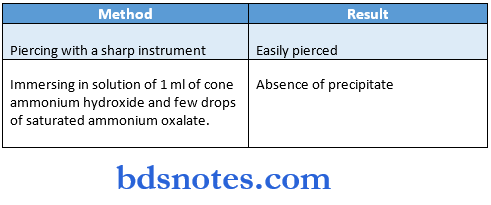
Tests:

Question 7. Xylene.
Answer:
Properties:
- Insoluble in water.
- Soluble in aromatic hydrocarbons.
- Flammable.
Uses:
- Result
- Easily pierced Absence of precipitate
- Used as a clearing agent to remove alcohol from the tissue. To remove paraffin from dried microscopic slides.
- After staining, slides are put in xylene prior to mounting. Used to dissolve gutta percha in endodontic treatment.
- As the solvent to remove synthetic immersion oil from the microscope.
Question 8. Alkaline phosphatase/Robinson’s Alkaline phosphatase.
Answer:
Alkaline phosphatase was first described by Robinson J.C.
- It is associated with the production of mineralized tissue.
- In hard connective tissue, it is found in the organic matrix.
- It hydrolyzes phosphate ions from organic radicals at alkaline pH.
- It cleaves pyrophosphate thereby permitting crystal growth to proceed.
- It is associated with osteogenesis and dentinogenesis.
- The osteoblasts and preosteoblasts exhibit alkaline phosphatase activity.
- It is seen in chondrocytes and cartilage matrix also.
- In human gingiva, the capillary endothelium of the lamina propria shows its activity.
- Alkaline phosphatase is involved in the mechanism of keratinization.
- Basement membrane associated with salivary gland acini and taste buds exhibits high alkaline phosphatase activity.
Question 9. Fixation of tissues:
Answer:
- It is an important step in tissue processing.
- It fixes the tissues to withstand subsequent treatment with various reagents with minimum loss of anatomy.
- The reagents used are called fixatives.
Purposes:
- Terminate ongoing biochemical reactions.
- Increases strength and stability of treated tissue.
- Preserves the structures
- Coagulate proteins.
- Prevents autolysis of cells.
Parameters:
- Minimal time between killing and fixation.
- The minimal size of the tissue
- Minimal tissue deformation.
Question 10. Frequently studied important enzymes.
Answer:
- Enzyme
- Alkaline phosphatize
- Acid phosphatize
- Oxidases and dehydrogenase
- Esterases
- Lysosomal sulfatase and adenyl cyclase

Question 11. Acid phosphatase.
Answer:
- Importance
- Related to the transfer of phosphate esters in the organic matrix of bone, enamel and dentin.
- Related to resorption of bone and dentin.
- Reflects metabolic activity of different oral tissues.
- Associated with the hydrolysis of carboxylic acid esters of alcohol Involved in the formation of cAMP.
- It is localized in membrane-bound vesicles called lysosomes.
- Types: Based on its interaction with substrates
- Para-nitro-phenyl phosphate
- Beta glycerophosphate.
Activity:
- In supranuclear and distal regions of secretory ameloblasts.
- During resorption, osteoclasts of bone and odontoblasts in resorbing dentin.
- Less activity occurs during the late secretory stage of amelogenesis.
Demonstration:
- Demonstrated cytochemical using naphthol ASBI phosphoric acid-fast red garnet GBC.
- In tissue sections using naphthol ASBI phosphoric acid pararosaniline.

Leave a Reply