Growth And Development Of Cranial And Facial Region
Question 1. Describe the modes by which the mandible grows from birth to adulthood.
Answer.
Postnatal Development of Mandible.
- Of all the facial bones, mandible undergoes large amount of post natal growth and undergo largest variability in its morphology.
- In postnatal life mandibular growth shows integration of both the periosteal and the capsular matrices of the functional matrix theory.
- Capsular matrix involves the oropharyngeal functional spaces and growth of the mandible occurs as per the functional needs of particular functional system. Process of surface remodeling involves the activity periosteal matrix. i.e. muscle fier.
During Birth
- During birth the size of mandible is very small.
- Condyles of the infant mandible are low and lies at position of occlusal plane.
- Ramus of mandible in infants is horizontal and consists of obtuse gonial angle.
Read And Learn More: Orthodontics Question And Answers
During First Year
- During fist year of life symphyseal suture grows and there is lateral expansion of symphyseal suture anteriorly for accommodating the erupting anterior teeth.
- Mental foramen is directed at 90° to surface of condyle.
- Posterior surface of mandible shows more deposition of bone.
- As the condyle and glenoid fossa are flt this lead to anteroposterior movement of mandible.
Growth of Mandible in an Adult/Concepts of V-Principle
As the child become adult mandible show various changes, i.e.
- Ramus of the mandible becomes long.
- Gonial angle becomes less obtuse.
- Condyles of mandible are well developed
- All over the bone become large.
V-Principle of Growth of Mandible
Growth of the mandible occurs in form of expanding V.

Length of the Mandible
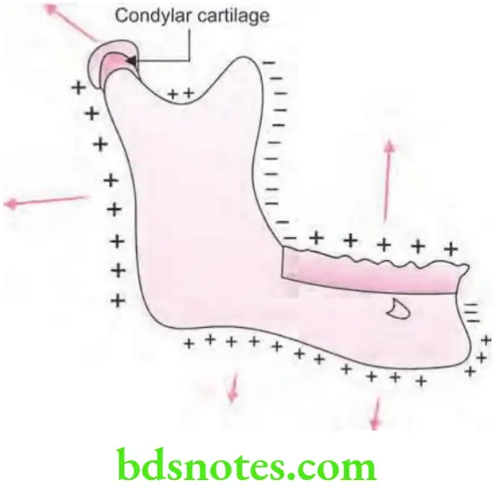
- Mandible grows in length anteroposteriorly by bone deposition at posterior surface of ramus and resorbing the leading edge at anterior surface.
- By this deposition and resorption of bone the mandible lengthens.
- Anterior part of ramus of mandible in future is occupied by posterior part of body of mandible.
- As the occupancy occurs permanent molars develop in it.
- Posterior growth of mandible causes its anterior displacement. This is because the articulation of condyle to glenoid fossa is constant.
- Anterior displacement brings about the change in mandible.
- Opening of mental foramen face backwards as there is anterior growth of mandible. This causes neurovascular bundle to leave foramen directed backward.
- Surface remodeling occurs at anterior border of the mandible.
- Bone deposition occurs at posterior surface of symphysis while bone resorption occurs at superior part of anterior surface. Bone deposition is also present at the inferior aspect.

Width of the Mandible
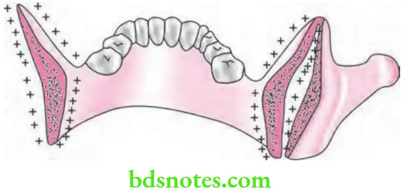
- Over the lateral surface of ramus of mandible deposition of bone occur while resorption of bone occurs towards the lingual surface below myelohyoid ridge.
- Coronoid process of mandible undergoes deposition of bone at mesial surface and resorption at lateral surface and leads to the expansion of mandible like V.
- Condyle of mandible undergoes resorption of bone over lateral aspect of neck of condyle and deposition of bone over medial aspect of neck of condyle. This leads to increase of neck of condyle in the length.
- Following V principle the distance between the ramuses of mandible is increased as mandible grows. In this way the mandible keep pace with growth of cranial base.
- Mandible establishes its orthognathic relation with maxilla in adult life as it grows in its length.
- Periosteal matrix influences the angle, coronoid and condylar processes.
Height of Mandible
- As the teeth erupts the height of alveolar process increases.
- At lower border of mandible deposition of bone occurs which increases the height of mandible.
Rotation of Mandible
- Bjork used implants to study growth pattrn of the mandible and found that mandible undergo growth rotation. It was seen that mandible undergoes the growth rotation, effcts seen are minimal because of external compensation.
- Conclusion was formed that growth of the mandible is largely influenced by functional matrices and the condylar cartilage has little influence on its overall growth.
Summary of postnatal growth of mandible
Increase in Length by:
- Surface apposition at posterior border of the ramus and resorption at the anterior border.
- Deposition at the bony part of chin
- Growth at the condylar cartilage
Increase in Height by:
- Surface apposition at alveolar border
- Apposition at lower border of mandible
- Growth at condylar cartilage
Increase in Width by:
- Sutural growth up to the fist year postnatally
- Lateral surface apposition at outer surface
Growth Sites in Mandible
- Condyle of the mandible
- Posterior border of ramus of mandible
- Alveolar process
- Lower border of the mandible
- At suture
Question 2. Defie growth and development. Write in detail about postnatal growth of mandible.
Or
Write long answer on postnatal growth of mandible.
Answer.
Growth
Growth was defied by many of the researchers which is as follows:
- It is defied as “self multiplication of living substance”.
- It is defied as “ increase in size, change in proportion over time”.
- It is defied as “Any change in morphology which lies within measurable parameter”.
- It is defied as “Quantitative aspect of biologic development per unit of time”
- It is defied as “Entire series of sequential anatomic and physiologic changes taking place from beginning of prenatal life to senility”
Development
It is explained as “Development can be considered as a continuum of causally related events from the fertilization of ovum onwards”. —Melvin Moss
Postnatal Growth of Mandible
Body of Mandible
- It undergoes anteroposterior growth.
- Length of body of mandible increases because of resorption of anterior border of ramus of mandible.
- Body of mandible shows transverse expanding V principle.
Ramus of Mandible
- Resorption of bone occurs at anterior border and deposition of bone at posterior border.
- Shifting of ramus occurs posteriorly and it becomes upright.
- Transverse deposition of bone occurs on mesial (inner) surface and resorption over lateral (outer) surface.
Coronoid Process
- Growth pattern of anterior border of ramus is followed.
- Growth pattern is influenced by activity of temporalis.
- Transverse deposition of the bone occur in medial surface while the resorption over lateral surface.
Condylar Process
- Condyle of mandible grows superiorly as well as posteriorly.
- This growth occurs when there is forward change in position of mandible by capsular matrix.
- As the resorption occur over anterior and posterior surfaces the neck of condyle flattens.
Symphysis of Mandible
- Symphyseal growth occurs because of resorption in alveolar bone just above the chin point at anterior border of mandible.
- Deposition of bone occurs at medial surface at chin.
Question 3. Enumerate various theories of growth. Discuss the functional matrix theory with special reference to the mandible.
Or
Enumerate various growth theories. Discuss in detail with diagrams the postnatal growth of mandible in context to functional matrix theory.
Answer.
Theories of Growth
- Theories depending on the area where growth center occur
- Genetic theory (Brodie)
- Sutural dominance theory (Sicher)
- Functional matrix theory (Melvin Moss)
- Cartilaginous theory (James Scott.
- Theories based on other concepts or hypothesis related to craniofacial growth
- Von Limborgh’s compromise theory
- Cybernetic theory (Petrovic)
- Hunter and Enlow’s growth equivalent concept.
Postnatal Growth of Mandible in Relation to Functional Matrix Theory
- Growth of the mandible is largely influenced by functional matrix.
- Growth of mandible in postnatal life shows the involvement of periosteal and capsular matrices of functional matrix theory.
- Capsular matrix includes the oropharyngeal functional spaces and growth of the mandible occurs according to the bfunctional needs of particular functional system.
- Process of surface remodeling includes activity of the periosteal matrix.
- Mandible shows the integrity of activity of both periosteal and capsular matrices.
- Mandibular matrix contains:
- Teeth
- Tongue
- Neurovascular triads
- All muscles attched to mandible
- Associated salivary glands
- Fat, skin and connective tissues
- Oral and pharyngeal matrix.
- Mandible is passively positioned, i.e. translated in space via the expansion of oral and pharyngeal spaces.
- Angle of mandible, condylar processes and coronoid processes act as micro-skeletal units. Core of the mandible act as macro-skeletal unit.
- The above micro-skeletal units are associated with periosteal matrices, i.e. masseter, temporalis and medial pterygoid muscle. Activity of these muscles remodel the micro-skeletal units.
- Growth of the mandible is equal to the sum of translation (change in position) which is carried out by expansion of capsular matrix plus changes in the form which is carried out by the activity of periosteal matrix.
Question 4. Describe in detail postnatal growth of mandible in human beings. Write importance of differential growth and growth rotations.
Answer.
Importance of Differential Growth
Growth of face is differential and follows a pattern. Knowledge of growth related changes is essential in planning orthodontic treatment. It is important to understand and anticipate the amount and relative rate of growth in diffrent parts of face especially in childhood and adolescence. The orthodontist needs to assess the developmental status of an individual and estimate remaining growth to plan treatment.
Importance of Growth Rotation
Growth rotations assume an important role in orthodontics because of its major impact on treatment strategies and outcome. Rotation of maxillary and mandibular jaw bases is the major factor in etiological assessment, determining the nature of anomaly, the prognostic evaluation, determining the possible forms of treatment, in choosing the principles of treatment and also in assessing the stability of treatment results.
Certain rotational pattrns of jaw bases can be manipulated effectively by means of functional and orthopedic devices while certain extreme rotations are very diffilt to treat and surgical correction has to be performed at a later stage.
Question 5. Defie growth and development. Describe in detail prenatal and postnatal growth of mandible.
Or
Describe in detail prenatal and postnatal growth and development of mandible.
Answer.
Prenatal Growth of Mandible
- Mandible develops from the first branchial arch or mandibular arch.
- On the lateral aspect of Meckel’s cartilage, during sixth week of embryonic development, a condensation of mesenchyme occurs in the angle formed by the division of the inferior alveolar nerve and its incisor and mental branches.
- At 7th week, intramembranous ossifiation begins in this condensation, forming the fist bone of the mandible.
- From this center of ossifiation, bone formation spreads rapidly anteriorly to the midline and posteriorly toward the point where the mandibular nerve divides into its lingual and inferior alveolar branches.
- The spread of new bone formation occurs anteriorly along the lateral aspect of Meckel’s cartilage, forming a trough that consists of lateral and medial plates that unite beneath the incisor nerve.
- This trough of bone extends to the midline, where it comes into approximation with a similar trough formed in the adjoining mandibular process. The two separate centers of ossifiation remain separated at the mandibular symphysis until shortly aftr birth.
- The trough soon is converted into a canal as bone forms over the nerve joining the lateral and medial plates.
- Similarly, a backward extension of ossifiation along the lateral aspect of Meckel’s cartilage forms a guttr, and converted into a canal that contains the inferior alveolar nerve.
- This backward extension of ossifiation proceeds in the condensed mesenchyme to the point where the mandibular nerve divides into the inferior alveolar and lingual nerves.
- From this bony canal, extending from the division of the mandibular nerve to the midline, medial and lateral alveolar plates of bone develop in relation to the forming tooth germs so that the tooth germs occupy a secondary trough of bone.
- This trough is partitioned, and thus the teeth come to occupy individual compartments, which finally are enclosed totally by growth of bone over the tooth germ. In this way body of mandible is formed.
- The ramus of mandible develops by rapid spread of ossifiation posteriorly into the mesenchyme of fist arch, turning away from Meckel’s cartilage.
- Thus by 10 weeks the rudimentary mandible is formed almost entirely by membranous ossifiation.
- Condylar process start forming at 10th week. As the condylar process is not completely formed malleus and incus bones forms a temporary joint with glenoid fossa and leads to mandibular movements.
- Meckel’s cartilage is replaced by bone and remnants of the meckel’s cartilage are malleus, incus and sof tissue of sphenomandibular ligament. Center of ossifiation lies at site of future mental foramen.
- Condyle of the mandible arises as separate mesenchymal condensation which is cone shaped at 10th week of IUL.
- As condyle is formed temporomandibular joint shift anteriorly.
- Ossifiation of ramus of mandible occurs and condyle is fused to mandible at 16th week of IUL.
- During 10 to 14th week of IUL coronoid process develops from secondary cartilage. During this period the intramembranous ossifiation causes fusion of coronoid process to ramus.
- Single or two cartilaginous fragments at mental foramen become ossified and fuse with mandible at 7th month of intrauterine life.
- Ossification center lies at future Meckel’s cartilage on both sides. Ossification commence anteriorly as well as posteriorly from this point and stops at site of future lingual.

Leave a Reply