Gingival Surgical Techniques
Question 1. Write indications, contraindications, and objectives of gingivectomy.
Answer. Gingivectomy means excision of the gingiva.
By removing the pocket wall gingivectomy provides visibility and accessibility for complete calculus removal and thorough smoothening of the roots, creating a favorable environment for gingival healing and restoration of a physiologic gingival contour.
Gingivectomy Indications
- Eliminate supra-alveolar pockets and pseudopockets.
- Remove fibrosed gingiva or edematous enlargement of the gingiva.
- Transform rolled or blunted margins to physiologic form.
- Create more esthetic form in cases in which exposure of the anatomic crowns has not fully occurred.
- Create bilateral symmetry.
- Correct gingival craters.
Read And Learn More: Periodontics Question And Answers
Gingivectomy Contraindications
- In the presence of thick alveolar ridge, interdentally craters or bizarre crystal bone form.
- When infrabony pockets are present.
- If pocket extends till/below the mucogingival junction.
- Inadequate oral hygiene maintenance by the patients.
- In un-cooperative patients.
- In medically compromised patients.
- Dentinal hypersensitivity before the surgical procedure.
- In cases where aesthetics is prime concern.
- In patients with high post-operative risk of bleeding.
Gingivectomy Objectives
Objective of the gingivectomy is the elimination of pocket.
Question 2. Write step-by-step procedure of gingivectomy surgery with diagram.
Or
Describe step-by-step procedure of gingivectomy and discuss the healing after gingivectomy periodontal surgery.
Or
Write short note on steps of gingivectomy procedure.
Answer. Gingivectomy means excision of the gingiva.
By removing the pocket wall-gingivectomy provides visibility and accessibility for complete calculus removal and thorough smoothening of the roots, creating a favorable environment for gingival healing and restoration of a physiologic gingival contour.

Surgical Gingivectomy
The step-by-step technique for gingivectomy is as follows:
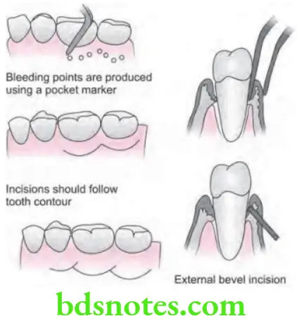
- Step 1: Periodontal pocket should be mapped out over the external gingival surface by inserting a probe to the bottom of pocket and puncturing external surface of gingiva at the depth of probe penetration.
- Step 2: Periodontal knives i.e. Kirkland knives are indicated for incisions on the facial and lingual surfaces. Orban periodontal knives are indicated for inter-dental incisions. Bard–Parker (BP) blades (#12 and #15), and scissors are indicated as an auxiliary instruments.
- External bevel incision should be started apical to the points marking course of pockets, and it is directed coronally to a point between base of the pocket and crest of the bone. This should be close to the bone without exposing it to remove soft tissue coronal to the bone.
- Exposure of bone is undesirable, if this occurs, healing presents minimal complications if the area is adequately covered by the surgical dressing. Either interrupted or continuous incisions can be used.
- Incision should be beveled at approximately 45 degrees to tooth surface, and it should re-create the normal festooned pattern of gingiva. Failure to bevel the incision will leave a broad, fibrous plateau which will delay development of physiologic contour.
- Step 3: Eliminate the excised pocket wall, irrigate the area and examine root surface.
- Step 4: Undergo Scaling and root planing.
- Step 5: Cover the area by surgical dressing.
Healing Following Gingivectomy
- Healing after surgical gingivectomy is basically by secondary intention:
- The initial response is the formation of a productive surface clot.
- The clot is then replaced by granulation tissue.
- Within 24 hours, there is an increase in new connective tissue cells mainly angioblasts and by third day numerous fibroblasts are located in this area.
- The highly vascular granulation tissues grow coronally, creating a new free gingival margin and sulcus.
- Capillary derived from blood vessels of periodontal ligament migrate into the granulation tissue, and with in two weeks they connect with gingival vessels.
- After 12 to 24 hours, epithelial cells at the margins of the wound start to migrate over the granulation tissue, separating it from the contaminated surface layer of clot. Epithelial activity at the margins reaches to maximum in 24 to 36 hours.
- The new epithelial cells arise from the basal and deeper spinous layers of the wound, edge epithelium and migrate over the wound over a fibrin layer that is later resorbed and replaced by connective tissue bed.
- The epithelial cells advance by tumbling action, with the cells becoming fixed to the substrate by hemidesmosomes and a new basement lamina.
- After 5 to 14 days, surface epithelialization is generally complete. During the first 4 weeks after gingivectomy, keratinization is less than it was before surgery. Complete epithelial repair take about 1 month.
- Vasodilation and vascularity begin to decrease after the fourth day of healing and appear to be almost normal by 16th day.
- Complete repair of the connective tissue take about 7 weeks.
Question 3. Write short note on gingivectomy.
Answer. Gingivectomy means excision of the gingiva.
By removing the pocket wall gingivectomy provides visibility and accessibility for complete calculus removal and thorough smoothening of the roots, creating a favorable environment for gingival healing and restoration of a physiologic gingival contour.
Rationale of Gingivectomy
- To provide the good visibility and accessibility for the complete calculus removal and thorough smoothening of the roots.
- To facilitate gingival healing by creating a favorable environment.
- To restore a physiological gingival contour.
Types of Gingivectomy
- Surgical gingivectomy
- Electrosurgical gingivectomy
- Laser gingivectomy
- Chemosurgical gingivectomy
Electrosurgical Gingivectomy
In this technique electric current is used for giving the incisions.
It uses high frequency current of 1.5 to 7.5 million cycles per second.
Here three classes of electrodes are used i.e.
- Single wire electrodes for both incising and excising
- Loop electrodes for planning the tissues
- Heavy bulkier electrodes for the coagulation.
Four Types of Electrosurgical Techniques are Present i.e.
- Electrosection: This is used to perform incising, excising and planning
- Electrocoagulation: This is used to prevent hemorrhage
- Electrofulguration: This uses high voltage current. This has limited usage in dentistry.
- Electrodessication: It uses the dehydrating current and is least used as dangerous technique. It is used in dermatological and the cancer surgeries.
Gingivectomy Procedure
- In gingivectomy, needle electrodes are supplemented by ovoid loop or diamond-shaped electrodes. In this fully rectified cutting and coagulation current are used.
- Activate the electrode and move it in the shaving motion.
- A needle electrode is used for giving the incision for drainage.
- Ball electrode should be used for hemostasis.
Laser Gingivectomy
- CO2 laser and Nd: YAG Laser are commonly used for the excision of gingival growths.
- In laser gingivectomy, healing is delayed as compared to surgical gingivectomy technique.
- Laser offers dry and bloodless surgery.
Chemosurgical Gingivectomy
- 5% paraformaldehyde or potassium hydroxide are the chemicals used to perform chemosurgical gingivectomy.
- This technique is not used now because of various disadvantages such as:
- Depth of chemical action cannot be controlled.
- Gingival remodeling cannot be accomplished effectively.
- Epithelialization and reformation of junctional epithelium, re-establishment of the alveolar crest fiber system are slow in chemically treated gingival wounds than in those produced by scalpel.
Question 4. Write short note on excisional new attachment procedure.
Or
Write short note on ENAP.
Answer. It was developed by US Naval Corps, yukna (1976).
ENAP is a definitive subgingival curettage procedure performed with a knife.
Indications
Suprabony pockets in the presence of convex root surfaces, normal gingival form and width.
Contraindications
- Soft tissue:
- Hyperplastic tissues.
- Inadequate keratinized gingiva.
- Hard tissue:
- Furcations
- Osseous defects.
- Malposed teeth.
- Close root approximation.
- Developmental tooth defects.
Excisional New Attachment Advantages
- Removal of epithelial pocket lining, junctional attachment and adjacent granulation tissue.
- Improved visualization and access to the root surfaces.
- Connective tissue attachment is left intact.
- Minimal trauma to gingiva.
- Does not affect the bone.
- Facilitates new attachment.
- Minimal postoperative recession.
Excisional New Attachment Disadvantages
- Limited application
- Technically exacting to determine the apical extent of the epithelial attachment, and epithelial-connective tissue junction.
Excisional New Attachment Procedure
- Deposition of few drops of the anesthetic solution into the lateral wall of pocket will help to reduce bleeding and make the tissue more firm.
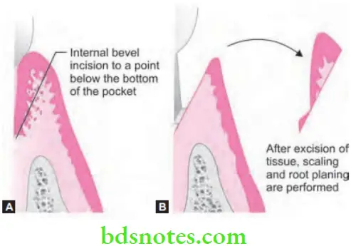
- An internal bevel incision is made from margin of free gingiva apically below the base of the pocket.
- Incision is carried all around the tooth surface. Care should be taken to retain as much interdental gingiva as possible.
- Excised tissue is removed with a curette and the root surface is planned to a smooth hard consistency. Pocket is flushed with normal saline.
- Wound edges are approximated with finger pressure. Interdental sutures and periodontal pack is placed.
- A long junction epithelium is formed in about 7 days.

Question 5. What are various objectives of periodontal therapy. Briefly describe various steps of gingivectomy.
Answer.
Objectives of Periodontal Therapy
- Eliminate pain.
- Eliminate gingival inflammation.
- Eliminate gingival bleeding.
- Eliminate infection.
- Reduces periodontal pockets and mobility of the teeth.
- Stops pus formation.
- Arrests the destruction of soft tissue and bone.
- Establishes optimal occlusal function.
- Restores tissue destroyed by disease.
- Re-establishes the physiologic gingival contour.
- Prevent the recurrence of disease.
- Reduces tooth loss.
Question 6. Define gingivectomy and gingivoplasty. Write its indications and contraindications and describe surgical gingivectomy in detail. Write brief note on healing after gingivectomy.
Answer. Gingivectomy means the excision of gingiva.Gingivoplasty is the reshaping of gingiva to create physiological gingival contour, with the sole purpose of recountouring the gingiva in the absence of periodontal pocket.
Question 7. Write short note on curettage.
Answer. Curettage is the scraping of gingival wall of periodontal pocket to separate diseased soft tissue.
Rationale
- To reduce the pocket depth.
- To improve the probing attachment levels
- To maintain the aesthetics
- To provide the periodontal therapy 1 systemically compromised patients.
Types of Curettage
Curettage is of three types, i.e.
- Gingival curettage: It is the removal of soft tissue lateral to pocket wall.
- Subgingival curettage: It is performed apical to epithelial attachment, serving the connective tissue attachment down to the osseous crest.
- Inadvertant curettage: It is the curettage which is done unintentionally when scaling and root planning procedure is performed.
Various gingival curettage techniques are conventional technique, ENAP, ultrasonic curettage, use of caustic drugs.
Conventional Technique
- Give local anesthesia before commencing with gingival curettage.
- Select a curette so that the cutting edge will be against the tissue (e.g., Gracey No 13-14 for mesial surfaces and Gracey No 11-12 for distal surfaces). Curettage can also be performed with a 4R-4L Columbia Universal curette.
- Insert the instrument to engage the inner lining of the pocket wall and is carried along the soft tissue, usually in a horizontal stroke.
- Pocket wall may be supported by gentle finger pressure on the external surface. The curette is then placed under the cut edge of the junctional epithelium to undermine it.
- In subgingival curettage, the tissues attached between the bottom of the pocket and the alveolar crest are removed with a scooping motion of the curette to the tooth surface.
- Flush the area to remove debris, and the tissue is partly adapted to the tooth by gentle finger pressure.
- In some of cases, suturing of separated papillae and application of a periodontal pack may be indicated.
Indications of Curettage
- In cases with moderately deep intrabony pockets which are located in accessible areas.
- In the patients in whom because of their age, systemic problems or other factors, when the more aggressive surgical techniques are contraindicated.
- It is performed as a maintenance therapy which is performed in the areas of recurrent inflammation and pocket depth on recall visits.
Contraindications of Curettage
- In presence of acute infections such as necrotizing ulcerative gingivitis.
- Fibrosis of soft tissue wall.
- Extension of base of pocket apical to the mucogingival junction.
- If patient is medically compromised, the benefits versus the risk of surgical procedure should be carefully weighed before committing the patient to procedure.
Question 8. Define gingivoplasty and gingivectomy and write indications and contraindications.
Answer.
Indications of Gingivoplasty
- In cases where there is need for correction of grossly thick gingival margins or gingival enlargement.
- In gingival cleft and carters caused due to acute necrotizing ulcerative gingivitis gingiva which interfere with normal food excursion and collect plaque and food debris.
- Sharply varying levels of gingival margin in adjacent areas.
- Saucer shaped deformities, buccolingual in interproximal lesions.
Contraindications of Gingivoplasty
- In patients with periodontal pockets.
- In patients with generalized thickened fibrous gingiva.

Leave a Reply