Extraoral Radiographic Techniques
Question 1. Write short note on lateral oblique view for body of mandible.
Answer. It is an extraoral radiographic technique for mandible.
Type of lateral oblique view are:
- Anterior body of the mandible view.
- Posterior body of the mandible view.
- Ramus view.
Anterior Body of the Mandible View
Structure shown
Anterior body of the mandible and teeth in same area.
Read And Learn More: Oral Radiology Question And Answers
Film Placement
The cassette is placed flat against the patient’s cheek and is centered over the body of mandible, overlying the canine teeth. Cassette should be positioned parallel to the body of mandible. Patient should hold the cassette position with the thumb placed under edge of palm against outer surface of cassette.
Position of Patient
Patient’s head is so adjusted that:
- The ala tragus line is parallel to the flor.
- Inferior border of the cassette should be parallel to the lower border of the mandible and below it.
- The sagittal plane is tilted so that it is 5° to the vertical and rotate 30° from the true lateral position.
Central Ray
- Central ray is directed under the mandible opposite side, from 2 cm behind the angle of the mandible.
- Beam is directed upward (–10° to –15°) and centered on the anterior body to mandible.
- Beam must be directed perpendicular to horizontal plane of fim.
Exposure Parameter
Using intraoral X-ray machine
kVp – 65–70
mA – 7–10
Seconds – 0.8
Using extraoral X-ray machine
kVp–40
mA–40
Second—1
Posterior Body of the Mandible View
Structure shown: Body of mandible, ramus oft he mandible, angle of the mandible, position of teeth in same area.
Film Placement
The cassette is placed flat against the patient’s cheek and is centered over the body of the mandible. The cassette should be positioned parallel to the body of mandible. Patient should hold the cassette position with the thumb placed under edge of palm against outer surface of cassette.
Position of Patient
- Ala tragus line is parallel to the flor.
- Inferior border of the cassette should be parallel to the lower border of the mandible and below it.
- The sagittal plane is tilted so that it is 5° to the vertical and the head is rotated 10° to 15° from the true lateral position.
Central Ray
- Directed from under the mandible opposite side, from 2 cm behind the angle of the mandible.
- Beam is directed upward (–10° to –15°) and centered on the anterior body to mandible.
Exposure Parameters
Using intraoral X-ray machine
kVp: 65–70.
mA: 7–10.
Seconds: 0.8.
Using extraoral X-ray machine
kVp: 40
mA: 40
Second:1
Ramus of Mandible
Structure shown: To evaluate third molars, large lesions, fracture into the ramus of mandible.
Film Placement
- Cassette is placed against the cheek of patient and is centered over the ramus of the mandible.
- The cassette should be positioned parallel to the ramus of the mandible.
Position of Patient
- Patient’s head is so adjusted that the ala tragus line is parallel to the flor.
- Inferior border of the cassette should be parallel to the lower border of mandible and below it.
- Sagittal plane is tilted so that it is 10° to the vertical and head is rotated 5° from true lateral position.
Central Ray
- It is directed from under the mandible of opposite side from behind the angle of the mandible to a point posterior to the third molar region on the side opposite to the cassette.
- The beam is directed upward (–10 to –15°) and centered on the ramus of the mandible.
- Beam must be directed perpendicular to horizontal plane of fim.
Exposure
Using intraoral, X-ray machine
kVp: 65–70.
mA: 7–10.
Seconds: 0.8.
Using extraoral, X-ray machine
kVp: 40
mA: 40
Seconds: 1
Ramus View
Structure shown
Impacted third molar, retromolar area, angle of the mandible, condyle and fractures that extend into the ramus oft he mandible.
Film Placement
The film placement should be such that the central beam is directed towards the center of the imaged ramus, from 2 cm below the inferior border of the opposite side of the mandible at the area of the fist molar.
Position of Patient
- Patient’s head is so adjusted that the ala tragus line is parallel to the flor.
- Mandible is protruded slightly. The cassette is placed over the patient’s cheek and centered over the area of interest usually over the ramus and far enough posteriorly to include the condyle.
- Lower border of the cassette is parallel and at least 2 cm below the inferior border of the mandible.
Position of Patient
The head is tilted toward the side being examined so that the condyle of the area of interest and the contralateral angle of the mandible form a horizontal line.
Exposure Parameters
KVp: 65–70
mA: 7–l0
Seconds: 0.8
Question 2. Write short note on cephalogram.
Answer. Cephalogram is an extraoral radiography of skull.
- This view is used to evaluate facial growth and development trauma, disease and developmental anomalies.
- This projection demonstrate the bones of face, skull as well as the sof tissue projection of the face.
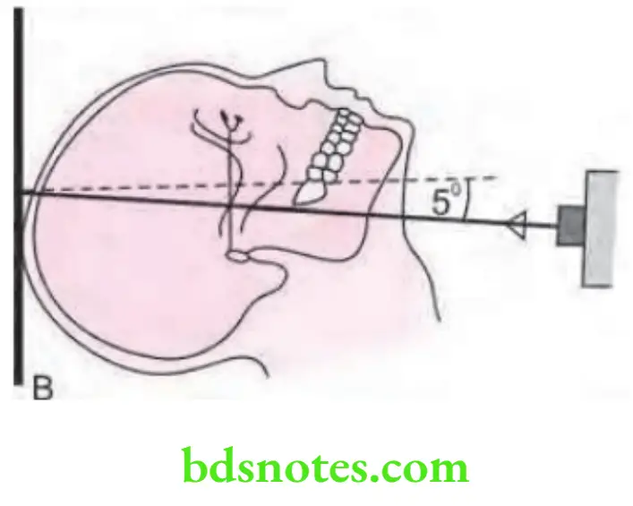
- In oral surgery and prosthesis, it is used to establish pretreatment and post-treatment records.
Film Position
Cassette is placed perpendicular to the flor with the long axis of the cassette placed vertically.
Position of Patient
- Left side of the patient’s head is positioned against the cassette.
- Mid-sagittal plane is perpendicular to the flor and parallel to the film.
- Patient’s head is stabilized with the help of the ear rods, nasion positioner and the orbital rod.
- Patient is asked to keep the teeth in occlusion.
Central Ray
- Central ray is directed perpendicular to the cassette through the portion.
- The distance between the X-ray source and the mid-coronal plane of the patient is 60 inches.
Exposure Parameter
kVp: 84.
mA: 13.
Seconds: 1.6.
Question 3. Write short note on transcranial view.
or
Write short note on transcranial technique of TMJ.
Answer.
Transcranial View
It is an extraoral radiographic view of temporomandibular joint.It is also known as Lindblom technique
- This technique is most useful in detecting arthritic changes in the articular surface.
- It helps to evaluate the joint bony relationship.
- Changes on the central and medial surface are not seen.
Film Position
Cassette is placed flat against the patient ear and centered over the TMJ of interest, against facial skin parallel to the sagittal plane.
Position of Patient
- Patient’s head is adjusted so that the sagittal plane is vertical.
- Ala tragus line is parallel to the flor.
- View is taken with the patient’s mouth in three position, i.e.
- Open mouth.
- Rest position.
- Closed mouth.
Central Ray
Point of entry of rays is different according to different techniques:
- Postauricular or Lindblom technique: Point of entry of central ray is ½ inches behind and 2 inches above the auditory meatus.
- Grewcock approach: Central ray enters through a point 2 inches above, the external auditory meatus.
- Gill’s approach: Central ray enters through a point ½ inches anterior and 2 inches above the external auditory meatus.
In all the above three techniques central ray is directed caudally at angle of +20° to + 25°.
Exposure Parameter
kVp: 70.
mA: 7.
Seconds: 1.5.
Uses of transcranial technique or transcranial View
- This view is very useful in detecting the articulating surface changes which are caused by the various forms of arthritis.
- Relationship of condyle to the articulating surface of the joint is viewed in this radiograph.
- Transcranial view shows the lateral oblique view of condylar head as well as articular fossa. It also shows very minute, subtle bony irregularities on the lateral bony surfaces.
Question 4. Write short note on technique and indication for PA Water’s view.
Answer.
PA Water’s View
Structure shown
This projection is used to demonstrate the maxillary sinus. It also shows frontal and ethmoidal sinus. The orbit, frontozygomatic suture, nasal cavity, coronoid process of the mandible and the zygomatic arch are also seen.
Film Placement
- Cassette is placed perpendicular to the flor in a cassette holding device.
- Long axis of the zygomatic arch is also seen.
Position of Patient
Position of patient should be such that the mid-sagittal plane should be vertical and perpendicular to the plane of film.
- Patient’s head is extended so that only the chin touches the cassette.
- Cassette is centered around the acanthion.
- The canthomeatal line should be at 37° to the plane of the fim external auditory meatus to the mental protuberance should be perpendicular to the fim.
- Water specified that the tip of the nose should be 0.5 to 1.5 mm away from the cassette.
- Patient’s head is extended as far as comfortable, to make lower border of mandible as parallel to cassette as possible. Only chin touches the cassette. Canthomeatal line should be approximately parallel to plane of the film.
Central Ray
Perpendicular and to the mid point of the fim.
Exposure Parameter
kVp: 65.
mA: 10.
Seconds: 2.3.
Indication
For demonstrating maxillary sinus, frontal and ethmoidal sinus.
Question 5. name the techniques used for temporomandibular joint radiography. Describe the technique of transcranial projection including its exposure parameters. Mention its indications.
Answer. Following are the techniques used for temporomandibular joint radiography:
- Transcranial projection
- Trans pharyngeal projection
- Trans orbital projection
- Reverse Towne’s projection
Transcranial Projection
Technique
Film Position
Cassette is placed flat against the patient’s ear and centered to TMJ against the facial skin parallel to sagittal plane.
Position of Patient
- Patient’s head should be adjusted so that sagittal plane is vertical.
- Ala tragus is parallel to the flor.
- View is taken with the patient’s mouth in three positions, i.e.
- Mouth in open position
- Mouth in rest position
- Mouth in closed position
Central Ray
Point of entry is different according to the technique used.
Postauricular or Lindblom technique
Technique
Point of entry of the central ray is 1/2 “behind and 2” above the auditory meatus.
According to Lindblom, the central ray should be directed from posteriorly so that it passes along long axis of condyle and the medial pole of condyle is more posterior to lateral pole.
Grewiock Approach
The central ray enters through a point 2” above the external auditory meatus.
Gill’s Approach
Central ray enters through a point 1/2” anterior and 2” above the external auditory meatus.
In all the three techniques, the central ray is directed caudally at an angle of + 20° to + 25°.
Point of exit is through TMJ of interest.
Exposure Parameters
Intra oral X-ray machine
kVp: 70
mA: 07
Seconds: 1.5
Indications
- This view is very useful in detecting the articulating surface changes which are caused by the various forms of arthritis.
- Relationship of condyle to the articulating surface of the joint is viewed in this radiograph.
- Transcranial view shows the lateral oblique view of condylar head as well as articular fossa. It also shows very minute, subtle bony irregularities on the lateral bony surfaces.
Question 6. Enumerate the various extraoral radiographic technique. Write in detail about Water’s view radiograph.
Answer. Following are the extraoral radiographic techniques:
- Radiography of Paranasal Sinuses
- Posteroanterior projection or occipitofrontal projection of nasal sinuses
- There are 2 methods for obtaining this projection.
- Posterior anterior or granger projection
- Modified method, inclined posterior anterior or cald well projection
- Radiography of the Maxillary Sinuses
- Standard occipitomental projection (0° OM)
- Modified method or 30° occipitomental projection
- Bregma-menton
- PA Water’s
- Radiography of the Mandible
- PA mandible
- Rotated PA mandible
- Lateral oblique
- Anterior body of mandible
- Posterior body of mandible
- Ramus of mandible
- Radiography of Base of the Skull
- Submento vertex projection
- Radiography of the Zygomatic Arches
- Jug handle view
- Radiography of the Temporomandibular Joint
- Transcranial projection
- Trans pharyngeal projection
- Trans orbital projection
- Reverse Towne’s projection
- Radiography of the Skull
- Lateral cephalogram
- True lateral
- PA cephalogram
- PA skull
- Towne’s projection
Question 7. Write short note on submentovertex view.
Answer. Radiography of base of skull is carried out by submentovertex view.
Structures shown
A complete axial view of base of the cranium shows following structures, i.e. symmetrical projection of the petrosa, mastoid process, foramen ovale, spinosum canals, carotid canals, sphenoidal sinuses, mandible, maxillary sinus, nasal septum, odontoid process of the atlas and the entire atlas, axial inclination of the mandibular condyles.

Film Placement
Cassette is placed perpendicular to the flor in a cassette-holding device. Long axis of the cassette should be placed vertically.
Position of Patient
Head of the patient is centered over the cassette, patient’s head and neck should be tipped back as far as possible, vertex of the skull touches the cassette. Mid-sagittal plane of both the sides is perpendicular to the plane of the film and the radiographic base line is parallel to the film.
Central Ray
Central ray is directed perpendicular to the film and through the mid-sagittal plane, between the angles of the mandible. Central ray should be perpendicular to an imaginary line joining the mandibular fist molars.
To view the petrous portion, the central ray should be directed at right angles to the film midway between the external auditory meatus.
Exposure Parameters
kVp: 50
mA: 20–30
Seconds: 0.4
Indications
Helps to study destructive/expansile lesions affecting the palate, pterygoid region or base of the skull, sphenoidal sinus.
Question 8. Write short note on various radiographic views to visualize temporomandibular joint pathologies.
or
Write short answer on transorbital views.
or
Write short answer on radiographic views used for TtMJ disorders.
Answer. Following are the various radiographic views to visualize temporomandibular joint:
- Transcranial projection
- Transpharyngeal projection
- Transorbital projection
- Reverse Towne’s projection.
Tranpharyngeal Projection
This view is a lateral projection which shows medial surface of condylar head and neck.
Film Placement
Cassette should be placed flat against the ear of patient and is centered to a point half inches anterior to external auditory meatus over temporomandibular joint of interest against the facial skin parallel to sagittal plane.
Patient’s Position
- Patient is positioned in such a manner that sagittal plane is vertical and parallel to the film and the temporomandibular joint of interest is adjacent to film.
- Occlusal plane should be parallel to transverse axis of film so that soft parts of nasopharynx should be in one line with temporomandibular joint.
- Patient is told to slowly inhale through the nose at the time of exposure to ensure filing of nasopharynx with air at the time of exposure.
- Patient should open his mouth.
Central Ray
- Directed from opposite side cranially from angle of –5 to –10° posteriorly.
- Ray is directed via mandibular notch in a window between coronoid, condyle and zygomatic arch of opposite side below base of skull to TMJ of interest.
Exposure Parameters
By Intraoral X-ray Machine
kVp: 65 to 70
mA: 7 to 10
Seconds: 0.8
By Extra-oral X-ray Machine
KVp: 40
mA: 40
Seconds: 1
Transorbital Projection (Zimmer Projection)
It is most successful in delineating the joint with minimum superimposition and lead to production of relatively true projection.
Structures shown
Anterior surface of TMJ, medial displacement of condyle and fractured neck of condyle.
Film Position
It is positioned behind the head of patient at an angle of 45° to sagittal plane.
Patient’s Position
- Positioning of patient should be in such a way that sagittal plane is vertical.
- Canthomeatal line is 10° to horizontal with head tipping downwards.
- Patient is asked to widely open the mouth.
Central Ray
- Head of the tube should be in front of the face of patient.
- Central ray is directed over the TMJ of interest at an angle of +20°, to strike the cassette at right angles.
- The point of entry may be taken at.
- Pupil of same eye, asking the patient to look straight ahead
- Medial canthus of same eye
- Medial canthus of opposite eye.
Exposure Parameters
By Intraoral X-ray machine
kVp: 65 to 70
mA: 7 to 10
Seconds: 0.8
By Extraoral X-ray Machine
kVp: 40
mA: 40
Seconds: 1
Reverse Towne’s Projection
It is used for viewing condylar head and neck, fracture of neck of condyle, intracapsular fracture of TMJ, condylar hypoplasia or hyperplasia.
Film Position
It is placed perpendicular to the flor in a cassette-holding device. Long axis of cassette is placed vertical.
Patient’s Position
- Sagittal plane should be vertical and perpendicular to the film.
- Film is adjusted in a manner such that lips centered over the film.
- Patient’s forehead should touch the film.
Central Ray
Directed through mid-sagittal plane at mandible and perpendicular to the film.
Exposure Parameters
By Extraoral X-ray Machine
KVp: 70–80
mA: 60–50
Seconds: 1.6 (bucky grid)

Leave a Reply