Environmental And Nutritional Diseases
Question 1. Write a short note on scurvy.
Or
Discuss in short scurvy.
Or
Write in brief on vitamin C deficiency.
Answer:
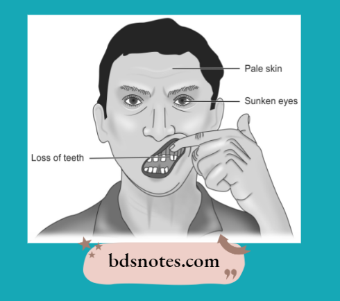
Vitamin C deficiency in the food or as conditioned deficiency results in scurvy.
The lesions and clinical manifestations are seen more commonly in two ages, i.e. early childhood and very aged.
Read And Learn More: Pathology Question And Answers
The lesions are:
1. Lesions in teeth and gums: Scurvy may interfere with the development of dentin. The gums are soft, and swollen and may bleed readily and get infected commonly. The gums are called scorbutic gums.
2. Hemorrhagic diathesis: A marked tendency to bleed is characteristic of scurvy. There may be hemorrhages of skin, mucus membranes, gums, muscles, joints, and under the periosteum.
3. Skeletal lesions: The most prominent change is the deranged formation of osteoid matrix and not deranged mineralization. Growing tubular bones and as well as flat bones are affected.
4. Delayed wound healing: There is a delay in healing wounds in scurvy due to:
- Deranged collagen synthesis
- Poor maturation and prevention of fibroblasts.
- Localization of infection in wounds.
5. Anemia: It is common in scurvy. It is most often Normocytic and Normochromic.
Scurvy Histopathology
The following are the histologic features of scurvy:
- In scurvy, osteoblasts fail to form osteoid on spicules of calcified cartilage matrix.
- Cartilage cells of the epiphyseal plate proliferate normally and salts are deposited in the matrix between columns of cartilage cells.
- A wide zone of calcified but non-ossified matrix known as scorbutic lattice develops in the metaphysis.
- As the scorbutic lattice increases in width more fragile zone develops which leads to a complete fracture of spicules with separation and deformity of the cartilage shaft junction.
- Fractures of calcified matrix material lead to the classic picture of scurvy known as the Trummerfeld zone.
- The area beneath the Trummerfeld zone is free of hematopoietic cells and is formed of connective tissue cells known as Gemestmark.
Question 2. Write a short note on rickets.
Or
Write briefly on rickets.
Answer:
Rickets refers to the disorder that occurs due to a deficiency of vitamin D.
- Primary defects in rickets are interference with the mineralization of bone and deranged endochondral and intramembranous bone growth.
- It develops in an area where sunlight is deficient.
- It also results from inadequate extracellular levels of calcium and inorganic phosphate, a mineral necessary for new bone to calcify. Osteoid builds in excessive amounts because it fails to mineralize properly.
Rickets Clinical Features
- It occurs in infants and children.
- Wrist and ankles are typically swollen. The changes in bone are found in the epiphyseal plates, metaphysis, and the shaft.
- Craniotabes, i.e. localized areas of thinning are present in the skull, when a finger pressure is applied it can produce indentation.
- The patient has a short stature and deformed extremities. Children with rickets show bowing of legs.
- Harrison’s sulcus is present, i.e. indentation of lower ribs at the site of attachment of the diaphragm.
- The rachitic rosary is the deformity of the chest due to cartilaginous overgrowth at the costochondral junction.
- Pigeon chest deformity is the anterior protrusion of the sternum due to the action of respiratory muscles.
- Frontal bossing is present.
- Knock knees are present due to enlarged ends of the femur, tibia, and fibula.
- Lumbar lordosis—the pelvis may be deformed. When an ambulatory child develops rickets, deformities are likely to affect the spine, pelvis, and long bones causing ’lumbar lordosis’.
Rickets Histopathology
- In long tubular bones
- The proliferation of cartilage cells at epiphysis is followed by inadequate provisional mineralization.
- Overgrowth of epiphyseal cartilage is present with deposition of the osteoid matrix in inadequately mineralized cartilage resulting in enlarged and expanded costochondral junction.
- Irregular overgrowth of small blood vessels in disorganized and weak bones.
- In flat bones
- Mesenchymal cells differentiate in osteoblasts with the laying down of osteoid matrix which fails to get mineralized resulting in weak and soft flat bones.
Rickets Biochemical Changes
- The lower level of active metabolites of vitamin D.
- Plasma calcium levels are slightly low or normal.
- Plasma phosphate levels are lowered.
- Plasma alkaline phosphatase is raised.
Question 3. Write a short note on diet and cancer.
Answer:
The human diet is a highly complex and variable mixture of naturally occurring synthetic chemicals. Naturally occurring chemicals consists of macronutrients, i.e. fat, carbohydrates, and protein; micronutrients, i.e. vitamins and trace metals; non-nutrient constituents.
Several carcinogens and anticarcinogens have been identified in the human diet.
Dietary Carcinogens
These are classified into four categories viz:
l. Naturally present carcinogens: Aflatoxin is a naturally occurring dietary carcinogen. This is a mycotoxin produced by fungus Aspergillus favus, it is implicated in the pathogenesis of hepatocellular carcinoma. Aflatoxicosis disease is caused by the intake of grains and nuts which are contaminated by the fungus.
2. Carcinogens forming during food preparation: Various burnt or barbecued foods consist of a group of carcinogenic substances known as polycyclic aromatic hydrocarbons. These polycyclic aromatic hydrocarbons are produced if food is overheated. High intake of fried and broiled foods such as meats can increase the risk of breast, colon, prostate, and pancreatic cancers.
3. Preservatives and coloring agents added to food: Artificial sweeteners (like saccharine and cyclamates) are known to cause bladder cancer. Cured, pickled or salty foods contain nitrates which have been implicated in gastric cancer.
4. Substances that are converted into carcinogens in the body: Sodium nitrite which may be present in drinking water and vegetables gets converted to nitrosamine, which is a carcinogen.
Cancer-Preventing Diets
- Fruits and vegetables in the diet lower the risk of cancer.
- Retinoic acid promotes differentiation of mucous-secreting epithelial cells so a diet having β-carotene and retinoic acid can reverse metaplastic and precancerous lesions of the respiratory tract.
- High fiber content with low animal fat in the diet prevents colonic carcinoma
- Folic acid, selenium, β-carotene, vitamin C, and vitamin E prevent free radical damage to cells and their DNA in this way it prevents cancer initiation. Vitamin A enhances immunity and may control free radical production by modulating inflammatory reactions.
Question 4. Write a short note on ascorbic acid.
Answer:
Ascorbic acid is also called vitamin C.
- Ascorbic acid is a white crystalline water-soluble substance with a sour taste.
- It is easily destroyed by cooking.
Ascorbic acid Absorption and Storage
- Ascorbic acid is easily absorbed from the small intestine, peritoneum, and subcutaneous tissues.
- It is not stored in any particular organ and is spread throughout the body.
Ascorbic acid Sources
Amla, citrus fruits, tomatoes, guava, green peppers.
Ascorbic Acid Physiological Functions
- It is involved in the oxidation reaction of cells.
- It requires the metabolism of amino acids, For Example. tyrosine and tryptophane.
- It is also involved in the conversion of folic acid into its active co-enzyme.
- It is required for iron absorption and also for the formation of collagen fibers and mucopolysaccharides of connective tissue.
Ascorbic acid Daily Requirement
Infants: 35 mg
Children: 40 mg
Adults: 45 mg
Pregnant and lactating women require 80 mg.
Ascorbic Acid Deficiency Manifestations
Severe ascorbic acid deficiency produces—scurvy.
Question 5. Write a short note on osteomalacia.
Answer:
Osteomalacia is the adult counterpart of rickets in which mineralization of the osteoid matrix fails.
Osteomalacia Etiology
- Dietary deficiency of vitamin D.
- Poor endogenous synthesis of vitamin D.
- Conditioned deficiency.
Osteomalacia Clinical Features
- Presence of vague bony pain.
- Muscular weakness.
- Fractures following mild trauma.
- Incomplete or green-stick fractures.
- Pseudofractures in weak places of bones.
Osteomalacia Histopathology
- H&E stained tissue shows widened and thickened osteoid seams.
- There is decreased mineralization between osteoid and bones.
- Increased osteoclastic activity is also appreciated.
- Fibrosis of bone marrow is present.
Osteomalacia Biochemical Changes
- Normal or low serum calcium levels.
- Low plasma phosphate levels.
- Raised alkaline phosphatase levels.
Question 6. Write a note on obesity.
Answer:
- Obesity is defined as abnormal or excessive fat accumulation that presents a risk to health.
- A crude population measure of obesity is the body mass index (BMI), i.e. a person’s weight (in kilograms) divided by the square of his or her height (in meters).
- A person with a BMI of 30 or more is generally considered obese. A person with a BMI equal to or more than 25 is considered overweight. In children, a healthy weight varies with age and sex.
Classification by World Health Organization and Published in 2000
The neurohumoral mechanisms that regulate body weight have three components:
- The afferent system generates humoral signals. It is constituted by leptin produced by adipocytes insulin produced by the pancreas and ghrelin produced by the endocrine cells of the stomach.
- The central processing unit is located primarily in the hypothalamus. It integrates afferent signals.
- The effector system carries out `orders’ from hypothalamic nuclei in the form of feeding behavior and energy expenditure.
Obesity Etiology
Obesity occurs when caloric intake exceeds utilization. This imbalance occurs in the following conditions:
- Due to overeating
- Due to inactivity and a sedentary lifecycle
- Genetic Predisposition to Obesity
- Diet derived from carbohydrates and fats than a protein-rich diet
- Secondary obesity occurs due to underlying diseases such as hypothyroidism, Cushing’s disease, insulinoma, etc.
Obesity Pathogenesis
Adipocytes are present in vascular and stromal compartments of the body. The most common function of adipocytes is to store fat, besides this, they release endocrine-regulating molecules.
The mass of adipose gets increased due to the enlargement of adipose cells because of excessive intracellular lipid deposition and also due to an increase in the number of adipocytes.
Obesity also occurs due to excessive consumption of nutrients. Recently two obesity genes are found, i.e. ob gene and db gene.
Obesity Metabolic Changes
- Hyperinsulinemia: Increased secretion of insulin is a feature of obesity. Most obese individuals have frank diabetes in spite of the presence of hyperinsulinemia. This occurs because of insulin resistance consequent to tissue insensitivity.
- Type 2 diabetes mellitus: Type 2 diabetes mellitus is associated with obesity
- Hypertension: Due to increased blood volume there is an association between hypertension and obesity.
- Hyperlipoproteinemia: Plasma cholesterol circulates in the blood as low-density lipoprotein which has circulating triglycerides. Obesity is associated with VLDL and LDL. Blood cholesterol levels are elevated in obesity.
- Atherosclerosis: Due to atherosclerosis and hypertension person is at risk of myocardial infarction and stroke in obese individuals.
- Nonalcoholic fatty liver disease: Obesity leads to nonalcoholic fatty liver disease which causes cirrhosis of the liver.
- Hypoventilation syndrome: There is hypersomnolence at night and day in obese individuals along with carbon dioxide retention, hypoxia, polycythemia, and right-sided heart failure.
- Osteoarthritis: These individuals develop degenerative joint disease which occurs due to wear and tear following trauma to joints due to high body weight.
- Cancer: Fatt diet derived from animal fat and meat shows a high chance of occurrence of cancer of the colon, breast, and prostate.
Question 7. Write a note on pathologic changes in vitamin A deficiency.
Answer:
Following are the pathologic changes in vitamin A deficiency:
- Ocular lesions: Night blindness is the first sign of vitamin A deficiency. There is also the presence of dry and scaly scleral conjunctiva. Lacrimal duct show hyperkeratosis.
Corneal ulcers can occur which may get infected and lead to keratomalacia. Bitot’s spots are seen which are the triangular areas of opacities that occur because of keratinized epithelium. Lastly, infection, scarring, and opacities lead to blindness. - Cutaneous lesions: There is the development of popular lesions on the skin which gives it a toad-like appearance. This results because of follicular hyperkeratosis and
keratin plugging in sebaceous glands. - Other lesions: Various other lesions are:
- Squamous metaplasia of the respiratory epithelium of the bronchus and trachea leads to respiratory infections
- Squamous metaplasia of pancreatic ductal epithelium can cause obstruction and cystic dilatation.
- Squamous metaplasia of the urothelium of the pelvis of the kidney can cause pyelonephritis.
- Metaplasia present for a long time can lead to anaplasia.
- There is immune dysfunction due to damage of barrier epithelium and compromised immune defenses.
Question 8. Write a note on pathologic changes in vitamin D deficiency.
Answer:
Following are the pathologic changes in vitamin D deficiency:
vitamin D deficiency In children
In children Skeletal Changes
These are as follows:
- Craniotabes is the earliest bony lesion occurring due to small round unossified areas in the membranous bones of the skull, disappearing within 12 months of birth. The skull looks square and box-like.
- Harrison’s sulcus appears due to the indrawing of soft ribs on inspiration.
- The rachitic rosary is a deformity of the chest due to cartilaginous overgrowth at the costochondral junction.
- Pigeon chest deformity is the anterior protrusion of the sternum due to the action of respiratory muscles.
- Bow legs occur in ambulatory children due to weak bones of the lower legs.
- Knocked knees may occur due to enlarged ends of the femur, tibia, and fibula.
- Lower epiphyses of the radius may be enlarged.
- Lumbar lordosis is due to the involvement of the spine and pelvis.
In children Biochemical Changes
These are as follows:
- Lowered levels of active metabolites of vitamin D.
- Plasma calcium levels are normal or slightly low.
- Plasma phosphate levels are lowered.
- Plasma alkaline phosphatase is usually raised due to osteoblastic activity.
vitamin D deficiency In adults
Osteomalacia is the adult counterpart of rickets in which there is the failure of mineralization of the osteoid matrix. It may occur following dietary deficiency, poor endogenous synthesis of vitamin D, or as a result of conditioned deficiency.
In adults Clinical Features
- Osteomalacia is characterized by:
- Presence of muscular weakness
- Presence of vague bony pains
- Fractures following trivial trauma
- Incomplete or greenstick fractures
- Looser zones or pseudofractures at weak places in bones.
In adults Histopathology
- H&E stained tissue shows widened and thickened osteoid seams.
- There is decreased mineralization between osteoid and bones.
- Increased osteoclastic activity is also appreciated.
- Fibrosis of bone marrow is present.
In adults Biochemical Changes
These are:
- Normal or low serum calcium levels
- Plasma phosphate levels lowered
- Raised serum alkaline phosphatase due to increased osteoblastic activity.
Leave a Reply