Orbit
Question 1. Enumerate the structures present in the orbit.
Answer.
These are:
- Eyeball
- Extrinsic muscles of eyeball
- Four recti
- Superior rectus (SR)
- Inferior rectus (IR)
- Medial rectus (MR)
- Lateral rectus (LR)
- Two obliques
- Superior oblique (SO)
- Inferior oblique (IO)
- Four recti
- Levator palpebrae superioris muscle
- Nerves
- Optic nerve
- Three divisions of ophthalmic nerve:
- Nasociliary
- Lacrimal
- Frontal
- Three motor nerves:
- Oculomotor (upper and lower divisions; CN 3)
- Trochlear (CN 4)
- Abducent (CN 6)
- Ciliary ganglion
- Ophthalmic artery
- Ophthalmic veins
- Lacrimal gland
- Orbital pad of fat
Read And Learn More: Selective Anatomy Notes And Question And Answers
Question 2. Write about the origin, insertion, nerve supply and actions of the extrinsic muscles of the eyeball.
Answer.
Extrinsic Muscles of Eyeball Origin
Four recti: From common tendinous ring.
Superior oblique: From the body of the sphenoid superomedial to the optic canal.
Inferior oblique: From a rough impression in the anteromedial part/angle of the floor of the orbit.
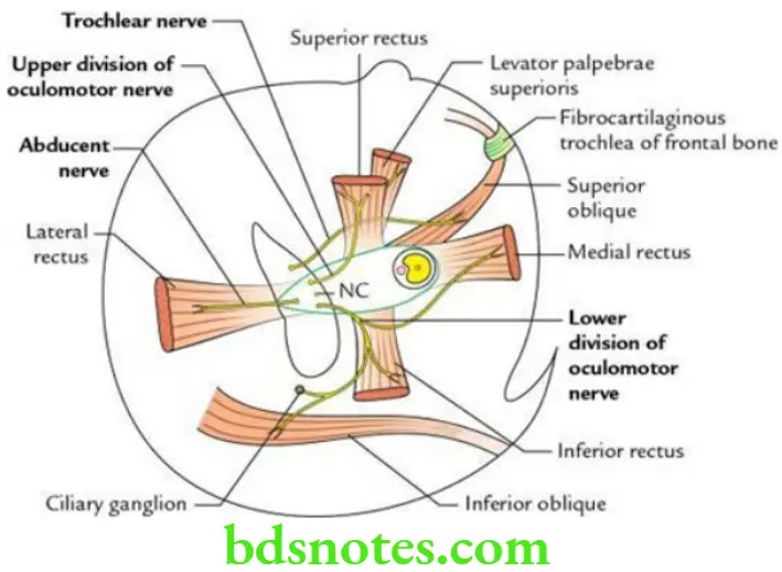
Extrinsic Muscles of Eyeball Insertion
Four recti: Into sclera, a little posterior to the limbus (i.e. corneoscleral junction). Distance from the cornea is as follows:
SR = 7.7 mm; IR = 6.5 mm; MR = 5.5 mm; LR = 6.9 mm
Superior oblique: Into sclera, behind the equator in the posterosuperior quadrant of the eyeball between SR and LR.
Inferior oblique: Into the sclera, behind the equator in the posteroinferior quadrant of the eyeball.
Extrinsic Muscles of the Eyeball Nerve supply
All the extrinsic muscles of the eyeball are supplied by CN 3, except the superior oblique, which is supplied by CN 4 and the lateral rectus which is supplied by CN 6.
Mnemonic: LR6SO4 (LR, lateral rectus; SO, superior oblique)
Extrinsic Muscles of Eyeball Actions
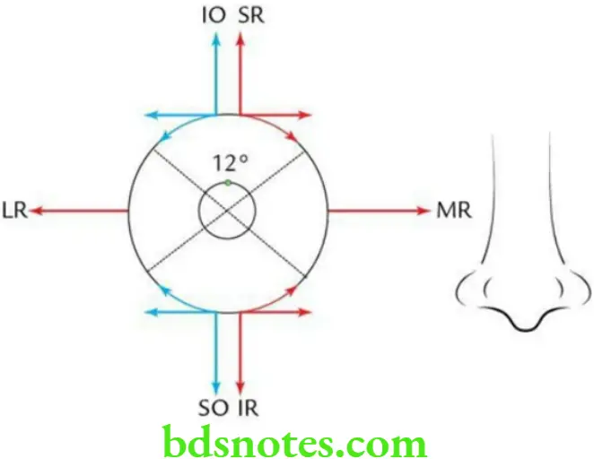
Actions of Extrinsic Muscles of the Eyeball

Question 3. Write about the origin, insertion, nerve supply and actions of the levator palpebrae superioris muscle.
Answer.
Levator Palpebrae Superioris Muscle Origin: From the undersurface of the lesser wing of the sphenoid near the apex of the orbit.
Levator Palpebrae Superioris Muscle Insertion
- Upper major skeletal part, into the skin of the upper eyelid.
- Lower minor smooth part (also called Müller’s muscle), into the superior border of a tarsal plate of the upper eyelid.
Levator Palpebrae Superioris Muscle Nerve supply
- The larger part of skeletal muscle, by the oculomotor nerve
- A smaller part of smooth muscle, by sympathetic fibres derived from superior cervical sympathetic ganglion
Levator Palpebrae Superioris Muscle Action: Elevation of the upper eyelid to open the eye.
Eyeball
The eyeball is the organ of sight (vision) located in the anterior two-thirds of the orbital cavity.
Question 1. Write a short note on Tenon’s capsule and mention its applied anatomy.
Answer.
Tenon’s Capsule General Features
- It is a membranous (fibrous) sac, which envelops the eyeball. It extends from the optic nerve to the corneoscleral junction.
- It is separated from the eyeball by an episcleral space.
- It forms a socket of the eyeball for its free movements.
Tenon’s Capsule Applied anatomy: During the enucleation of the eyeball, the Tenon’s capsule is left intact to allow the movements of the implanted artificial eyeball.
Question 2. Enumerate the coats of eyeball.
Answer.
The eyeball consists of the following three coats from superficial to deep these are:
- An outer fibrous coat called sclera in its posterior five-sixths and cornea in its anterior one-sixth
- The middle vascular coat called the uveal tract (consisting of the choroid, ciliary body and iris)
- The inner neural coat called the retina
Question 3. Write a note on the microscopic structure of the retina.
Answer.
The cells of the retina are arranged in the following four layers, from outside to inside:
- Pigment cell layer
- Layer of rods and cones
- Layer of bipolar cells
- Layer of ganglion cells
Question 4. What are the compartments of eyeballs?
Answer.
The interior of the eyeball is divided into two compartments by the lens within the eyeball.
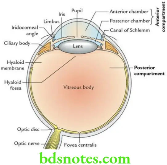
Anterior compartment: It is small and lies in front of the lens. It is filled with aqueous humour. It is further subdivided into two parts:
- A smaller anterior chamber, between the iris and cornea.
- A larger posterior chamber, between the iris and lens.
These two portions of the anterior chamber communicate with each other through a circular aperture in the iris, the pupil.
Posterior compartment: It is large (four-fifths) and lies behind the lens. It is filled with a colourless, transparent jelly-like material called vitreous humour/vitreous body.
- The vitreous humour is enclosed in a delicate hyaloid membrane.
- Anteriorly, the vitreous body presents a shallow depression (hyaloid fossa) to accommodate the lens.
Question 5. Enumerate the various refractive media of the eyeball.
Answer.
The four refractive media in the eyeball, from before backwards are as follows:
- Cornea
- Aqueous humour
- Lens
- Vitreous body
Question 6. What are eyelids? Enumerate the layers of the eyelid and discuss the related applied anatomy.
Answer.
The eyelids are mobile curtains of soft tissue to close and open the eyes. They protect the eye from dust particles and help in its moistening.
The layers of the eyelid: From outside to inside, these are as follows:
- Skin
- Superficial fascia
- Orbicularis oculi
- Tarsal plate of palpebral fascia
- Conjunctiva
Eyelids Applied anatomy
- Ptosis (drooping of upper eyelid): It occurs due to paralysis of the levator palpebrae superioris muscle.
- Stye (hordeolum): It is suppurative inflammation of glands of Zeis (i.e. sebaceous glands present in the eyelashes).
- Chalazion: It is the inflammation of the tarsal (meibomian) gland (a modified sebaceous gland in the tarsal plate).
Question 7. Enumerate the branches of the ophthalmic artery.
Answer.
The ophthalmic artery is a branch of the internal carotid artery. It gives rise to the following branches within the orbit:
- The central artery of the retina
- Lacrimal artery
- Posterior ciliary arteries
- Supraorbital artery
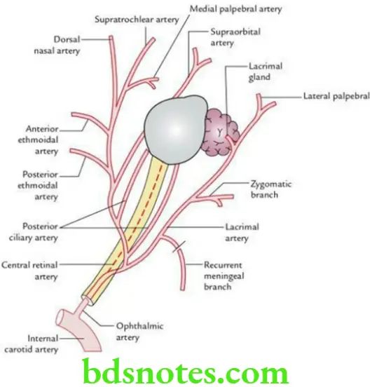
Question 8. Write a short note on the central retinal artery.
Answer.
It is the first and most important branch of the ophthalmic artery. It pierces the optic nerve 1.25 cm behind the eyeball reaches the optic disc through the central part of the disc and divides into four branches, one for each quadrant. These branches are superior nasal, inferior nasal, superior temporal and inferior temporal. It supplies the optic nerve and the inner six or seven layers of the retina.
Central Retinal Artery Applied Anatomy: Any blockage of a central retinal artery leads to loss of vision.
Question 9. Write about the origin, course and distribution of Ophthalmic Nerve.
Answer.
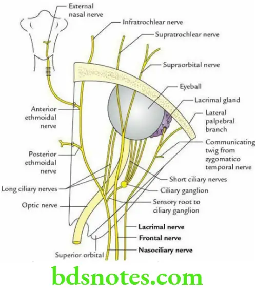
- The ophthalmic nerve is the first and smallest division of the trigeminal nerve. It arises from the trigeminal ganglion and enters the lateral wall of the cavernous sinus. Here it divides into three branches, nasociliary, frontal and lacrimal, which enter the orbit through the superior orbital fissure.
- The nasociliary nerve runs forward and medially crosses the optic nerve obliquely from the lateral to the medial side, then runs along the medial wall of orbit to terminate by dividing into anterior ethmoidal and infratrochlear nerves.
It gives rise to the following branches:- Sensory root to the ciliary ganglion
- Two or three long ciliary nerves
- Posterior ethmoidal nerve
- Anterior ethmoidal nerve
- Infratrochlear nerve
- The frontal nerve (largest branch) runs forward and terminates by dividing into supraorbital and supratrochlear nerves.
- The lacrimal nerve (smallest branch) runs along the lateral wall of the orbit end and ends in the lacrimal gland.
It gives rise to the following branches:- Branches to the lacrimal gland
- The lateral palpebral branch to the skin of the lateral part of the upper eyelid
Question 10. Write about the location, roots and distribution of the ciliary ganglion.
Answer.
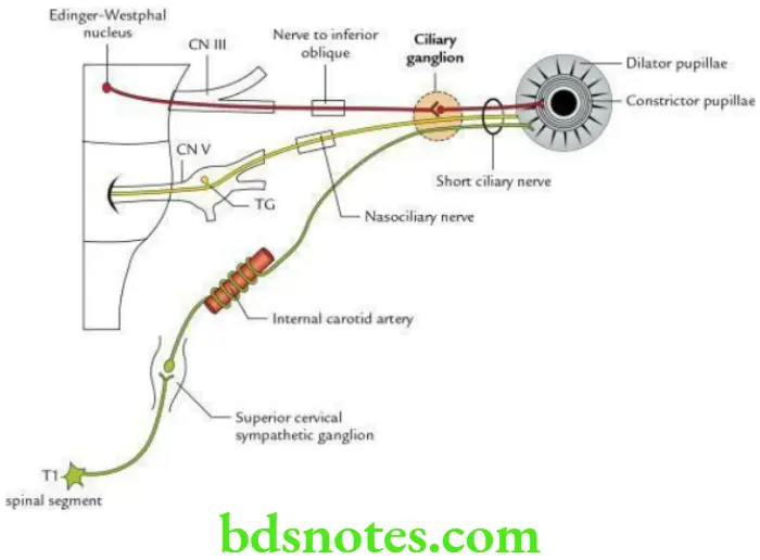
Ciliary Ganglion Location: It is a small peripheral parasympathetic ganglion of pin-head size (about 2 mm in diameter), located near the apex of orbit, between the optic nerve and lateral rectus muscle.
Ciliary Ganglion Roots The roots are 3 in number as follows:
- Parasympathetic root: It is derived from the inferior division of the oculomotor nerve. The preganglionic fibres arise from Edinger–Westphal nucleus, run in the inferior division of the oculomotor nerve and pass through the nerve to the inferior oblique to relay in the ciliary ganglion. The postganglionic parasympathetic fibres arise from ganglion and run through short ciliary nerves.
- Sympathetic root: It is derived from the sympathetic plexus around the internal carotid artery. The preganglionic sympathetic fibres arise from the T1 spinal segment and relay in the superior cervical sympathetic ganglion. The postganglionic fibres arise from this ganglion, form a plexus around the internal carotid artery and pass through the ciliary ganglion without a relay to enter into short ciliary nerves.
- Sensory root: It is derived from the nasociliary nerve – a branch of the ophthalmic nerve that passes through the ganglion without relay.
Ciliary Ganglion Distribution The distribution of its branches is as follows:
- Parasympathetic fibres: They supply sphincter pupillae and ciliary muscles through short ciliary nerves.
- Sympathetic fibres: They supply dilator pupillae and blood vessels of the eyeball through short ciliary nerves.
- Sensory fibres: They provide sensory innervation to the whole eyeball, except the conjunctiva, through short ciliary nerves. The sensory supply of conjunctiva is derived from frontal, lacrimal and infraorbital nerves.

Leave a Reply