Diseases Of Bone And Joints
Question.1. Write short note on cleidocranial dysplasia
Answer. It is also called as cleidocraniodysostosis or “Marie and Santon disease.”
Cleidocraniodysostosis Etiology
- It appears as true dominant Mendelian characteristic.
- It is transmitted as an autosomal dominant trait with complete penetrance and variable expressivity.
Oral Manifestation Of Cleidocranial Dysplasia
- Maxilla and paranasal air sinuses are underdeveloped resulting in maxillary micrognathia.
- Maxilla is underdeveloped in relation to mandible.
- There is prolonged retention of primary dentition.
- There is complete absence of cementum.
- Disorganization of developing permanent dentition.
- There is presence of supernumerary teeth usually in anterior region.
- High narrow arched palate and cleft palate is common.
- Roots of teeth are often short and thinner than the normal.
- The crown may be pittd as a result of enamel hypoplasia.
Read And Learn More: Oral Pathology Question And Answers
Clinical Features Of Cleidocranial Dysplasia
- There is complete absence of clavicle.
- It primarily affects skull, clavicle, and dentition.
Treatment For Cleidocranial Dysplasia
- Not specific
- Dental care should be taken.
Question 2. Write note on osteogenesis imperfecta.
Answer. It is also called brittle bone or lobstein disease or fragility ossium or osteopsathyrosis.
This is an autosomal dominant condition affecting bone formation.
It presents a hereditary autosomal dominant trait.
Types Of Osteogenesis Imperfecta.
- Congenital type
- Lobstein type.
Clinical Features Of Osteogenesis Imperfecta.
- It usually occur in infants.
- There is extreme fragility or porosities of bone with proneness of fracture.
- There is occurrence of pale blue sclera which is thin and pigmented choroids shows through and produces blue color.
- There is deafness due to osteosclerosis, laxity of ligaments and peculiar shape of skull.
- Increase tendency for capillary bleeding.
Oral Manifestations Of Osteogenesis Imperfecta.
- Osteogenic imperfecta is associated with dentinogenesis imperfecta.
- There is hypoplasia of teeth.
- Deciduous teeth are poorly calcified and semi-translucent or waxy.
- Teeth appear as faintly dirty pink, half normal size, with globular crown and relative short roots, in proportion to other dimensions.
Histopathological Features Of Osteogenesis Imperfecta.
- Osteoblastic activity appears as retarded and imperfect.
- Failure of fetus to be transformed with mature collagen.
- The trabeculae of cancellous bone are delicate and often show fracture.
Treatment Of Osteogenesis Imperfecta.
No known treatment.
Question.3.Enumerate firoosseous lesion. Describe in detail monostotic firous dysplasia.
Answer. In fibrous dysplasia, normal bone is replaced by the benign fibrous tissue showing varying amounts of mineralization.
Classification of Fibro-osseous Lesions
- Developmental:
- Solitary bone cyst
- Gigantiform cementoma
- Cherubism
- Reactive/Reparative:
- Aneurysmal bone cyst
- Central giant cell granuloma
- Garre’s osteomyelitis
- Osseous dysplasia
- Florid osseous dysplasia
- Cemental osseous dysplasia
- Focal osseous dysplasia or sclerosing osteomyelitis
- Osseous keloid
- Traumatic periostitis
- Neoplasms:
- Benign cementoblastoma
- Ossifying fibroma
- Conventional
- Juvenile trabecular
- Juvenile psammomatoid
- Osteoma
- Osteoid osteoma
- Osteoblastoma
- Endocrinal/Metabolic: Brown tumor of hyperparathyroidism
- Idiopathic:
- Fibrous dysplasia
- Paget’s disease.
Monostotic Fibrous Dysplasia
Monostotic fibrous dysplasia is a condition in which single bone is involved and is replaced by abnormal fibrous connective tissue which undergoes osseous metaplasia and the bone is transformed into dense lamellar bone.
Clinical Features Of Fibrous Dysplasia
- It involves only one bone and pigmented skin lesions are present.
- The sites which affected are maxilla, mandible, ribs and femur.
- It occurs in children younger than 10 years and both the sexes are equally affected.
Oral Manifestations Of Fibrous Dysplasia
- Maxilla is more commonly affected than the mandible and most of the changes occur in posterior region. The most common area involved is premolar area.
- There is presence of unilateral facial swelling which is slow growing with intact overlying mucosa.
- Swelling is painless but patient may feel discomfort.
- There is presence of enlarging deformities of alveolar process mainly buccal and labial cortical plates.
- In mandible, there is protrusion of inferior border of mandible.
Teeth present in the affected area are malaligned and tipped or displaced. - Supernumerary teeth are often impacted and affect the eruption of the teeth.
Histopathology Of Fibrous Dysplasia
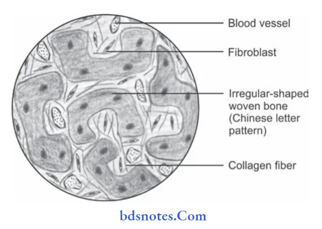
- Lesion is essentially a fibrous bone made up of proliferating firoblasts in compact stroma of interlacing collagen fiers.
- Irregular trabeculae of bone are scattered throughout the lesion.
- Some of the trabeculae are Cshaped and described as Chinese character-shaped.
- There is permanent maturation arrest in woven bone stage.
Treatment Of Fibrous Dysplasia
- Surgical removal of lesion
- Osseous countering is necessary for characterizing deformity for aesthetic or pre esthetic purposes.
Question.4.Write note on firous dysplasia.
Or
Write short note on firous dysplasia.
Answer. Fibrous dysplasia is an idiopathic condition in which an area of normal bone is gradually replaced by abnormal fibrous connective tissue which then again undergoes osseous metaplasia and eventually the bone is transformed into dense lamellar bone.
Types Of Fibrous Dysplasia
- Monostotic fibrous dysplasia: Only one of the bone is involved.
- Polyostotic fibrous dysplasia: More than one bone is involved.
- Jaff’s type: Variable number of bones are involved accompanied by pigmented lesions of skin or cafeaulait spots.
- Albright syndrome: It is a severe form of fibrous dysplasia involving all the bones in the body, accompanied by pigmented lesions of the skin and endocrine disturbances of various types.
Clinical Features Of Fibrous Dysplasia
- It is seen during the fist and second decades of life.
- Disease is more common among the females
- Polyostotic firous dysplasia commonly involve skull,facial bone, clavicle, pelvic bone, etc. and monostotic firous dysplasia involve maxilla and mandible.
- Monostotic form of disease can never be transformed into polyostotic form.
Histopathology Of Fibrous Dysplasia
- Fibrous dysplasia reveals presence of highly cellular, proliferating well vascularized fibrillar connective tissue which replaces the normal bone.
- Within fibrous tissue the firoblasts cells are arranged in “Whorled pattern”.
- Trabeculae represent the Chinese lettr pattrn.
- Osteoblastic and osteoclastic activity may be present in relation to some trabeculae of bone.

Treatment Of Fibrous Dysplasia
Surgical removal of lesion.
Question.5. Write note on Paget’s disease of bone.
Or
Write notes on histopathology of Paget’s disease.
Or
Write in detail on Paget’s disease.
Or
Write short note on Paget’s disease.
Answer. Paget’s disease is a relatively uncommon bony disorder which is characterized by the excessive uncoordinated phases of resorption and deposition of osseous tissue in single or multiple bones.
It is also known as “Osteitis Deformans.”
Etiology Of Paget’s disease.
- Inflammatory
- Circulatory disturbance
- Genetic and environmental factors
- Others: Vasculitis, trauma, hormonal balance and degenerative neurological disorders.
Clinical Features Of Paget’s disease.
- It occurs during 5th, 6th and 7th decades of life.
- Males are more commonly affcted.
- It is prone to occur in axial skeleton especially in skull,femur, sacrum and pelvis.
- Most of the patient complain initially of the deep and aching bone pain with bilaterally symmetrical swelling of bone.
- Enlargement of skull, bowing of legs and curvature of spine are commonly seen.
Oral Manifestations Of Paget’s disease.
- Maxilla is more commonly involved than the mandible.
- There is movement and migration of affcted teeth and malocclusion.
- As the disease progresses, the mouth remains open exposing the teeth.
- Extraction site heals slowly and incidence of osteomyelitis is higher.
Histopathology Of Paget’s disease.
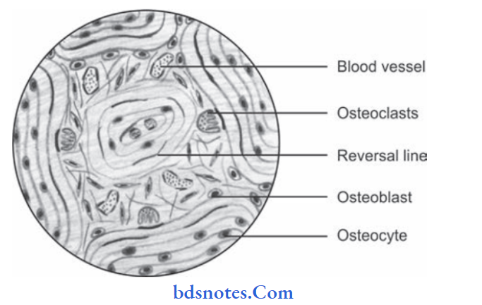
- Osteoclastic bone resorption occurs and the bone is replaced by highly vascularized cellular connective tissue.
- Osteoclasts are usually larger and may contain over 100 nuclei.
- Deposition of new lamellar or woven bone within the connective tissue by osteoblast cells occurs and fatty hemopoietic bone marrow is replaced by the fibrous stroma.
- Newly formed bone may again resorbed by osteoclast causing loss of normal architecture of bone.
- Chronic inflammatory cells and dilated blood capillaries are present within the bone.
- Bone resorption and deposition results in irregular fragments of bone formation characterized by prominent basophilic reversal and resting lines, and they produce mosaic pattern in bone.

Treatment Of Paget’s disease.
- Bisphosphonate therapy should be given.
- Calcitonin is administered.
- Surgical correction for bone deformities, fractures and severe degenerative arthritis
Question.6. Write note on cementoma.
Or
Write short note on cementoma.
Answer. It is a true neoplasm of functional cementoblasts which forms large masses of cementum-like tissue or tooth root.
Recent Concept
Cementoma these days is known as periapical cemento osseous dysplasia, this is because cementoma is an unorganized productivity of bone, PDL membrane cementum complex.
Clinical Features of Cementoma
- It occurs most frequently under the age of 25 years.
- Mandible is affected more commonly than maxilla.
- Mandibular fist molar is most commonly affected tooth.
- It produces a slow enlarging bony hard swelling of jaw which causes expansion of jaw and displacement of regional teeth.
- Expansion of buccal and lingual cortical plates.
- Low-grade intermittent pain is present which felt more when area is palpated.
- A dull sound is produced when tooth is percussed.
Histopathology of Cementoma
- It presents a large mass of amorphous cemental tissue which shows the presence of multiple reversal line.
- Intervening connective tissue stroma contains many cementoblasts and cementoblasts cells.
- PDL adjacent to normal cementum becomes integrated with capsule and separate the neoplasm from surrounding bone.
- Multinucleated cells are present in the large number in central area and are associated with active resorption.
- Root of the involved tooth extends up to the center of lesion and neoplastic cemental tissue is continuous with normal cemental layer.
Treatment of Cementoma
Surgical excision is done.
Question.7. Give a classification of bone disorders of face and jaw. Write shortly about the clinical features and histopathology of fibrous dysplasia.
Answer. Bone disorders of face and jaw:
1. Developmental defects of bone formation of face and jaw:
- Agnathia
- Micrognathia
- Macrognathia
- Facial hemiatrophy
- Facial hemihypertrophy
- Cleft palate.
2. Benign and malignant lesions of bone:
- Osteoma
- Osteosarcoma
- Ewing’s sarcoma.
3. Fibrosseous lesions
- Fibrous dysplasia of bone
- Ossifying fibroma
- Cementifying fibroma
- Paget’s disease of bone
- Cherubism
- Osteogenesis imperfecta
- Cleidocranial dysplasia
- Hurler’s syndrome
- Garre’s osteomyelitis
- Jaw lesions in hyperparathyroidism
- Aneurysmal bone cyst.
Question.8. Write short note on cherubism.
Or
Describe in brief cherubism
Or
Write short essay on cherubism.
Answer. Cherubism is an autosomal dominant firo-osseous lesion of the jaw which involves more than one quadrant that stabilizes after growth period leading to facial deformity and malocclusion.
Clinical Features Of Cherubism
- Children of age 1 to 3 years are more commonly affected.
- Males are more commonly affected than females.
- Affected children are normal at birth but as soon the child’s growth take place selflimited bone growth begins to slow down till patient reaches 5 years of age and stop at the age of 12–15 years.
- There is deforming mandibular and maxillary overgrowth with respiratory obstruction and impairment of vision and hearing.
- Enlargement of cervical lymph nodes contributes to patient’s full-faced appearance.
- A rim of sclera is may be beneath the iris, giving classic eye-to-heaven appearance.
Oral Manifestations Of Cherubism
- Agenesis of 2nd and 3rd molars of mandible.
- Displacement of the teeth
- Premature exfoliation of primary teeth
- Delayed eruption of permanent teeth
- Transposition and rotation of teeth
- In severe cases tooth resorption occurs.
Histopathology Of Cherubism
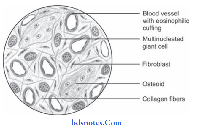
- Lesion presents a highly cellular and vascular connective tissue stroma, which is often arranged in a “whorled pattrn”.
- Numerous proliferating firoblasts and variable numbers of multinucleated giant cells are also found within the stroma.
- Giant cells are relatively smaller in size and they often aggregate around the thinwalled blood capillaries.
- A hallmark of the disease is the presence of an “eosinophilic perivascular cuffing” of collagen fibers, which often surrounds the blood capillaries.
- Within the connective tissue extravasated blood and deposits of hemosiderin pigments are sometimes seen.
- Lymph nodes exhibit reactive hyperplasia, fibrosis and chronic inflammatory cell infiltration.

Treatment Of Cherubism
No treatment is required as cherubism is a selflimiting disease.
Question.9. Classify firoosseous lesions of oral cavity. Discuss clinical features, radiographic differential diagnosis, and management of fibrous dysplasia.
Or
Classify firoosseous lesions of jaw. Discuss the clinical and histopathological features of fibrous dysplasia.
Or
Classify firoosseous lesions of jaws. Describe in detail fibrous dysplasia.
Or
Classify firoosseous lesions. Discuss in detail about the etiology, clinical, radiographic, and histopathologic features of fibrous dysplasia.
Or
Enumerate firoosseous lesions of jaws and describe fibrous dysplasia in detail.
Or
Enumerate firoosseous lesions. Describe in detail the clinical radiological and histopathological features of firous dysplasia.
Or
Classify firoosseous lesions affcting the jaws.Write in detail about the etiopathogenesis, clinical features,radiographic features of firous dysplasia.
Or
Enumerate the firoosseous lesions involving orofacial region. Describe firous dysplasia in detail with diagram.
Or
Classify firoosseous lesions. Write in detail about firous dysplasia.
Answer.
Fibrous dysplasia is a skeletal developmental anomaly of the bone forming mesenchyme which manifests as a defect osteoblastic differentiation and maturation.
Etiology/Etiopathogenesis
- Genetic: Fibrous dysplasia is caused by postzygotic mutation in GNAS1 gene which encodes a Gprotein which stimulates production of cAMP. Mutation leads to excessive secretion of cAMP which leads to hyperfunction of endocrine organs, increase proliferation of melanocytes, and affect differentiation of osteoblasts. If mutation occur in early pre-embryonic life polystotic fibrous dysplasia
occur. If mutation occur in early postembryonic life monostotic firous dysplasia occur. - Developmental: As the disease begins in early life, it was considered to be developmental.
- Endocrine: Some of the investigators thought that it occurs due to local endocrinal disorders.
Clinical Features
- Monostotic Fibrous Dysplasia
- It involves only one bone and pigmented skin lesions are present.
- The sites which affcted are maxilla, mandible, ribs and femur.
- It occurs in children younger than 10 years and both the sexes are equally affcted.
- Maxilla is more commonly affected than the mandible and most of the changes occur in posterior region. The most common area involved is premolar area.
- There is presence of unilateral facial swelling which is slow growing with intact overlying mucosa.
- Swelling is painless but patient may feel discomfort.
- There is presence of enlarging deformities of alveolar process mainly buccal and labial cortical plates.
- In mandible, there is protrusion of inferior border of mandible.
- The teeth present in the affcted area are malaligned and tipped or displaced.
- Supernumerary teeth are often impacted and affcted the eruption of teeth.
- Polyostotic Fibrous Dysplasia
- It is seen during first and second decades of life.
- Female predilection is seen with male to female ratio of 1:3.
- Most common sites to be involved are skull, facial bones, clavicle, thighs, shoulder, chest and neck.
- Caféaulait spots are seen over the skin which are irregular in shape and are light brown melanotic spots.
- Patient complains of recurrent bone pain.
- There is expansion of jaws and asymmetry of facial bones which leads to enlargement and deformity.
- Teeth eruption is not proper.
Radiographic Features
Lesions Representing Dominance of Fibrous Tissue Or Osteolytic Stage of Fibrous Dysplasia
- Such lesions are generally radiolucent with ill defined borders. Defect in bone is unilocular but at times bony septa presents a picture of multilocular activity.
- Margins of lesion are well defied.
- Loss of lamina dura is evident.
- Resorption of roots is evident.
Lesions presenting mixed radiolucent and radiopaque picture Or Mixed stage of firous dysplasia
- The lesion got both the firous and osseous tissue so, i.e.
the lesion presents both mixed radiopaque and radiolucent picture. - Newly formed bone represents multiple small opacities of poor density. As they increases in size they appear granular.
Mature Lesions with Radiopacity or Mature Stage of Fibrous
Dysplasia
- In this stage, bone is prominent, i.e. they are referred to as mature lesions with radiopacity.
- Radiograph reveals orange peel appearance.
- In the affected area; teeth are bodily displaced and tilted.
- Bony expansion is seen on both distal and buccal aspects.
- As mandible get involved, vertical depth of bone is increased. Inferior border of mandible appears ribbon-like cortex. In various cases, area over cortex is lost, there is a curved downward projection of inferior margins of mandible. This appearance is called as thumbprint appearance.
- It also shows ground-glass appearance.
Radiographic Differential Diagnosis
Osteolytic Stage of Fibrous Dysplasia
- Central Giant cell granuloma: Trabeculae in central giant cell granuloma are faint while in firous dysplasia internal calcifiations are seen which are stippled and granular.
- Traumatic bone cyst: Cortical bulging, as well as displacement of teeth, is present in traumatic bone cyst while it is absent in firous dysplasia.
Mixed Stage of Fibrous Dysplasia
- Osteosarcoma: It presents its typical features as sunburst pattern and Codman’s triangle while fibrous dysplasia show granular appearance.
- Lymphoma of bone: Lymphoma of bone appears irregular as well as bizarre in radiographs while firous dysplasia is smooth and has well contoured boney margins.
- Cemento ossifying fibroma: Shape of cemento ossifying fibroma is rounded while of fibrous dysplasia is rectangular.
- Margins in cemento ossifying firoma are sharply defied while in firous dysplasia they are indistinct.
Mature Stage of Fibrous Dysplasia
- Paget’s disease: Ground glass appearance is present in both Paget’s disease and firous dysplasia. But in Paget’s disease complete effct is rarefaction.
Management
- Surgical removal of the lesion should be done.
- Osseous contouring is done to correct the deformities so that esthetics of patient should be improved.
Question.10. Describe in detail the serum investigations done in bone disorders and give detailed account of fibrous dysplasia.
Answer. Serum investigations in bone disorders.
Following are the serum investigations done in bone disorders:
Serum Calcium
- In circulation, the calcium is present in three forms namely ionized calcium (48–50%) protein-bound fraction (40%) and rest as complex calcium.
- The estimated normal value of calcium is around 9 to 10.6 mg/dL and because of diurnal variation it reaches its peak value in midday and lowest in early morning.
It is essential to correct serum calcium values to serum albumin levels as per formula.
Free Serum Calcium
Serum calcium (mg/dL) + [4–serum albumin (g%) × 0.8].
It is advisable to avoid tourniquet while drawing blood for serum calcium estimation irrespective of fasting or nonfasting state.
Ionized Calcium
- It comprises 48 to 50% of total serum calcium, and is primarily responsible for physiological functions like muscle contraction, coagulation and bone mineralization.
The normal value of ionized calcium is around 4.5 to 5.2 mg/dL in fasting state (1.l2 to 1.3 micromoles/L) with maximum at 10:00 hours and minimum at 18:00 to 20:00 hours. - Serum calcium decreases in osteomalacia, hypoparathyroidism, secondary hyperparathyroidism, and increases in primary hyperparathyroidism.
Serum Phosphate
- Out of total phosphorus in plasma, Inorganic phosphate or phosphorus constitutes 1/3 fraction measurement of which is clinically useful for assessment of metabolic bone disorders.
- The normal range of inorganic phosphate is 2.5–4.5 mg/dL with higher values in elderly male and postmenopausal women.
The inorganic phosphate is 20% protein bound and rest is free ionic form.
The serum phosphorus level is higher in infants and children.
Phosphate is essential to most biological systems.
High levels are found in renal failure and hypoparathyroidism, while low levels are associated with primary hyperparathyroidism, hypophosphatemic rickets and osteomalacia.
Serum Magnesium
Magnesium is present mostly in ionized form (55%), protein-bound (30%) and rest in complex form.
The normal value of serum magnesium is 1.7 to 2.6 mg/dL. The hypomagnesemia is observed in hypoparathyroidism.
Increase in serum magnesium level is seen in hemolysis.
Severe and prolonged hypomagnesemia inhibits parathyroid hormone (PTH) release and induces resistance to PTH action on bones.
Serum 25-hydroxyvitamin D
Vitamin D status is best assessed using serum 25(OH)D3, as 1,25(OH)D3 has a short half-life and does not accurately reflect true vitamin D status.
Levels are only measured if disorders of vitamin D metabolism are suspected. Whilst rickets and osteomalacia occur with vitamin D deficiency.
The deficiency is suspected at levels below <25 nmol/L (10 mg/mL) and insufficiency is suspected at <75 nmol/L (30 mg/mL).
Serum Alkaline Phosphatase
The normal value in adults is 40–150 IU/L and in children 11-306 IU/L. The bone-specific serum alkaline phosphatase (ALP) a marker of osteoblastic activity is raised in Paget’s disease of bones, metastatic bone disease, osteomalacia,osteoporosis.
However, it is important to exclude hepatobiliary disease where alkaline phosphatase level is also high, especially in patients with cholestasis.
The bone specific alkaline phosphatase fraction has better predictive value in evaluating bone disorders.
Question.11. Write oral manifestations of cherubism.
Answer. Following are the oral manifestations of cherubism:
- The most commonly affected site is angle of mandible bilaterally.
- Patient complains of problems in speech, mastication, deglutition and have restricted jaw movements.
- Presence of enlargement of mandible bilaterally which leads to the rounded appearance of the lower face. This is followed by bilateral enlargement of maxilla.
- Alveolar process become wide, this is because to occupy whole roof of mouth.
- Palate becomes narrow fissure-like between alveolar process.
- Rapid expansion of mandible is seen at 7–8 years of age. After this age, lesion progresses very slowly till puberty. As puberty is over maxillary lesions regress.
- Mandibular lesions start regressing after 20 years of age before that lesion regresses slowly. Face of the patient becomes normal in 4th and 5th decades of life.
- Since the disease occur in childhood some of the primary teeth at time are absent. This leads to gapping between the erupted deciduous teeth.
- Premature loss of primary teeth too is seen.
- Permanent dentition is often defective with absence of numerous teeth and displacement and lack of eruption of those present.
- Malocclusion is also present.
Question.12. Write short note on trisomy 21.
Or
Write short note on Down’s syndrome.
Answer. It is also called as Down’s syndrome or Mongolism
- It occurs due to excessive chromosomal material involving a part of chromosome 21 or complete chromosome 21.
- It is the form of a mental retardation which is associated with morphological features and many somatic abnormalities due to number of chromosomal aberrations.
- Trisomy 21 is one of the cytologic variants of Down’s syndrome and occurs in 95% of patients with Down’s syndrome.
- There is typical trisomy 21 with 47 chromosomes.
Clinical Features Of Down’s Syndrome.
- It occurs in children.
- Mental retardation is present.
- Head appears small, i.e brachycephaly.
- Flat facies are present with hypertelorism.
- Nasal bridge is depressed and there is presence of broad,short neck.
- Presence of narrow, upward and outward slanting of palpebral fisures.
- Ocular abnormalities are seen such as strabismus, cataract and retinal detachment.
- Skeletal abnormalities such as short stature, broad and short hands, feet as well as digits; dysplasia of pelvis; wide gap between fist and second toes.
- Protuberant abdomen, hypogenitalism and delayed and incomplete puberty.
Oral Manifestations Of Down’s Syndrome.
- Macroglossia is present, scrotal tongue is seen.
- Maxilla is hypoplastic.
- Tooth eruption is delayed. Partial anodontia and enamel
hypoplasia can also be seen. - Higharched palate is present.
- Cleft lip or cleft palate can be seen.
- Juvenile periodontitis is present.

Leave a Reply