Diencephalon And Third Ventricle
Question 1. What is a diencephalon? List its subdivisions.
Answer.
The diencephalon is the part of the brain that lies between the brainstem and cerebrum. The cavity within it is called 3rd ventricle.
Diencephalon Subdivisions The major subdivisions of diencephalon are as follows:
- Thalamus
- The metathalamus, consisting of medial and lateral geniculate bodies
- Epithalamus, consisting of pineal body and habenular nuclei
- Hypothalamus
Read And Learn More: Selective Anatomy Notes And Question And Answers
Question 2. What is thalamus? Enumerate its major nuclei.
Answer.
Anatomically, the thalamus is a large, egg-shaped mass of grey matter lying above the brainstem, from which it is separated by a small amount of neural tissue called the subthalamus. Functionally, the thalamus is the great sensory relay station (gateway) to the cerebral cortex.
It receives sensory impulses from the opposite half of the body and transmits them to the sensory cortex. There are two thalami situated one on each side of a slit-like cavity called the 3rd ventricle.
Thus, the medial surface of each thalamus forms the side wall of the 3rd ventricle. The anterior end of the thalamus is narrow and forms the tubercle of the thalamus. Its posterior end is expanded and free, which is called pulvinar.
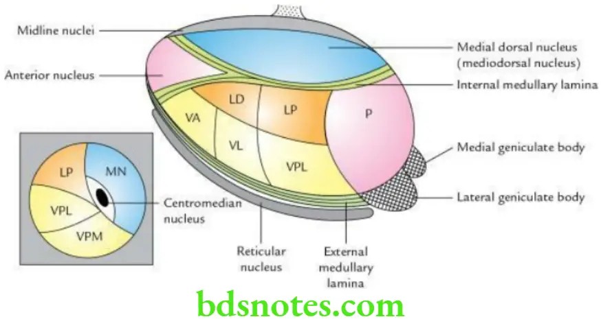
Major nuclei of the thalamus The major nuclei of the thalamus are as follows:
- Anterior nucleus
- Mediodorsal (dorsomedial) nucleus
- Pulvinar
- Ventral tier of nuclei:
- Ventral anterior nucleus (VA)
- Ventral lateral nucleus (VL)
- Ventral posterior nucleus (VP)
- Ventral posterolateral nucleus (VPL)
- Ventral posteromedial nucleus (VPM)

Question 3. Write a short note on the metathalamus.
Answer.
The metathalamus consists of medial and lateral geniculate bodies, which are rounded elevations on the inferior aspect of the pulvinar.
Lateral geniculate body: The lateral geniculate body (LGB) is a small ovoid swelling that projects downwards and laterally forms the pulvinar of the thalamus.
- It is visible at the terminal end of each optic tract. It is the thalamic visual nucleus. It receives fibres from the optic tract and gives fibres (optic radiation) to the visual cortex of the occipital lobe (areas 17, 18 and 19). It is the last relay station on the visual pathway.
- Structurally, the LGB consists of six laminae, numbered 1–6 from the ventral to the dorsal side. Laminae 1, 4 and 6 receive fibres from the contralateral retina and laminae 2, 3, and 5 from the ipsilateral retina.
Medial geniculate body: The medial geniculate body is a small oval swelling on the inferior surface of the pulvinar and is more prominent than the LGB. It is the thalamic auditory nucleus.
It receives the fibres from the lateral lemniscus through the inferior colliculus and inferior brachium and gives fibres (auditory radiation) to the auditory cortex in the temporal lobe (areas 41 and 42). It is the last relay station on the auditory pathway.
Question 4. Write a short note on the pineal gland.
Answer.
The pineal gland is a small cone-shaped body (only 3 × 5 mm in size) projecting downwards in the midline from the posterior wall of the 3rd ventricle. It is located in the groove between the superior colliculi below the splenium of the corpus callosum.
Its stalk divides into two laminae – ventral and dorsal. The ventral lamina is continuous with the posterior commissure, while the dorsal lamina is continuous with the habenular commissure.
Pineal Gland Functions
- Produces melatonin – a hormone that inhibits the secretion of gonadotrophins (GnRH) from the hypothalamus. Melatonin probably holds back the development of reproductive organs till a suitable age (i.e. puberty). In other words, the melatonin is believed to regulate the onset of the puberty.
- Acts as a biological clock and is responsible for circadian rhythm.
- Regulates the secretion of all other endocrine glands.
Pineal Gland Applied anatomy
- Tumours of the pineal gland cause precocious puberty.
- Calcification of pineal gland: The calcareous concretions appear in the pineal gland after 17 years of age and form aggregations called brain sand or corpora arenacea.
The calcification of the pineal gland is seen as a radiopaque shadow in 50% of individuals in the midline and provides a useful landmark to detect any shift of the brain due to a tumour, etc.
Question 5. Describe the hypothalamus under the following headings:
- Introduction,
- Boundaries,
- Regions/parts and nuclei,
- Functions and
- Applied anatomy.
Answer.
Hypothalamus Introduction The hypothalamus is a small part of the diencephalon that lies below the thalamus. It forms the floor and lower part of the lateral walls of 3rd ventricle.
The hypothalamus controls the various autonomic activities of the body. The sympathetic activities are controlled by its posterior part, while parasympathetic activities are controlled by its anterior part. Hence, it is also called the head ganglion of the autonomic nervous system.
Hypothalamus Boundaries The hypothalamus is bounded:
- Anteriorly by the lamina terminalis
- Posteriorly by the subthalamus
- Laterally by the internal capsule
- Medially by the cavity of 3rd ventricle
- Superiorly by the thalamus
- Inferiorly by the structures in the floor of the 3rd ventricle, i.e. tuber cinereum, a stalk of the infundibulum and mammillary bodies (these are actually parts of the hypothalamus)
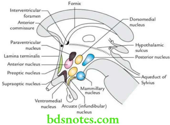
Regions/parts and nuclei The various regions of the hypothalamus and the nuclei present in them are as follows:
- Preoptic region adjoining the lamina terminalis: It contains a preoptic nucleus.
- Supraoptic region (above the optic chiasma). It contains:
- Supraoptic nucleus
- Anterior nucleus
- Paraventricular nucleus
- The tuberal region includes the tuber cinereum, infundibulum and area around it. It contains:
- Arcuate (infundibular) nucleus
- Ventromedial nucleus
- Dorsomedial nucleus
- Mammillary region (includes mammillary bodies and the area around it). It contains:
- Posterior nucleus
- Mammillary nuclei

Pineal Gland Functions
- Autonomic control: The anterior part controls the parasympathetic nervous system, while the posterior part controls the sympathetic nervous system.
- Endocrine control: By producing and releasing hormones or release-inhibiting hormones, it controls the functions of the endocrine glands of the body.
- Neurosecretion: The oxytocin and antidiuretic hormone (ADH)/vasopressin are synthesized in the supraoptic and paraventricular nuclei, respectively, and are transported to the posterior pituitary via the hypothalamo-hypophyseal tract.
- Regulation of food and water intake: The medial zone is responsible for hunger, thirst and drinking, while the lateral zone acts as a ‘satiety centre’.
- Temperature regulation: The anterior portion prevents the rise in temperature, while the posterior portion promotes heat production and conservation.
- Control of sexual behaviour and reproduction: By influencing the secretion of gonadotrophin (GnRH) by the pituitary gland.
- Control of emotional behaviour like laughing, crying, sweating and flushing.
- Acts as a biological clock, i.e. regulates the cyclic activities of the body (sleep and wake cycle called circadian rhythm).
Pineal Gland Applied Anatomy The impaired secretion of ADH/vasopressin leads to diabetes insipidus characterized by polyuria and polydipsia. The absence of glycosuria differentiates it from diabetes mellitus.
Question 6. Describe the 3rd ventricle under the following headings:
- Gross anatomy,
- Boundaries,
- Recesses and
- Applied anatomy.
Answer.
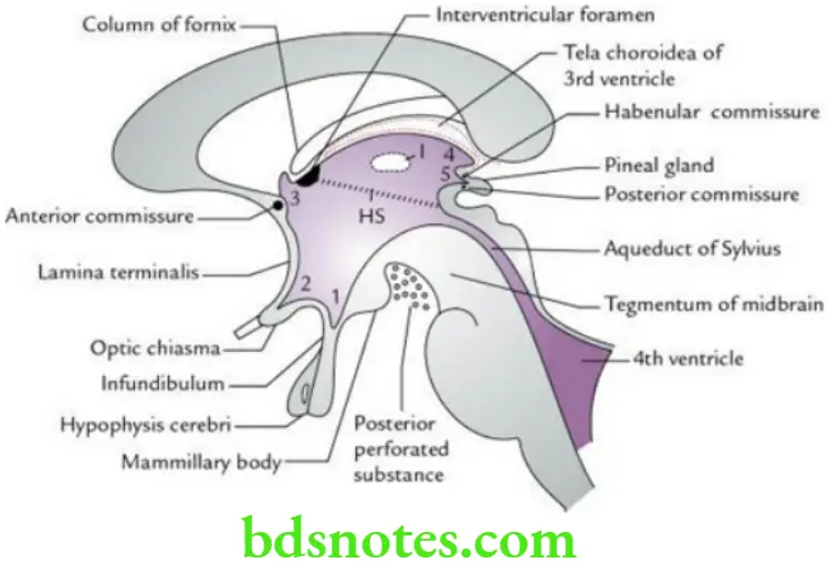
The 3rd ventricle is a midline slit-like cavity of the diencephalon, which extends from the lamina terminalis anteriorly to the upper end of the cerebral aqueduct posteriorly. The 3rd ventricle communicates with the lateral ventricles through interventricular foramina (of Monro) and with the 4th ventricle through the cerebral aqueduct (of Sylvius).
3rd Ventricle Boundaries The 3rd ventricle presents anterior wall, posterior wall, floor, roof and lateral walls.
- Anterior wall is formed from above downwards by:
- Anterior column of fornix
- Anterior commissure
- Lamina terminalis
- The posterior wall is formed from above downwards by:
- Pineal gland
- Posterior commissure
- Commencement of cerebral aqueduct
- The floor is formed from before backwards by:
- Optic chiasma
- Tuber cinereum
- Infundibulum (stalk of pituitary gland)
- Mammillary bodies
- Posterior perforated substance
- Tegmentum of midbrain
- The roof is formed by the ependyma stretching across the upper limits of the two thalami.
- The lateral wall is formed by:
- The medial surface of the anterior two-thirds of the thalamus (forms the lateral wall above the hypothalamic sulcus)
- The medial surface of the hypothalamus (forms the lower part of the lateral wall below the hypothalamic sulcus)

3rd Ventricle Recesses These are extensions (pocket-like protrusions) of the cavity of 3rd ventricle into the surrounding structures:
- Suprapineal recess: Above the stalk of the pineal gland
- Pineal recess: Between the superior and inferior laminae of the stalk of the pineal gland
- Infundibular recess: Into the stalk of the pituitary gland
- Anterior recess (also called vulva of the ventricle): Between the diverging anterior columns of the fornix in front of the interventricular foramen and behind the anterior commissure
3rd Ventricle Applied Anatomy
- The obstruction of 3rd ventricle leads to the accumulation of excessive CSF in it and two lateral ventricles, which causes an increase in intracranial pressure in adults and hydrocephalus in infants.
- The ventriculography is often done to visualize the obstruction/dilatation of 3rd ventricle.

Leave a Reply