Diagnostic Cytopathology
Question 1. Write a short note on the microscopic examination of urine.
Answer:
Microscopic Examination
Fresh or properly preserved sample is used. 1.5 mL of urine is centrifuged at 2000 rpm for 10 minutes. The supernatant is separated leaving a few drops to resuspend the sediment.
A drop of this sediment is put on a clean glass slide, covered with a clean coverslip.
The preparation is examined microscopically with reduced illumination, first under low power and then under high power. The following should be noted.
1. Microscopic examination of urine Cells
- Red cells: They appear as pale yellow refractile discs and disintegrate with the addition of 2% acetic acid. The significant number indicates hematuria.
- Pus cells: These are round cells with lobulated nuclei and round cytoplasm. The nuclei become prominent with the addition of a drop of acetic acid. They are seen in small numbers in glomerulonephritis and renal infarction. A large number indicates pyogenic infection of the kidney, urinary or genital tract.
- Epithelial cells: They may be round, caudate, or squamous in nature. Round renal cells may be found in various renal lesions and nephrotic syndrome.
Read And Learn More: Pathology Question And Answers
2. Microscopic examination of urine Casts
Casts are cylindrical structures with parallel edges and are formed by the coagulation of proteins within DCT and CT when the urine is likely to reach its maximum concentration and acidity—both favoring the precipitation of proteins. All casts are hyaline to start with. The casts are:
- Hyaline casts: They are colorless, semitransparent, and homogeneous, and may be resent normally. Large numbers are seen in acute and chronic glomerulonephritis.
- Epithelial casts: They are seen in acute glomerulonephritis and renal infarcts.
- Granular casts: They are formed by the disintegration of epithelial cells.
- Waxy casts: They are colorless, refractile, and homogeneous.
- Pus cell casts and red cell casts indicate acute nephritis.
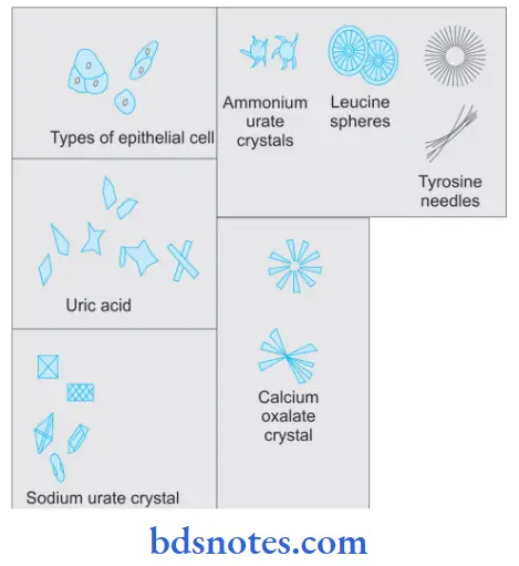
3. Crystals and Amorphous Materials
- Crystals of Acidic urine are:
- Uric acid
- Urates
- Calcium oxalate.
- Crystals of alkaline urine are:
- Triple phosphates
- Amorphous phosphates
- Calcium carbonates
- Ammonium biurate.
- Abnormal crystals include:
- Cystine
- Cholesterol
- Leucine
- Tyrosine.
4. Microscopic examination of urine Parasites
- Trichomonas vaginalis
- Microfilaria
- Hooklets or scolices of Echinococcus granulosus
- Ova of Schistosoma haematobium.
5. Microscopic examination of urine Cytological Examination
Smears are made from fresh urine samples, fi immediately in an ether-alcohol mixture, and stained by Papanicolaou stain which is helpful in early diagnosis of urinary tract malignancies.

Question 2. Write a short note on fine needle aspiration cytology.
Answer:
It is a type of interventional cytology in which samples are obtained by aspiration.
It is used for diagnostic cytopathology.
Application of FNAC
It is used for the diagnosis of palpable mass lesions like breast masses, enlarged lymph nodes, enlarged thyroids, and superficial soft tissue masses.
FNAC Materials Required
A syringe with a well-fitting needle, some microscopic glass slides, and a suitable fixative.
FNAC Advantages
- No hospitalization is required.
- No anesthesia is required
- The procedure is quick, safe, and painless.
- Results are obtained rapidly in a matter of hours.
- It is a low-cost procedure.
FNAC Complications
- Hematoma at the puncture site
- Infection
- Pneumothorax—transcutaneous aspiration of the lung causes pneumothorax.

Leave a Reply