Development And Growth Of teeth
Question 1. Describe the detail about stages of tooth development.
Answer:
Stages of tooth development:
- The development of a tooth is divided into several stages that are named after the shape of the enamel organ.
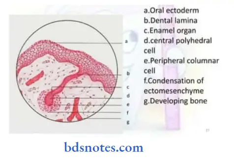
1. Bud stage:
Read And Learn More: BDS Previous Examination Question And Answers
- The epithelium of the dental laminae is separated from the underlying ectomesenchyme by a basement membrane.
- Round or avoid swellings develop from the basement membrane of 10 different points corresponding to tire 10 deciduous teeth.
- These are the primordial of the enamel organs, the tooth buds.
- In the bud stage, the enamel organ consists of peripherally located low columnar cells and centrally located polygonal cells.
- The supporting ectomesenchymal cells are packed closely beneath and around the epithelial bud.
- These cells undergo mitosis.
- As a result of it and the migration of the neural crest cells into it condensation of the tooth bud occurs.
- Condensation is immediately subjacent to the enamel organ in dental papillae.
- It forms tooth pulp and dentin.
- Similarly, ectomesenchymal condensation around tooth buds and dental papillae called dental sacs forms cementum arid periodontal ligament.
2. Bud to cap transition:
- The transition from bud to cap marks the onset of morphologic differences between tooth germs that give rise to different types of teeth.
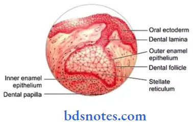
3. Cap stage:
- As the tooth bud continues to proliferate, due to unequal growth in different parts of the tooth bud, it leads to the cap stage.
- The peripheral cells covering the convexity of the cap are cuboidal called the outer enamel epithelium.
- The cells in the convexity of the cap called the inner enamel epithelium are tall, and columnar.
- Between these two layers are star-shaped cells called stellate reticulum.
- The outer enamel epithelium is separated from the dental sac and the inner enamel epithelium from the dental papilla by a basement membrane.
- The inner enamel epithelium meets the outer enamel epithelium at the rim of the enamel organ called the cervical loop.
4. Bell stage:
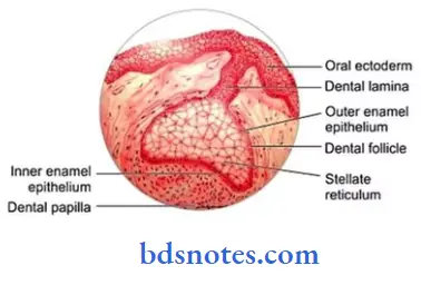
- The continued growth of tire tooth germ leads to the bell stage as the tire enamel organ resembles a bell as the undersurface of the epithelial cap deepens.
- During the tills stage, the tooth crown assumes its final shape.
- Four different types of epithelial cells can be identified.
- Inner enamel epithelium:
- Consists of a single layer of cells of 4 – 5 pm in diameter and about 40 um high.
- These cells differentiate into ameloblast.
- Stratum intermedium
- It is essential for enamel formation.
- Cells in this layer are closely attached by desmosomes.
- Stellate reticulum
- It consists of star-shaped cells which help for attachment between the cells.
- It collapses to reduce the distance between the centrally situated ameloblasts and the nutrient capillaries near the outer enamel epithelium.
- Outer enamel epithelium
- The cells of this layer are cuboidal.
- Its smooth surface is laid down in folds between which capillary loops provide a rich nutritional supply for intense metabolic activity.
- Before the inner enamel epithelium begins to produce enamel, the peripheral cells of dental papilla differentiate into odontoblasts forming dentin.
- Remnants of dental lamina
- Outer enamel epithelium
- Ameloblasts
- Stellate reticulum Dental follicle Stratum intermedium Odontoblasts
- Collasped stellate reticulum Ameloblasts
- Inner enamel epithelium:
5. Advanced bell stage:
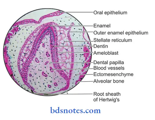
- It is characterized by mineralization and root formation.
- The boundary between inner enamel epithelium and odontoblasts outlines the dentin enamel junction (DEJ).
- the dentin formation begins at this point and then proceeds pulpal and apically.
- Over it, ameloblast by down enamel which then proceeds coronally and cervically.
- In the cervical portion, the enamel organ gives rise to the epithelial root sheath of Hertwig.
Question 2. Enumerate stages of tooth development. Describe the bell stage in detail.
Answer:
Stages of tooth development:
- Bud stage
- Cap stages
- Bell stage
- Advanced bell stage.
Question 3. Write briefly about Hertwig’s root sheath.
Answer:
Hertwig’s root sheath:
- Epithelial cells of the inner and outer dental epithelium proliferate from the cervical loop of the enamel organ to form a double layer of cells known as Hertwig’s epithelial root sheath
- It consists of only inner and outer enamel epithelium excluding stratum intermediate and stellate reticulum.
- It establishes the future dentioccmental junction.
- This sheath of epithelial cells extends around the denial pulp between it anil the dental follicle.
- The aim of this root sheath, the epithelial diaphragm, encloses the primary apical foramen.
- As the inner epithelial cells of the root sheath progressively enclose more and more expanding dental pulp, they initiate the differentiation of odontoblasts and eventually form the dentin of the root.
- The cells of the inner layer remain short and normally do not produce enamel.
- The proliferation of the cells of the diaphragm is accompanied by a proliferation of the cells of the connective tissue of the pulp.
- The free end of the diaphragm does not grow into the connective tissue but proliferates coronal to the epithelial diaphragm.
Fate:
- If Hertwig’s epithelial root sheath is lost prior to the cementum formation, accessory canals are formed.
- If it persists, it leads to the formation of epithelial pearls or enamel pearls
- After the epithelial rest cells form the dentin of the root, it loses its structural continuity and its close relation to the surface of the root.
- Its remnants are found in the periodontal ligament of erupted teeth and are called the rest of molasses.
Question 4. Enumerate the stages of tooth development and write about the formation of the root.
Answer:
Stages of tooth development:
- Bud stage
- Cap stage
- Bell stage
- Advanced bell stage
Root formation:
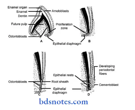
- Hertwig’s epithelial root sheath (HFRS) plays an important role in root formation.
- HERS consists of the inner and outer epithelial layers.
- It establishes the future cementoenamel junction.
- Root formation occurs after the shape of the crown has been established.
- The layers of HERS bend at the future cementoenamel junction at a right angle to narrow the cervical opening of the developing tooth.
- The rim of the HERS the epithelial diaphragm encloses the primary epithelial layer and initiates the differentiation of odontoblasts from ectomesenchymal cells to the form dentin of the root.
- The connective tissue of the dental sac surrounding the root sheath proliferates and invades 5 rd dividing it into a network of epithelial strands.
- The epithelium is moved away from the dentinal surface.
- Due to it connective tissue cells come into contact with the outer surface of the dentin and differentiate into cementoblast to deposit a layer of cementum over the dentin.
- The Aside apical foramen is reduced first to that of diaphragmatic opening and later reduced apposition of dentin and cementum to the apex of the root.
- In this way, a single root is formed.
For multirooted teeth:
- Expansion of the cervical opening of the enamel organ occurs which results in tongue-like extension cr the horizontal diaphragm.
- Before division, the free ends of the horizontal epithelial flaps grow towards each other and fuse.
- The single cervical opening of the enamel organ is then divided into two or three openings.
- On the pulpal surface of the dividing epithelial bridges, dentin formation starts and on the periphery reel development occurs in a similar manner as that of single-rooted teeth.
Question 5. Dental lamina.
Answer:
Development:
- During 6 weeks of intrauterine life, a continuous band of thickened epithelium forms around the mouth occupying the upper and lower jaw which is roughly horseshoe-shaped called a primary epithelial band.
- At about 7th week, this band divides into a lingual process called dented lamina and a buccal process called the vestibular lamina.
Question 6. Advanced bell stage of tooth development
Answer:
Features:
- Formation of future dentin enamel junction (DEJ)
- It is the boundary between the inner enamel epithelium and odontoblasts
The first layer of dentin is laid down in the region of future cusps
↓
Proceeds pulpal and apically
↓
Ameloblast lies down enamel over dentin in the incisal and cuspal area
↓
It proceeds coronally and cervically
2. Commencement of mineralization
3. Formation of HERS
- The cervical portion of the enamel organ contributes to Hertwig’s epithelial root sheath formation
- It consists of only inner enamel epithelium and outer enamel epithelium
- Inner enamel epithelium induces differentiation of dental papilla into odontoblast to lie down dentin in the radicular portion
- HERS is responsible for root formation
4. Root formation
- Root formation occurs after the shape of the crown has been established
- The layers of HERS bend at the future CEJ at a right angle to narrow the opening of the developing tooth
- The rim of the HERS the epithelial diaphragm encloses the primary epithelial layer to initiate differentiation of odontoblasts from ectomesenchyme cells to form dentin of the root
- The connective tissue of the dental sac surrounding the root sheath proliferates and invades HERS and divides it into a network of epithelial strands
- The epithelium is moved away from the dentinal surface
- Due to it connective tissue cells come into contact with the outer surface of the dentin and differentiate into cementoblast to deposit a layer of cementum over the dentin
- The wide apical foramen is reduced first to that of diaphragmatic opening and later reduced by apposition of dentin and cementum to the apex of the root
- In this way, a single root is formed
Question 7. Cap stage
Answer:
- After the proliferation of tooth bud, unequal growth occurs in different parts of tooth byd
- This results in cap-stage development
- It is characterized by shallow invagination on the deep surface of the bud
Layers:
1. Inner enamel epithelium
- The cells in the concavity of the cap become tall, and the columnar cell represents the inner enamel epithelium
- It is separated from the dental papilla by a delicate basement membrane
2. Outer enamel epithelium
- The peripheral cells covering the convexity of the cap are cuboidal
- They represent outer enamel epithelium
- It is separated from the dental sac by a basement membrane
3. Stellate reticulum
- It is a layer of polygonal cells between the inner and outer enamel epithelium
- Due to the entry of water into the enamel organ, these cells become star-shaped
- These cells maintain contact with each other through their cytoplasmic process
- They act as shock absorbers to support and protect the delicate enamel-forming cell
Structures formed during this stage:
- Enamel knot
- Enamel cord
- Enamel septum
- Enamel navel
Question 8. Root formation
Answer:
- Hertwig’s epithelial root sheath (HERS) plays an important role in root formation
- HERS consists of the inner and outer epithelial layer
- It establishes the future cementoenamel junction
- Root formation occurs after the shape of the crown lias been established
- The layers of HERS bend at the future CEJ at a right angle to narrow the opening of the developing tooth
- The rim of the HERS the epithelial diaphragm encloses the primary epithelial layer to initiate differentiation of odontoblasts from ectomesenchyme cells to form dentin of the root
- The connective tissue of the dental sac surrounding the root sheath proliferates and invades HERS and divides it into a network of epithelial strands
- The epithelium is moved away from the dentinal surface
- Due to it connective tissue cells come into contact with the outer surface of the dentin and differentiate into cementoblasts to deposit a layer of cementum over the dentin
- The wide apical foramen is reduced first to that of diaphragmatic opening and later reduced by apposition of dentin and cementum to the apex of the root
In this way, a single root is formed For multi-rooted teeth:
- Expansion of the cervical opening of the enamel organ occurs which results in tongue-like extension of the horizontal diaphragm
- Before division, the free ends of the horizontal epithelial flaps grow toward each other and fuse
- The single cervical opening of the enamel organ is then divided into two or three openings
- On the pulpal surface of the dividing epithelial bridges, dentin formation starts and on the periphery, root development occurs in a similar manner as that of single-rooted teeth
Question 9. Epithelial-mesenchymal interaction
Answer:
- Epithelial-mesenchymal interaction governs the development of epidermal organs like teeth
- During the early stages of tooth development, a local ectodermal thickening expressing severe signaling molecules appear
- These signals to the underlying mesenchyme trigger mesenchymal condensation and tooth development
Example:
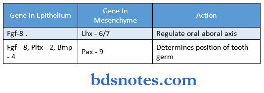
Genes and their action:
- Pax – 9 – determines morphogenesis
- LHX – Mesenchymal marker for initiation
- Fgf – 8 – a marker of the first branchial arch
- Bmp – 2, Bmp – 4 – patterning of tooth
- Bmp – 5 – endochondrogenesis
- Bmp – 7 – dentinogenesis
- Msx 1, Msx 2 – expresses incisor in mandible
- Msx 1, Msx 2 &Dlx – 2 – express canine
- Dlx 2 &Barx 1 – expresses molar
Question 10. Dental lamina.
Answer:
- It results from the dividing of the primary epithelial band at around the 7th week of intrauterine life.
- It represents the lingual process of this band.
- It later breaks to form discrete epithelial islands which are named epithelial pearls.
- It serves as the primordium for the ectodermal portion of the deciduous teeth,
- It has a surface of squamous cells and a basal layer of columnar cells.
Question 11. Dental sac.
Answer:
- It is the condensed ectomesenchyme limiting the dental papilla and encapsulating the enamel organ.
- It gives rise to the supporting tissues of the tooth.
- Before the formation of dental tissues begins, the dental sac shows a circular arrangement of its fibers and resembles a capsular structure.
- With the development of the root, the fibers of the dental sac differentiate into the periodontal fibers that become embedded in the developing cementum and alveolar bone.
Question 12. Histophysiological stage of tooth development.
Answer:
1. Initiation:
- Specific cells within the dental laminae have the potential to form the enamel organ that initiates tooth development.
- Different teeth are initiated at definite times.
- Dental papilla mesenchyme can induce tooth epithelium and even non-tooth epithelium to form enamel.
2. Proliferation:
- Proliferation occurs at the points of initiation and results in the bud, cap, and bell stages.
- It causes regular changes in the size and proportions of the growing tooth germ.
3. Histodifferentiation:
- The developed formative cells of the tooth germ undergo definite morphologic as well as functional changes.
- The cells differentiate and give up their capacity to multiply.
- Inner enamel epithelium on the mesenchyme causes the differentiation of the adjacent cells of the dental papilla into odontoblasts to form dentin.
- The cells of the inner enamel epithelium differentiate into ameloblast to form an enamel matrix.
4. Morpho differentiation:
- It results in the establishment of the basic form and relative size of the future tooth.
- The ameloblasts, odontoblast, and cementoblast deposit enamel, dentin, and cementum, thus giving the tooth its final form and size.
5. Apposition:
- It is the deposition of the matrix of the hard dental structures.
- Appositional growth of enamel and dentin is a layer-like deposition of an extracellular matrix.
Question 13. Hertwig’s root sheath.
Answer:
- Epithelial cells of the inner and outer dental epithelium proliferate from the cervical loop of the enamel organ to form a double layer of cells known as Hertwig’s epithelial root sheath.
- It consists of only inner and outer enamel epithelium.
- Its inner epithelial cells initiate the differentiation of odontoblasts from ectomesenchymal cells to form dentin of the root.
- The aim of this sheath encloses the primary apical foramen,
- The remnants of this sheath are named epithelial pearls or enamel pearls.
Question 14. Vestibular lamina.
Answer:
- It results from the dividing of the primary epithelial band at around 7th week of intrauterine life,
- It represents the buccal process of this band.
- The vestibule forms as a result of the proliferation of the vestibular lamina into the ectomesenchyme.
- Its cells rapidly enlarge and then degenerate to form a cleft that becomes the vestibule between the cheek and the tooth-bearing area.
Question 15. Stratum intermedium.
Answer:
- It is a layer of squamous cells present between the inner enamel epithelium and the stellate reticulum.
- These cells are closely attached by desmosomes and gap junctions.
- The well-developed cytoplasmic organelles, acid monopoly saccharides, and glycogen deposits indicate a high degree of metabolic activity.
- It is essential for enamel formation.
Question 16. Bell stage.
Answer:
During this stage, crown shape is determined layers seen during this stage are:
1. Outer enamel epithelium:
- Low cuboidal cell layer present at the periphery of the enamel organ.
2. Inner enamel epithelium:
- Short columnar cell layer bordering the dental papilla.
- These epithelia meet at the rim of the enamel organ called the cervical loop.
3. Stellate reticulum:
- Consists of star-shaped cells with long anastomotic processes
- They collapse reducing the distance between the ameloblasts and the nutrient capillaries near the outer enamel epithelium.
4. Stratum intermedium:
- It is present between the inner enamel epithelium and the stellar reticulum.
- Its cells are characterized by the high activity of the enzyme alkaline phosphate.
Question 17. Enamel organ.
Answer:
- The enamel organ originates from the stratified epithelium of the native oral cavity.
- It consists of
Epithelium layers:
- Outer enamel epithelium
- Stellate reticulum
- Stratum intermedium
- Inner enamel epithelium
Dentinoenamel junction:
- It is borderline between the inner enamel epithelium and the connective tissue of the dental papilla.
Cervical loop:
It is the region where the inner enamel epithelium reflects onto the outer enamel epithelium at the border of the wide basal opening of the enamel organ.
Question 18. Development of root.
Answer:
- The development of the roots begins after enamel and dentin formation has reached the future cementoenamel junction.
- The radicular dental papilla cells differentiate under the influence of HERS cells into odontoblasts which lay down the first layer of dentin.
- The epithelium is moved away from the surface of the dentin allowing connective tissue cells of the dental sac to come into contact with the outer surface of the dentin which then differentiates into cementoblasts and deposits a layer of cementum.
- The epithelial root sheath loses its structural continuity and disintegrates.
- Differential growth of the epithelial diaphragm in multirooted teeth causes tire division of the root trunk into two or three roots.
Question 19. Stellate reticulum.
Answer:
- It is located between the outer and inner enamel epithelium.
- It consists of star-shaped cells with a long cytoplasmic process.
- This gives the stellate reticulum a cushion-like consistency and acts as a shock absorber that may support and protect tire delicate enamel-forming cells.
- Desmosomal junctions are observed between cells of the stellate reticulum, stratum intermedium and outer enamel epithelium.
- Before enamel formation begins, the tire stellate reticulum collapses, reducing the distance between the centrally situated ameloblasts and tire nutrient capillaries near the tire outer enamel epithelium.
Question 20. Cell rest of series.
Answer:
- As the tooth develops, it loses its continuity with tire dental lamina.
- Later mesenchymal invasion occurs which leads to the breaking up of dental lamina.
- These remnants of the dental lamina persist as epithelial islands and are referred to as the cell rest of the series.
- Location within the jaw, in tire gingiva.
Question 21. Epithelial rests
Answer:
- It is a remnant of Hertwig’s epithelial root sheath
- Present near and parallel to root surfaces and Attached to one another by desmosomes
- During disease conditions they undergo proliferation
- Persists as a network strand, island, or tubule
- Exhibits tonofilaments
Question 22. Dental papilla
Answer:
- Under the influence of proliferating epithelium of the enamel organ, tire ectomesenchyme surrounding the inner enamel epithelium proliferates
- It condenses to form dental papilla
- Dental papilla shows active budding of capillaries and mitotic figures
- Its peripheral cells adjacent to the inner enamel epithelium enlarges and later differentiate into odontoblasts
Structures derived from dental papilla:
- Dentin
- Pulp

Leave a Reply