Dentin
Question 1. Describe various types of dentin.
Answer:
Types of dentin:
1. Primary dentin:
- Dentin which is formed before root completion is known as primary dentin.
- There are two types of primary dentin.
- Mantle dentin:
- It is the first formed dentin in the crown.
- It lies immediately below the dentin enamel junction.
- It is formed before a tooth erupts into the oral cavity
- It is the outermost, peripheral part of primary dentin.
- Mantle dentin:
Read And Learn More: BDS Previous Examination Question And Answers
- Thickness:
- It is 20 Jim thick.
- Function:
- As it is soft, it provides a cushioning effect to the tooth.
- Fibers:
- It contains larger diameter fibers of about 0.1 – 0.2 pm in diameter.
- They are argyrophilic.
- They are known as von Kroff’s fibers.
- They mainly contain type II collagen.
- They are directed perpendicular to DEJ.
- Mineralization:
- Mantle dentin is less mineralized
- It involves globular mineralization.
- Circumpulpal dentin:
- It is the primary dentin that outlines the pulp chamber.
- Fibers:
- Smaller in diameter (0.05 Jim)
- They are closely packed.
- Mineralization:
- Circumpupal dentin is more mineralized
- It undergoes globular or linear mineralization.
2. Secondary dentin:
- It is a narrow band of dentin bordering the pulp
- It is the dentin that is formed after root formation.
- It represents continued lower deposition of dentin by odontoblasts.
- It is not deposited evenly around the periphery of the pulp chamber.
- There is greater deposition on the roof and floor of the chamber than other parts.
Tubules:
- Secondary dentin contains fewer tubules.
- They are less regular
- At the boundary between primary and secondary dentin, tubules change their direction and show a wavy course.
- These tubules sclerose more readily.
- This reduces the overall permeability of the dentin thereby protecting the pulp.
Synonym:
- Regular secondary dentin – due to the regular arrangement of dentinal tubules.
3. Tertiary dentin:
- It is that dentin is produced by odontoblasts in response to irritation of the pulp caused by chemical, thermal, or microbial stimuli.
- It contains very few irregular tubules.
- The quality and the quantity of tertiary dentin produced depends on the intensity and duration of the stimulus.
- Tertiary dentin is produced only by those cells directly affected by the stimulus.
- The cells forming tertiary dentin line its surface or become included in the dentin called osteopontin.
- It is subclassified as
- Reactionary dentin:
- It is deposited by pre-existing odontoblasts.
- Reparative dentin:
- It is deposited by newly differentiated odontoblast-like cells.
- Reactionary dentin:
Question 2. Define dentin. Write about the physical and chemical properties of dentin.
Answer:
Dentin;
- Dentin is the hard tissue portion of the pulp-dentin complex which forms the bulk of the tooth.
Physical properties:
1. Color:
- Dentin is usually light yellow in color.
- It becomes darker with age.
2. Hardness:
- Dentin is slightly harder than bone and softer than enamel.
- Dentin hardness varies slightly between tooth types and between crown and root dentin.
- Dentin is harder in its central part than on its periphery.
- The dentin of primary teeth is slightly less hard than that of permanent teeth.
- Dentin appears more radiolucent than enamel and more radiopaque than pulp.
3. Elasticity:
- Dentin is viscoelastic and subject to slight deformation.
- Dentin’s elastic property is important for the proper functioning of the tooth.
- The elasticity of dentin provides flexibility and prevents the fracture of the overlying brittle enamel.
4. Permeability:
- Dentin permeability depends upon the potency of dentinal tubules.
- Tubular occlusion, smear layer formation, and lack of tubular communication between primary and secondary dentin will result in reduced permeability.
- This lessens the sensitivity of dentin.
- Dentin permeability increases as the pulp chamber is approached as the number and diameter of the tubules are more per unit area towards the pulp than towards the periphery.
Chemical properties:
- Dentin consists of
- It chiefly consists of collagen fibers with fractional inclusion of Jipids and noncollagenous matrix proteins.
- Collagen fibers contain mainly type I collagen with small amounts of type III and V.
- These fibers are embedded in ground substances of mucopolysaccharides.
- Noncollagenous matrix proteins pack the space between collagen fibrils and accumulate along the periphery of dentinal tubules.
- These proteins regulate mineral deposition and can act as promoters or inhibitors.
- The main content of the ground substance is proteoglycans which prevent the premature mineralization of the organic matrix.
- The matrix also contains growth factors.
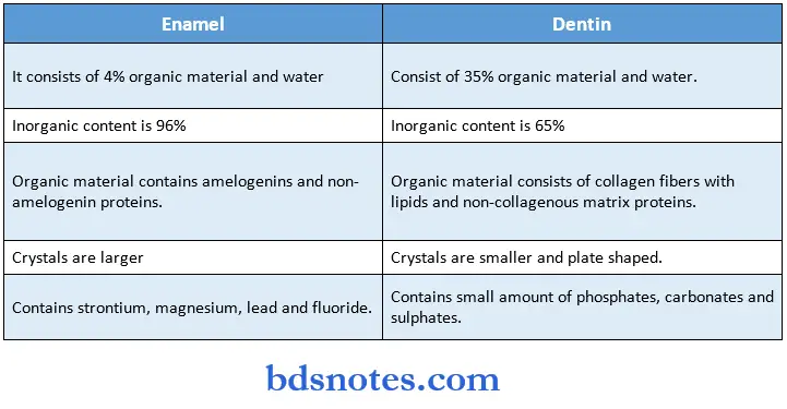
- Inorganic material – 65%.
- The inorganic content consists of hydroxyapatite which is composed of several thousand unit cells.
- The crystals are plate-shaped and smaller than enamel crystals.
- These crystals are poor in calcium but rich in carbon.
Dentin also contains small amounts of phosphates, carbonates, and sulfates.
Question 3. Describe hypocalcified structures of dentin. Add a note on the histology of dentinal tubules.
Answer:
Hypocalcified structures of dentin:
1. Counterlines of Owen:
- These lines result from a coincidence of the secondary curvature between neighboring dentinal tubules.
- They are caused by accentuated deficiencies in mineralization.
- These are easily recognized in ground sections.
- These lines are accentuated because of disturbances in the matrix and mineralization process.
2. Neonatal line:
- Interglobular dentin is used to describe areas of unmineralized or hypo mineralized dentin where globular zones of mineralization have failed to fuse into a homogenous mass within mature dentin.
- It is prevalent in persons with Vit. D deficiency or who had exposure to high levels of fluoride at the time of dentin formation.
- It is mostly seen in circumpolar dentin.
- The tubular pattern remains unchanged.
- Dentinal tubules run uninterrupted.
- No peritubular dentin exists.
- Frequently seen regions area
- In crown:
- Cervical and middle third followed by a cuspal and coronal third.
- In root:
- Highest in the cervical third followed by the middle third.
4. Predentin:
- It is first formed dentin and is not mineralized.
- It lines the innermost i.e., the pulpal portion of the tooth.
- It consists principally of collagen and non-collagenous components.
- It is 2-6 pm wide.
- It gradually mineralizes into dentin as various noncollagenous matrix proteins are incorporated at the mineralization front.
- Its thickness remains constant because the amount that calcifies is balanced by the addition of a new unmineralized matrix.
- It is thickest during active dentinogenesis and diminishes in thickness with age.
Histology of dentinal tubules:
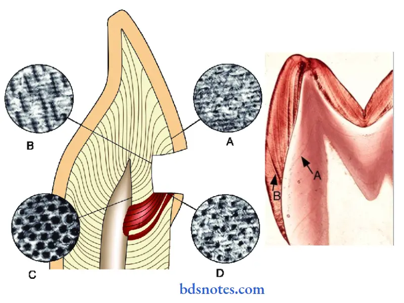
- Dentinal tubules extend through the entire thickness of the dentin from the dentin enamel junction to the pulp.
- Their configuration indicates the course taken by the odontoblasts during dentinogenesis.
- They follow an S-shaped path from the outer surface of the dentin to the perimeter of the pulp.
- This curvature is least pronounced beneath the incisal edges and cusps.
- The tubules are longer than die dentin and are thick because diet curves through dentin.
- The tubules end perpendicular to the dentin enamel and dentino cementum junction.
- The curvatures result from the crowding of and path followed by odontoblasts as they move toward the center of the pulp.
- In root dentin, little or no crowding results from a decrease in surface area, and tubules run in a straight course.
- The tubules are farther apart in the peripheral layers and are more closely packed near the pulp.
Size:
- 2.5 pm in diameter near the pulp.
- 1.2 pm in diameter in the midportion of the dentin.
- 900 nm near DEJ.
Number:
- There are more tubules per unit area in the crown than in the die root.
- Near the die pulpal surface of die dentin the number per square millimeter varies between 50,000 and 90,000.
Density:
- Reduction in the average density of tubules occurs in radicular dentin compared with cervical dentin.
- Higher tubule density occurs on the lingual and buccal walls of the pulp than on the mesial and distal walls.
Branches:
- Major branches occur more frequently in root dentin than in coronal dentin.
- Branches of the dentinal tubules near the terminals are referred to as terminal branches.
- The dentinal tubules have lateral branches throughout the dentin called canaliculi.
- These canaliculi are ppm or less in diameter and originate more or less at right angles to the main tubule.
Question 4. Describe in detail the microscopic structures of dentin.
Answer:
Microscopic structures of dentin:
1. Dentinal tubules:
- Odontoblast processes run in canaliculi that transverse the dentin layer and are referred to as dentinal tubules.
- They extend through the entire thickness of the dentin from the DEJ to the pulp.
- These tubules are perpendicular to DEJ and DCJ.
- Near the root tip and along incisal edges and cusps, tubules are almost straight.
- These are tapered structures.
- They are larger in diameter near the pulpal cavity and smaller at their outer ends.
- There are more tubules per unit area in the crown than in the root.
- The dentinal tubules have terminal and lateral branches.
- Lateral branches extend throughout the dentin and are referred to as canaliculi.

2. Peritubular dentin:
- The dentin that immediately surrounds the dentinal tubules is termed peritubular dentin.
- It is hypermineralized.
- It contains little collagen
- It is twice as thick in the outer dentin than in the inner dentin.
- It constricts the dentinal tubules to a diameter of 1 |im near the DEJ.
- This is described as a thin organic membrane, high in glycosaminoglycan.
- Between the odontoblastic process and the peritubular dentin, a space known as a periodontoblastic space is present.
- In decalcified sections, the tubule diameter appears similar in the inner and outer dentin because of the loss of peritubular dentin.
3. Sclerotic dentin:
- It describes dentinal tubules that have become occluded with calcified material.
- It can be observed in the teeth of elderly people, especially in the roots.
- It may be found under slowly progressing caries, common in the apical third of the root and in the
crown midway between the DEJ and the surface of the pulp. - It is harder than normal dentin- with reduced fracture toughness.
- Its crystals are smaller than normal dentin.
- It reduces the permeability of dentin which help to prolong pulp vitality.
4. Intertubular dentin:
- It is the dentin located between the dentinal tubules.
- It represents the primary secretory product of the odontoblasts.
- It consists of a network of type I collagen fibrils in which apatite crystals are deposited.
- The ground substance consists of non-collagenous proteins.
- It is highly mineralized.
- Apatite crystals are formed along the fibers with their long axes oriented parallel to the collagen fibers.
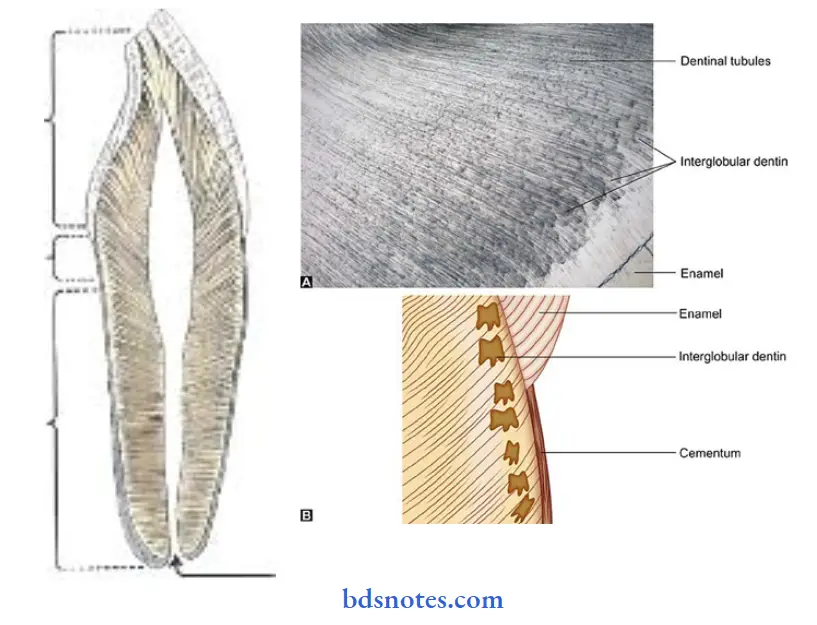
5. Interglobular dentin:
- It is unmineralized or hypo-mineralized dentin.
- It is prevalent in Vit.
- deficiency and high exposure to fluoride.
- It is mostly seen in circumpolar dentin.
- Dentinal tubules run uninterrupted as no peritubular dentin exists.
- It occurs due to a defect of mineralization and not of matrix formation.
6. Incremental growth lines:
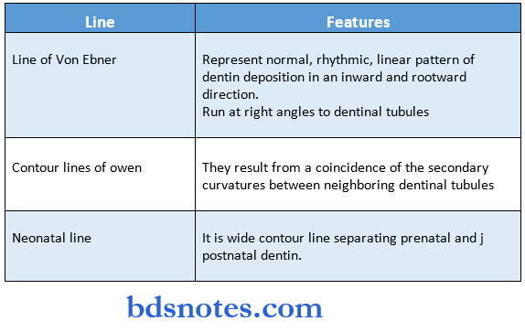
- The incremental lines of Von Ebner appear as fine lines or striations in dentin.
- They run at a right angle to dentinal tubules.
- They represent a normal rhythmic, linear pattern of dentin deposition in an inward and rootward direction.
- Its course indicates the growth pattern of dentin.
- The distance between lines varies from 4-8 mm in the crown to much less in the root.
- The daily increment decreases after a tooth reaches functional occlusion.
- Another line called contour lines of owen which results from a coincidence of the secondary curvatures between neighboring dentinal tubules.
- A wide contour line called the neonatal line is seen which separates the dentin formed before birth and that formed after birth.
7. Granular layer of tomes:
- A granular appearing area seen in transmitted light in ground sections of root dentin is called a granular layer of tomes.
- It is present just below the surface of the dentin where the root is covered by cementum.
- It increases from CEJ to the apex of the tooth.
Question 5. Age changes in dentin.
Answer:
1. Color:
- Dentin becomes darker with age.
2. Dentinal tubules:
- There is a reduction in the diameter of dentinal tubules with age due to the continued deposition of intratubular dentin.
- Gradually, there is a complete closure of the tubule.
- This obliterates the pulp chamber.
3. Sclerotic dentin:
- It appears in older people.
- The dentinal tubule becomes occluded with calcified material.
- It is harder than normal dentin.
- It reduces the permeability of dentin.
4. Development of dead tracts:
- Dentinal tubules are emptied by complete retraction of the odontoblast process from the tubule or through the death of the odontoblast with age.
- The dentinal tubules then become sealed off at their pulpal ends.
- The tubules are filled with fluid or gas.
- In-ground sections, it may entrap air and appear black in transmitted light and white in reflected light.
5. permeability:
- As age advances, dentin becomes less permeable.
- It occurs due to tubular occlusion, smear layer formation, and lack of tubular communication between primary and secondary dentin.
6. Sensitivity:
- Reduction in dentin permeability would lessen the sensitivity of dentin.
Question 6. Structure of dentin.
Answer:
1. Dentinal tubules:
- They extend through the entire thickness of the dentin from the dentin enamel junction to the pulp.
- They follow an S-shaped path.
- They are longer than the dentin and are thick because they curve through the dentin.
- The tubule ends are perpendicular to the DEJ and DCJ.
- There are more tubules per unit area in the crown than in the tire root.
- They have terminal and lateral branches.
2. Peritubular dentin:
- The dentin that immediately surrounds the dentinal tubules is called peritubular dentin.
- It is a hypermineralized structure.
- This is described as a thin, organic membrane high in glycosaminoglycan.

Intertubular dentin:
- It is the dentin located between the dentinal tubules.
- It is highly mineralized.
- It forms the main body of dentin.
- About one-half of the volume is organic matrix mainly collagen fibers, which are randomly oriented around dentinal tubules and ground substances of non-collagenous proteins.
Presenting:
- It is first formed by dentin.
- It lines the pulpal portion of the tooth.
- It mainly consists of collagen and non-collagenous components.
- It is 2-6 pm wide.
Odontoblast process:
- They are the cytoplasmic extensions of the odontoblasts.
- It extends into the dentinal tubules They are largest in diameter near the pulp.
- They are narrow to about half the size of the cell as they enter the tubules.
- They are composed of microtubules of 20 pm in diameter and small filaments of 5 – 7.5 pm in diameter.
- These odontoblastic processes are divided near the dentin enamel junction and extend into enamel in the enamel spindles.
- Their lateral branches extend laterally into adjacent tubules.
Question 7. Curvatures in dentinal tubules.
Answer:
- Dentinal tubules are found throughout normal dentin.
- They are longer than the dentin and are thick because they curve through the dentin.
- The course of tubules follows a gentle curve in the crown but is less curved in the root.
- Thus, it resembles a gentle S-shape curve.
- These curvatures are called “primary curvatures”.
- Starting at right angles from the pulpal surface first convexity of this curve is directed towards the root apex.
- Tubules end at right angles to DEJ and DCJ.
- Tubules also exhibit “secondary curvatures”.
- These curvatures are minute, regular, and sinusoidal in shape.
- They are present over the entire length of the tubules.
Question 8. Incremental lines in dentin.
Answer:
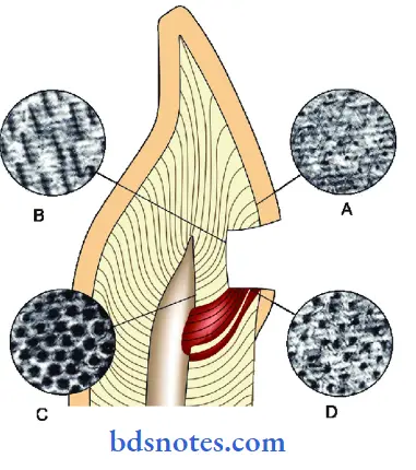
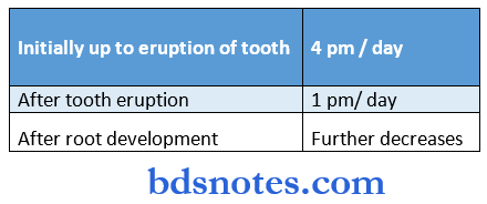
1. Incremental lines of Von Ebner:
- They appear as fine lines or stations in dentin.
- They run at right angles to dentinal tubules.
- They represent a normal, rhythmic, linear pattern of dentin deposition in an inward rootward direction.
- Its course indicates the growth pattern of dentin.
- The distance between lines varies from 4-8 mm in the crown to much less in the root.
- The daily increment decreases after a tooth reaches functional occlusion.
- They are seen in conventional and ground sections.
2. Contour lines of Owen:
- These lines result from a coincidence of the secondary curvatures between neighboring dentinal tubules.
- They are caused by accentuated deficiencies in mineralization.
- They are demonstrated in ground sections.
- They represent hypocalcified bands.
- They are accentuated because of disturbances in the matrix and mineralization process.
3. Neonatal line:
- It is a wide contour line.
- It separates the dentin formed before birth and that formed after birth.
- It reflects the disturbance in mineralization created by the physiologic trauma of birth.
- The dentin matrix formed prior to birth is usually of better quality than that formed after birth.

Question 9. Briefly explain dentinal sensitivity.
Answer:
Theories of dentinal sensitivity:
1. Direct nerve innervations theory:
- It states that the dentin contains nerve endings that respond when it is stimulated.
- Some nerves penetrate a short distance into some tubules.
This theory is rejected because. - The dentinal nerves do not extend beyond the inner dentin.
- Some nerves occur within some tubules but dentin sensitivity does not depend on the stimulation of such nerve endings.
- A newly erupted tooth does not possess such nerve endings and yet it is sensitive.
- Application of local anesthetics or silver nitrate to exposed dentin does not eliminate dentin sensitivity.
2. Transduction theory:
- It states that the odontoblasts serve as receptors and are coupled to nerves in the pulp.
- It was supported because the odontoblast is of neural crest origin, it retains an ability to transducer and propagate an impulse.
- But it was rejected because.
- Absence of synaptic relationship between the odontoblast and pulpal nerves.
- There are no neurotransmitter vesicles in the odontoblastic process to facilitate the synapse
- The membrane potential of odontoblasts measured is too low to permit transduction.
- Local anesthetic and protein precipitants do not abolish sensitivity.
3. Fluid or hydrodynamic theory:
- It states that the tubular nature of dentin permits fluid movement to occur within the tubule when a stimulus is applied.
- This fluid movement either inward or outward stimulates the pain mechanism in the tubules by mechanical disturbance of the nerves closely associated with the odontoblast and its process.
- When a stimulus is applied to dentin, there is fluid displacement within the tubule and this distorts the tire’s local pulp environment and is sensed by the free nerve endings in the plexus of Raschkow.
- These nerve endings may act as mechanoreceptors as they are affected by the mechanical displacement of fluid.
- This theory is supported because.
- When dentin is first exposed, small bells of fluid can be seen on the cavity floor.
- When a cavity is derived with air or cotton wool, a greater loss of fluid is induced, leading to more movement and more pain.
- Profuse branching of the tubules at the DEJ is responsible for increased sensitivity at the DEJ.
- Pain is produced by thermal change, mechanical probing of hypertonic solutions, and dehydration.

Question 10. Dentinogenesis
Answer:
- It is a two-phase sequence of dentin formation
- Collagen is formed and calcified
- The next layer of prevention is formed
- It is not a continuous process, period of rest and accentuation is denoted by incremental lines
- It becomes at the cusp tips after the odontoblasts have differentiated
Steps:
- Differentiation of odontoblast due to the involvement of fibronectin, decorin, laminin, and chondroitin sulfate
- TGF, IGF & BMP are released from inner enamel epithelium
- Taken up by pre odontoblast
- The shape of the odontoblast changes from ovoid to a columnar shape
- Length of odontoblast increases upto 40pm
- Proline is released into the extracellular collagenous matrix of the prevention
- Proteins get involved in mineralization
- Alkaline phosphatase present in matrix vesicles increases the concentration of phosphates
- This combines with calcium and forms apatite crystals
- As a result of matrix formation in this way, the odontoblast process lengthens Thus prevention is formed
- Each increment of prevention is formed and gets calcified than the next increment of presenting forms the Rate of dentin formation

Question 11. Primary, secondary, and tertiary dentin/Types of dentin
Answer:
1. Primary dentin
- Dentin which is formed before root completion is known as primary dentin
- There are two types of primary dentin
- Mantle dentin
- It is first formed dentin in the crown
- It is the outermost part of the primary dentin
- It contains larger von Korff s fibres
- It provides a cushioning effect to the tooth
- Circumpulpal dentin
- It is the primary dentin that outlines the pulp chamber
- Mantle dentin
2. Secondary dentin
- It is a narrow band of dentin bordering the pulp
- It is the dentin that is formed after root formation
- It contains fewer tubules
- This reduces the overall permeability of dentin
- It represents continued, slower deposition of dentin by odontoblasts
3. Tertiary dentin
- It is dentin produced by odontoblasts in response to pulpal irritation caused by chemical, thermal, or microbial stimuli
- It contains very few irregular tubules
- It is subclassified into
- Reactionary dentin
- It is deposited by pre-existing odontoblasts
- Reparative dentin
- It is deposited by newly differentiated odontoblast-like cell
Question 12. Osteodentin
Answer:
- It is cellular inclusions within the matrix of reparative dentin.
- It is seen in vitamin A deficiency and in conditions where ameloblasts fail to differentiate properly.
- When a stimulus is a carious lesion, there is extensive destruction of dentin and pulpal damage
- To repair it, tertiary dentin is deposited rapidly with sparse and irregular tubular patterns and some cellular including
- This is referred to as osteopontin
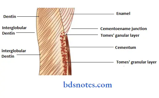
Question 13. Tome’s granular layer
Answer:
- A granular appearing area has been seen in the transmitted light-granular layer ;
- It is present just below the surface of the dentin where Features:
- It increases from CEJ to apex of the tooth D Remains unmineralized
- Consists of high concentrations of calcium and phosphorous
- Have a special arrangement of collagen and noncollagenous matrix protein at the interface between dentin and cementum
Cause of development:
- Branching and looping
Question 14. Denticles
Answer:
- Calcifications within pulp are called pulp stones or denticles Dystrophic calcification is most commonly seen in pulp. (AIIMS -99)
Types:
- True pulp stones
- They have dentinal tubules and odontoblastic processes
- False pulp stones
- They do not have dentinal tubules
- Common in coronal pulp
- Free pulp stones
- Entirely surrounded by pulp tissue
- Attached pulp stones
- They are partly fused with dentin
- Embedded pulp stones
- They are entirely surrounded by dentin
Question 15. Dentinal tubules.
Answer:
- Dentinal tubules extend through the entire thickness of the dentin from the dentin enamel junction to the pulp.
- They follow an S-shaped path from the outer surface of the dentin to the perimeter of the pulp.
- The tubules are longer and thicker than dentin because they curve through dentin.
- They end perpendicular to the DEJ and DCJ.
Size:
- 2.5 pm in diameter near the pulp.
- 1.2 pm in diameter in the midportion of the dentin.
900 nm near DEJ.
Number:
- There are more tubules per unit area in the crown than in the root.
Branches:
- Terminal branches – near the terminals.
- Lateral branches/ canaliculi – throughout the dentin.
Question 16. Types of dentin.
Answer:
1. Primary dentin:
- Dentin which is formed before root completion is known as primary dentin.
- There are two types of primary dentin.
- Mantle dentin:
- It is the first formed dentin in the crown.
- Circumpulpal dentin:
- It is the primary dentin that outlines the pulp chamber.
2. Secondary dentin:
- It is a narrow band of dentin bordering the pulp
- It is the dentin that is formed after root formation.
3. Tertiary dentin:
It is that dentin is produced by odontoblasts in response to pulpal irritation caused by chemical, thermal, or microbial stimuli.
Question 17. Secondary dentin.
Answer:
- It is a narrow band of dentin bordering the pulp.
- It represents continued, slower deposition of dentin by odontoblasts.
- There is greater deposition on the roof and floor of the chamber than on other parts.
- It contains fewer tubules which sclerose more readily.
- This reduces the overall permeability of the dentin.
- Due to the regular arrangement of dentinal tubules, it is also called regular secondary dentin.
Question 18. Mantle dentin.
Answer:
- It is first formed dentin in the crown before a tooth erupts into the oral cavity.
- It is the outermost part of primary dentin.
- It is 20 pins thick.
- It provides a cushioning effect to the tooth.
- It contains larger diameter Von Korff’s fibers.
- It is less mineralized.
- It involves globular mineralization.
Question 19. Neonatal line.
Answer:
- It is a wide contour line.
- It separates the dentin formed before birth and that formed after birth.
- It is found in those teeth mineralizing at birth.
- It reflects the disturbance in mineralization created by the physiologic trauma of birth.
- The dentin matrix formed prior to birth is usually of better quality than that formed after birth.
Question 20. Interglobular dentin.
Answer:
- It is unmineralized or hypomineralised dentin, and It is prevalent in Vit.
- deficiency and with high exposure to fluoride, It is mostly seen in circumpolar dentin,
- It occurs due to a defect of mineralization and not of the matrix, Thus, dentinal tubules run uninterrupted.
- It is frequently seen in the cervical and middle third followed by a cuspal and coronal third in the crown,
- It is also seen highest in the cervical third followed by the middle third in the root.

Question 21. Tomes granular layer.
Answer:
- A granular appearing area seen in transmitted light in ground sections of root dentin is called tomes’ granular layer.
- It is present just below the surface of the dentin where the root is covered by cementum.
- This zone increases from the cementoenamel junction to the apex of the tooth.
- They represent sections made through the looped terminal portions of dentinal tubules.
- This layer has a special arrangement of collagen and non-collagenous matrix proteins at the interface between dentin and cementum.
- Interglobular—— dentinft

Question 22. Dead tracts.
Answer:
- Dentinal tubules are emptied by complete retraction of the odontoblast process from the tubule or through the death of the odontoblast.
- The dentinal tubules then become sealed off so that in the ground section air-filled tubules appear by transmitted light as black dead tracts.
- They are most often in coronal dentin.
- Frequently bound by bands of sclerotic dentin.
- These areas demonstrate decreased sensitivity.
- They are the initial step in the formation of sclerotic dentin.
Question 23. Odontoblasts.
Answer:
- Odontoblasts are columnar cells forming dentin.
- They have oval nuclei present in the basal part of the cell.
- Adjacent to the nucleus, rough-surfaced endoplasmic reticulum and the Golgi apparatus are present.
- Between adjacent cells, there is the presence of tight and desmo.
- soma junctions.
- They are approximately 5-7 pm in diameter and 25-40 pm in length.
- They are present adjacent to the present.
- Their number corresponds to the number of dentinal tubules.
- The odontoblast morphology and its organelles vary with the functional activity of the cell.
- The odontoblast’s cell bodies remain external to dentin, but their processes exist within tubules in the dentin.
- The odontoblasts secrete both collagen and other components of the extracellular matrix.
Question 24. Incremental/structural lines in dentin.
Answer:
Question 25. Theories of dentinal sensitivity.
Answer:
1. Direct nerve innervations theory:
- It states that the dentin contains nerve endings that respond when it is stimulated.
- This theory is rejected because the dentinal nerves do not extend beyond the inner dentin.
2. Transduction theory:
- It states that the odontoblasts serve as receptors and are coupled to nerves in the pulp.
- It was rejected because of the absence of a synaptic relationship between tire odontoblast and pulpal nerves,
- There are no neurotransmitter vesicles in the odontoblastic process to facilitate tire synapse.
3. Fluid or hydrodynamic theory:
- It is the only accepted one.
- It states that the tubular nature of dentin permits fluid movement to occur within the tubule when a stimulus is applied.
- This fluid movement stimulates the pain mechanism in the tubules by mechanical disturbances of the nerves closely associated with the odontoblast and its process.
Question 26. Tonofilaments.
Answer:
- Tonofilaments are the filamentous strands present in the epithelial cells.
- They are fibrous proteins synthesized by the ribosomes.
- They appear as long filaments with a diameter of approx 8 nm.
- They belong to a class of intercellular filaments.
- They consist of intracellular proteins known as cytokeratins.
- When they become aggregated to form bundles of filaments, they are known as tonofibrils.
- These tonofilaments are not contractile but are important in the maintenance of cell shape and contact between adjacent cells and the extracellular matrix.
Question 27. Reparative dentin.
Answer:
- It is a type of tertiary dentin formed by a newly differentiated odontoblast-like cell.
- It is produced by odontoblasts in response to pulpal irritation caused by chemical, thermal, or microbial stimuli.
- It is formed only in localized areas in reaction to trauma such as caries or restorative procedures.
- It has few and more twisted dentinal tubules.
- The formation of the reparative dentin seals off the zone of injury.
- Due to the irregular nature of the dentinal tubules, it is also known as irregular secondary dentin.
Question 28. Curvatures in dentin.
Answer:
1. Primary curvatures:
- They are sigmoidal curves.
- They resemble a gentle S-shape curve.
- They are formed as the dentinal tubules follow a gentle curve in the crown which is less curved in the root.
2. Secondary curvatures:
- They are minute, regular and sinusoidal in shape.
- They are present over the entire length of the tubules.
Question 29. Osteodentin.
Answer:
- The cells forming tertiary dentin become included in the dentin and are referred as osteopontin.
- It is produced in response to rapidly progressing caries.
- It has irregular nature of the dentinal tubules.
- BMP is involved in its production.
- It develops due to a deficiency of vitamin A.
Question 30. Difference between the composition of enamel and dentin.
Answer:
Question 31. Physical properties of dentin
Answer:
- Color
- Light yellow in color
- Hardness
- Harder than bone softer than enamel
- Harder in its central part than its periphery
- Elasticity
- Dentin is viscoelastic
- Permeability
- Dentin permeability depends upon the potency of dentinal tubules
- Tubular occlusion, smear layer formation, and lack of tubular communication between primary and secondary dentin will result in reduced permeability
Question 32. Contour lines of Owen
Answer:
- These lines result from a coincidence of the secondary curvature between neighboring dentinal tubules
They are caused by accentuated deficiencies in mineralization - These are easily recognized in ground sections
- These lines are accentuated because of disturbances in the matrix and mineralization process
Question 33. Incremental lines of von Ebner
Answer:
- Incremental lines of von Ebner appear as fine lines or striations in dentin
- They run at a right angle to dentinal tubules
- They represent a normal rhythmic, linear pattern of dentin deposition in an inward and rootward direction
- Its course indicates the growth pattern of dentin.
- The distance between lines varies from 4-8 mm in the crown too much less in the root
- The daily increment decreases after the tooth reaches functional occlusion

Leave a Reply