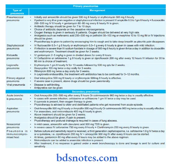
Treatment Of Community Acquired Pneumonia or Management of Pneumonia.

Question 1. Outline the investigation and management of community acquired pneumonia.
Or
Discuss briefly the management of community acquired pneumonia.
Answer:
- Community-acquired pneumonia is also known as primary pneumonia.
- Community-acquired pneumonia is defined as an infection that begins outside the hospital or is diagnosed within 48 hours after admission to the hospital in a patient who has not resided in a long-term facility for 14 days or more before the onset of symptoms.
Investigations pneumonia.
Blood tests
- WBC count is marginally raised or is normal in patients with pneumonia caused by atypical microorganisms.
- Absolute neutrophil count is more than 15 × 109/L and this is indicative of bacterial etiology.
- A very high or low WBC count can be seen in severe pneumonia.
- Creactive protein is typically elevated.
Read And Learn More: General Medicine Question And Answers
Radiological Examination
- A confident diagnosis should always need a chest X-ray.
- In pneumococcal pneumonia, a homogeneous opacity is seen localized to the affected lobe or segment which appears within 12 to 18 hours from the onset of illness.
Microbiological Investigations
- Most of the cases of community acquired pneumonia are successfully managed, if they are not severe.
So the need for microbiological investigation is based on clinical circumstances. - In severe cases of community-acquired pneumonia, a full range of microbial tests should be carried out.
- If the patient does not respond to the initial therapy, microbiological tests provide proper modification of therapy.
Assessment Of Gas Exchange
- Pulse oximetry is a noninvasive method that measures arterial oxygen saturation and monitors the response to oxygen therapy.
- An arterial blood gas is sampled in patients with low arterial oxygen saturation or having features of severe pneumonia to assess whether the patient has evidence of ventilator failure or acidosis
Diagnose and Manage a Case of Pneumococcal Pneumonia?
Answer. It is defined as the inflammation of the lung parenchyma.
Pneumococcal pneumonia is caused by pneumococcus.
Diagnosis of Pneumococcal Pneumonia
- Physical signs:
- At the time of early stage of illness, there are decreased respiratory movements.
- Impairment of percussion node
- Breadth sounds are diminished.
- A pleural rub is present on the affcted side.
- Three days after the disease, signs of consolidations are seen, i.e. highpitched bronchial breathing and increased vocal resonance.
- At the time of resolution, numerous coarse crackles/crepitations are heard indicating liquefaction of alveolar exudates.
- Blood test: It reveals marked neutrophil leucocytosis.
- Blood culture: It shows the presence of Streptococcus pneumonia.
- Examination of sputum: Gram staining of sputum may demonstrate pneumococci.
- Chest radiograph: In pneumococcal pneumonia, a homogeneous opacity is seen localized to the affected lobe or segment which appears within 12–18 hours from the onset of illness.
- Serological test: It can detect pneumococcal antigens in the serum.
- In some cases, fiber-optic, bronchoscopic aspiration, or transthoracic needle aspiration is required.
Treatment Of Community-Acquired Pneumonia or Management Pneumococcal pneumonia
- Initially, oral amoxicillin should be given 500 mg 8 hourly or erythromycin 500 mg 6 hourly.
- If the patient is very ill or gramnegative or staphylococcal infection is present IV ampicillin 0.5–1 g 6 hourly + flcoxacillin 250–500 mg IV 6 hourly + gentamycin 6080 mg every 8 hourly IV is given.
- Antibiotic therapy should be given for 7–10 days.
- The choice of antibiotics depends on the causative microorganisms.
- Oxygen therapy is given to seriously ill patients. Oxygen should be delivered at a very high rate.
- Analgesics such as mefenamic acid 250–500 mg pethidine 50–100 mg or morphine 10–15 mg IM or IV injections should be given.
- Physiotherapy is given to the patient by encouraging him to cough and to take deep breaths as pleuritic pain disappears
Lobar Pneumonia Symptoms
Mention the complications of lobar pneumonia.
Answer. Complications of Pneumonia
In lung: lobar pneumonia.
- Pleural effusion
- Pneumothorax
- Empyema
- Lung abscess
- Respiratory failure.
In cardiac involvement: lobar pneumonia.
- Pericarditis
- Endocarditis
- Myocarditis
- Cardiac failure.
GIT complications: lobar pneumonia. They are uncommon and may be in the form of:
- Acute dilatation of the stomach
- Paralytic ileus
- Peritonitis.
Another complication is lobar pneumonia.
- Pneumococcal meningitis
- Arthritis
- Parotitis
- Otitis media
- Nephritis
- Venous thrombosis.

Leave a Reply