Blood supply of cerebrum
Question 1. Write a short note on the blood supply of the brain.
Answer.
The proper blood supply of the brain is the most essential because consciousness is lost within 10 seconds of the cessation of the blood supply and if this state continues, irreversible brain damage starts at 4 minutes and is completed within 10 minutes.
Read And Learn More: Selective Anatomy Notes And Question And Answers
Question 2. Write a short note on the circle of Willis.
Answer.
Formation and location
The brain is supplied by two pairs of major arteries:
- A pair of vertebral arteries and
- A pair of internal carotid arteries.
The branches of these arteries anastomose in the region of interpeduncular fossa at the base of brain to form somewhat circular anastomosis called circle of Willis. The two vertebral arteries unite at the lower border of pons to form the basilar artery, which divides at the upper border of the pons into right and left posterior cerebral arteries.
The internal carotid artery of each side terminates in the region of anterior perforated substance by dividing into a smaller anterior cerebral artery and a larger middle cerebral artery. The posterior communicating artery (a branch of the internal carotid artery) on each side communicates with the posterior cerebral artery.
A single short anterior communicating artery connects the two anterior cerebral arteries.
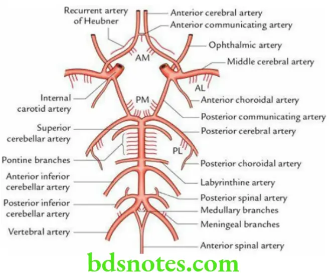
The circle of Willis (circulus arteriosus) is thus formed by the anterior communicating artery and the anterior cerebral arteries anteriorly, the termination of the internal carotid artery and the posterior communicating artery on each side, and bifurcation of basilar artery including posterior cerebral arteries posteriorly.
Function Provides various alternate routes for collateral circulation.
Applied anatomy The sites where two arteries unite to form circle of Willis may dilate to form berry shaped dilatations called berry aneurysms. The rupture of these aneurysms leads to subarachnoid haemorrhage at the base of brain in the region of interpeduncular fossa.
Question 3. Write a short note on basilar artery. Give its branches.
Answer.
Formation and termination
- It is formed by the two vertebral arteries at the lower border of the pons.
- It runs upwards and forwards in the midline groove on the ventral aspect of pons.
- On reaching the upper border of pons, it divides into right and left terminal branches – the posterior cerebral arteries.
Branches The branches of the basilar artery:
- Anterior inferior cerebellar arteries
- Labyrinthine arteries
- Pontine branches
- Superior cerebellar arteries
- Posterior cerebral arteries (terminal branches)
Question 4. Describe briefly arterial supply of the superolateral surface of the cerebral hemisphere.
Answer.
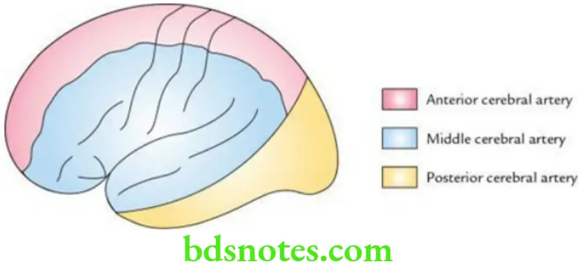
- The narrow strip of about an inch breadth along its superomedial border as far as the parieto-occipital sulcus is supplied by the anterior cerebral artery.
- The occipital lobe and narrow strip along the lower border of temporal lobe excluding the temporal pole (the inferior temporal gyrus) is supplied by the posterior cerebral artery.
- The rest of the superolateral surface is supplied by the middle cerebral artery.
The arterial supply of the superolateral surface of the cerebral hemisphere.
Question 5. Describe briefly arterial supply of the inferomedial surface of the cerebral hemisphere.
Answer.
- Medial surfaces of the frontal and parietal lobes are supplied by the anterior cerebral artery.
- Medial surfaces of the occipital and most of the temporal lobes (except temporal pole) are supplied by the posterior cerebral artery.
- The medial surface of the temporal pole is supplied by the middle cerebral artery.
- Medial part (one-third) of the orbital surface (of inferior surface) is supplied by the anterior cerebral artery. The lateral part (two-third) of the orbital surface as well as the temporal pole on inferior surface is supplied by the middle cerebral artery.
- Rest of the tentorial surface (of inferior surface) is supplied by the posterior cerebral artery.
Question 6. Write a short note on great cerebral vein of Galen.
Answer.
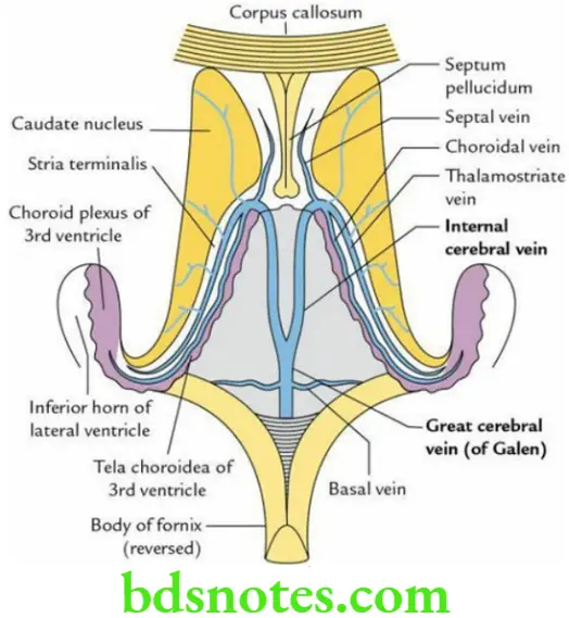
The great cerebral vein of Galen is formed by the union of two internal cerebral veins below the splenium of the corpus callosum. It joins the inferior sagittal sinus to form the straight sinus. The tributaries of the great cerebral vein are as follows:
- Internal cerebral veins
- Basal veins
Question 7. Enumerate the deep cerebral veins.
Answer.
The deep cerebral veins are:
- Thalamostriate veins
- Choroid veins
- Septal veins
The White Matter Of The Cerebrum
Question 1. Define white matter and discuss its various types.
Answer.
The white matter of cerebrum is made up of myelinated nerve fibres, which connect various parts of cerebral cortex on the same side, with opposite side and also with the other parts of the CNS.
Types of white fibres There are three types of white fibres in the cerebrum: association fibres, commissural fibres and projection fibres.
Association fibres (intrahemispheric): These fibres connect different areas of cerebral cortex of the same hemisphere. They are further classified into two types:
- Short association fibres, which connect the adjacent gyri and
- Long association fibres, which connect the distant gyri.
Commissural fibres: These fibres connect the cortical areas of one cerebral hemisphere with the corresponding cortical areas of the opposite hemisphere. The bundles of such fibres are called commissures. The important commissures in the brain are
- Corpus callosum,
- Anterior commissure and
- Posterior commissure.
Projection fibres: These fibres connect the cortical areas with the subcortical centres such as corpus striatum, thalamus, hypothalamus, brainstem and spinal cord. They include both motor and sensory fibres of long tracts. The most important bundles of projection fibres are
- Internal capsule and
- Fornix.
Question 2. Enumerate the various commissures of the brain.
Answer.
The important commissures of the brain:
- Corpus callosum
- Anterior commissure
- Posterior commissure
- Hippocampal commissure
- Habenular commissure
Question 3. Describe the corpus callosum under the following headings:
- Definition,
- Parts,
- The course of fibres,
- Functions and
- Applied anatomy.
Answer.
Corpus Callosum Definition The corpus callosum is the largest commissure of the brain. It is 10 cm long, which is nearly half of the anteroposterior length of the cerebrum. It consists of 300 million fibres. The corpus callosum connects all the parts of neocortex, except for the lower and anterior parts of temporal lobes, which are connected by the anterior commissure.
Corpus Callosum Parts In the sagittal section, the corpus callosum is divided into four parts, from before to backwards:
- Rostrum
- Genu
- Body/trunk
- Splenium
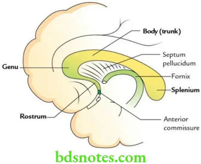
The corpus callosum begins at the anterior commissure in the upper part of the lamina terminalis, which passes upwards and forwards as the rostrum, then bends sharply upwards and backwards to form the genu and finally, it extends backwards as the body and ends posteriorly as a thick massive extremity called splenium.
- Its inferior surface is connected in the midline with the upper surface of the fornix by septum pellucidum, which lies between two lateral ventricles.
- Its superior surface is related to indusium griseum and medial and lateral longitudinal striae.
The course of fibres of the corpus callosum
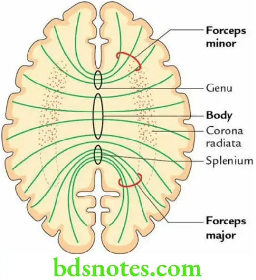
- The fibres of the genu curve forwards on each side towards the frontal cortex forming the forceps minor.
- The fibres of the body spread out laterally on each side to form the roof of the central part of the lateral ventricle. These fibres are intersected by the vertically running fibres of the corona radiata.
- The fibres of the splenium curve backwards on each side towards the occipital cortex forming the forceps major. Its fibres form the upper part of the medial wall of the posterior horn of the lateral ventricle.
- The fibres of the posterior part of the body together with some fibres from the splenium extend laterally to form the roof of the posterior horn of the lateral ventricle and then turn downwards to form the lateral wall of both the posterior and inferior horns.
Functions
- Interhemispheric transfer of learned/visual memory.
- Interhemispheric transfer of speech function.
- Coordination of activities of two cerebral hemispheres for proper bilateral coordination and responses.
Applied anatomy
- Patients with lesion of corpus callosum respond as if they have two separate brains – a condition called Split-brain effect/syndrome.
- A surgical section of corpus callosum has been attempted in the past to prevent spread of seizures.
Question 4. Write a short note on anterior commissure.
Answer.
The anterior commissure is a small, round bundle of commissural fibres that crosses the midline in the upper part of the lamina terminalis. It consists of two components:
- A large, posterior neocortical component, which interconnects the cortical areas of the lower and anterior parts of the temporal poles.
- A smaller, anterior paleocortical component, which interconnects the olfactory regions, such as olfactory bulbs and olfactory tubercles, of two sides.
Question 5. Describe the internal capsule under the following headings:
- Definition and location,
- Gross anatomy,
- Fibres,
- Arterial supply and
- Applied anatomy.
Answer.
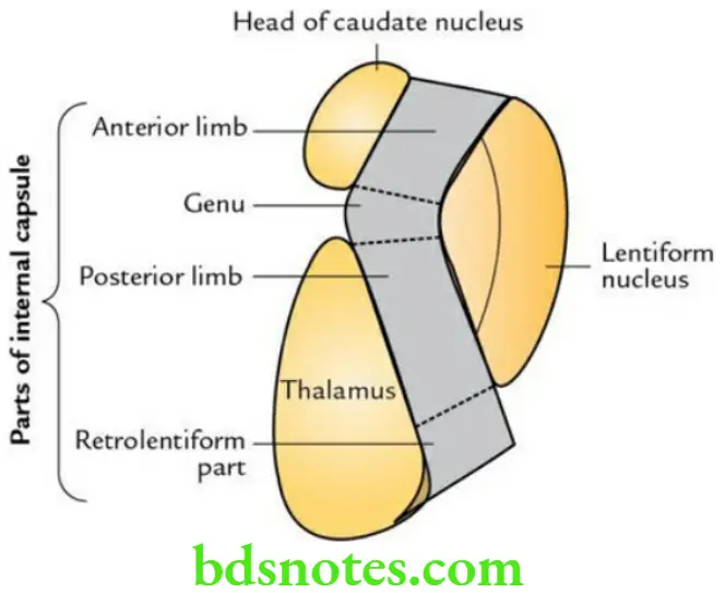
Internal Capsule Definition and Location The internal capsule is a compact bundle of projection fibres lying in the inferomedial part of the cerebral hemisphere. Hence, it lies in a narrow space between the lentiform nucleus laterally, and the caudate nucleus and thalamus medially.
The internal capsule connects the cerebral cortex to the brainstem and spinal cord.
It contains important fibres belonging to pyramidal tract and sensory fibres from the opposite half of the body.
Internal Capsule Gross anatomy
Shape: In horizontal section, it appears as a ‘V’-shaped mass of white fibres.
Internal Capsule Parts: It consists of five parts:
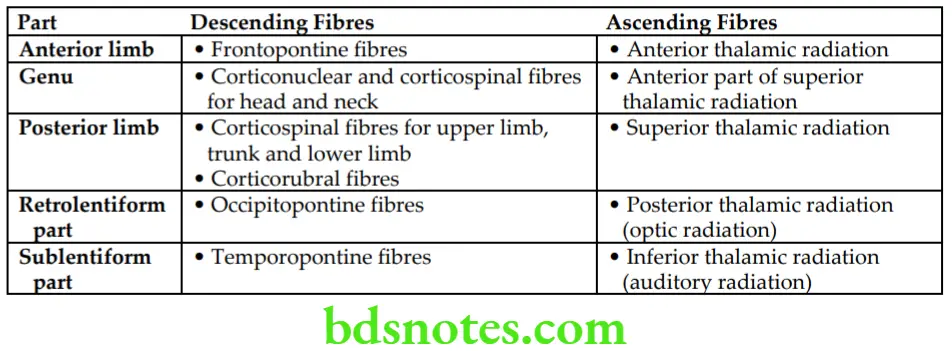
- Anterior limb: Lies between the lentiform nucleus and the caudate nucleus.
- Genu (bend of internal capsule): Lies in the angle between the caudate nucleus, thalamus and lentiform nucleus.
- Posterior limb: Lies between the lentiform nucleus and the thalamus.
- Retrolentiform part: Lies behind the lentiform nucleus.
- Sublentiform part: Lies deep to the lentiform nucleus.
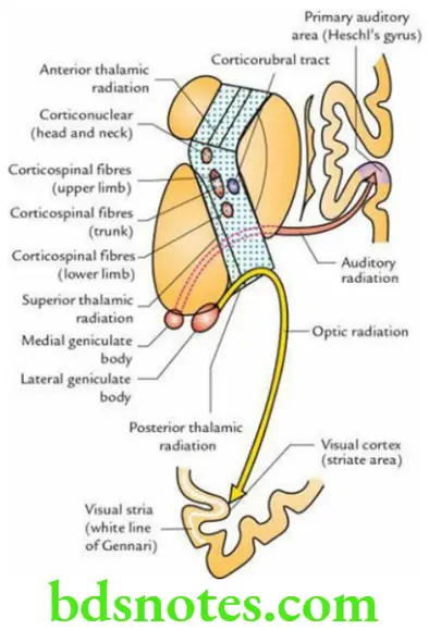
Internal Capsule Fibres The constituent fibres in the different parts of the internal capsule.
Constituent Fibres in the Different Parts of the Internal Capsule
Internal Capsule Arterial supply The arterial supply of different parts of the internal capsule is as follows:
- Anterior limb is supplied by the middle and anterior cerebral arteries.
- Genu is supplied by the middle cerebral artery and recurrent artery of Heubner.
- Posterior limb is supplied by Charcot’s artery of cerebral haemorrhage and anterior choroidal artery.
- The sublentiform part is supplied by the anterior choroidal artery.
- Retrolentiform part is supplied by the posterior cerebral artery.
Internal Capsule Applied anatomy
- A small lesion in the internal capsule results in widespread paralysis and sensory loss because huge number of motor and sensory fibres are packed densely in the internal capsule.
- The commonest lesion in the internal capsule occurs due to cerebral haemorrhage or cerebral thrombosis. It causes complete hemiplegia on the opposite side (i.e. contralateral hemiplegia).
- The most common cause of cerebral haemorrhage is the rupture of Charcot’s artery of cerebral haemorrhage and it usually involves posterior limb of internal capsule leading to contralateral hemiplegia.
Question 6. Write a short note on thalamic radiation.
Answer.
With few exceptions, all the afferent fibres to brain relay in the thalamus. From thalamus, the thalamocortical fibres radiate in different directions to reach the widespread area of the cerebral cortex (thalamic radiation). The important thalamocortical fibres (or thalamic radiations) are as follows:
- Sensory radiation from the thalamus to the sensory cortex in the postcentral gyrus.
- Auditory radiation from the medial geniculate body to the auditory cortex in the temporal lobe.
- Optic radiation from the lateral geniculate body to the visual cortex in the occipital lobe.

Leave a Reply