Cerebellum And Fourth Ventricle
Question 1:Define the cerebellum and list its functions.
Answer. The cerebellum (Latin, cerebellum = little brain) is the largest part of the hindbrain and the second-largest part of the brain as a whole. It weighs about 150 g and is located in the posterior cranial fossa underneath the tentorium cerebelli, behind the pons and medulla oblongata.
Read And Learn More: Selective Anatomy Notes And Question And Answers
Functions Of Cerebellum The functions of the cerebellum include:
- Maintenance of equilibrium
- Maintenance of muscle tone
- Maintenance of posture
Question 2: What are the parts of the cerebellum?
Answer.
Parts Of Cerebellum The cerebellum consists of three parts:
- Two large lateral hemispherical parts are called cerebellar hemispheres.
- A narrow median worm-like part which unites two cerebellar hemispheres called vermis.
Question 3:What are the anatomical lobes of the cerebellum?
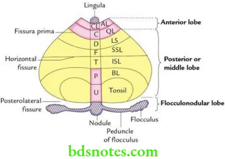
Answer. Anatomically, the cerebellum is divided into three lobes:
- Flocculonodular lobe: It is the smallest lobe that comprises two flocculi and their peduncles and the nodule. Together with the lingula of the vermis, it forms the vestibular part of the cerebellum (archicerebellum).
- Anterior lobe: It lies on the superior surface of the cerebellum anterior to the fissura prima excluding the lingula; together with the pyramid and uvula of vermis, it forms the spinal part of the cerebellum (paleocerebellum).
- Middle (posterior) lobe: It is the largest lobe and lies between the fissure prima on the superior surface and the posterolateral fissure on the inferior surface excluding the pyramid and uvula of the vermis. It forms the cerebral part of the cerebellum (neocerebellum).
Question 4: Enumerate the intracerebellar nuclei and discuss their connections.
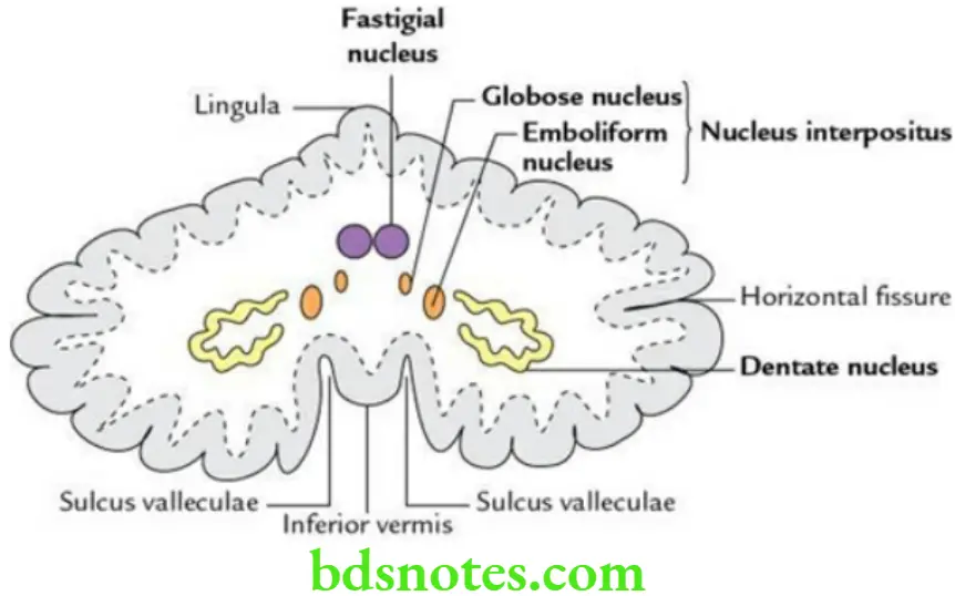
Answer. There are four intracerebellar nuclei on either side of the midline. From lateral to medial side, these are as follows:
- Dentate nucleus
- Nucleus emboliformis
- Nucleus fasting
- Nucleus globosus
Dentate nucleus: It is the largest and has the shape of a crumpled bag (crenated crescent) with its hilum facing anteromedially. It receives afferents from the neocerebellum and sends efferents through the superior cerebellar peduncle to the red nucleus of the opposite side, which in turn projects the spinal cord.
Nucleus emboliformis and nucleus globosus: The nucleus emboliformis is oval in shape, whereas the nucleus globosus is round in shape. They receive afferents from the paleocerebellum and send efferents through the superior cerebellar peduncle to the red nucleus of the opposite side. These two nuclei together represent the ‘nucleus interpositus’ of lower mammals, e.g. monkeys.
Nucleus fastigius: It lies near the midline in the region of vermis. It receives afferents from the archicerebellum and sends efferents through the inferior cerebellar peduncle to the vestibular nuclei and the reticular formation of the medulla oblongata.
Question 5: What are cerebellar peduncles? Enumerate their constituent fibres.
Answer. The cerebellar peduncles are large bundles of afferent and efferent fibres that connect the cerebellum with other parts of the brain. They are three in number:
- The superior Cerebellar Peduncle is the most medial and connects the cerebellum with the midbrain.
- The Middle Cerebellar Peduncle is the largest and connects the cerebellum with the pons.
- The inferior Cerebellar Peduncle connects the cerebellum with the medulla oblongata.
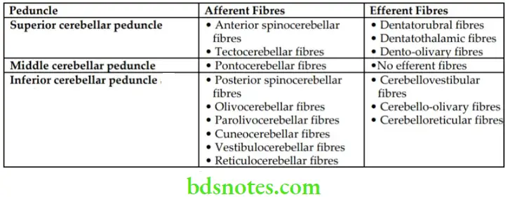
The constituent fibres of three cerebellar peduncles.
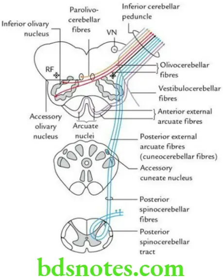
Main Constituent Fibres Of Cerebellar Peduncles
Question 6:Enumerate the important signs and symptoms seen in the cerebellar lesions.
Answer.
- Asthenia: Muscular hypotonia due to loss of muscle tone.
- Asynergia: Jerky movements due to incoordination of muscles.
- Ataxia: Staggering gait and inability to walk in a straight line due to incoordination of different muscle groups in the lower limb.
- Adiadokokinesis (disdiadokokinesis): Inability to perform alternate movements in rapid succession such as pronation and supination.
- Dysmetria: Loss of ability to measure the distance for reaching the intended goal.
- Dysarthria (or scanning speech): Loss of incoordination of muscles concerned with speech.
- Rebound phenomenon: Inability to check the action of agonist muscles by the corresponding antagonist muscles.
- Intention tremor: Due to dysmetria. The tremors are evident during purposeful movements and are diminished or absent at rest.
Question 7: Define the 4th ventricle and discuss its boundaries, communications and recesses.
Answer.
The 4th ventricle is a tent-like cavity of the hindbrain lined with ependyma. It lies behind the pons and medulla and in front of the cerebellum.
4th Ventricle Boundaries
Lateral (on each side):
- Superolaterally by the superior cerebellar peduncle
- Inferolaterally by the inferior cerebellar peduncle
Floor: It is formed by the posterior surfaces of the pons and the upper part of the medulla oblongata. It is rhomboidal in shape; hence, it is often called ‘rhomboid fossa’.
Roof: It is formed by
- Superior medullary velum, in the upper part
- Inferior medullary velum, in the lower part
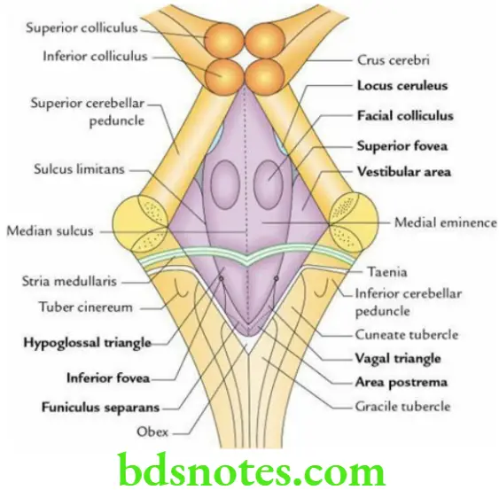
Communications Of 4th Ventricle
It communicates above with 3rd ventricle through the cerebral aqueduct (aqueduct of Sylvius) and below with the central canal of the spinal cord. Posterolaterally, it communicates with the subarachnoid space through three foramina in its roof (foramen of Magendie and foramina of Luschka).
Recesses Of 4th Ventricle They are five in number:
- Two lateral recesses
- Median dorsal recess
- Two lateral dorsal recesses
Question 8: Discuss the features of the rhomboid fossa in brief.
Answer.
The features of the rhomboid fossa are as follows:
- It is rhomboidal in shape and has four angles: rostral, caudal and two lateral.
- The floor is divided into two equal halves by a median sulcus. Each half of the floor is further divided into two parts (pontine and medullary) by fibres of stria medullaris, which run from the median sulcus to the lateral boundary.
Features Above The Medullary Striae (I.E. In The Pontine Part)
- Presence of facial colliculus, on either side of median sulcus. It is an oval elevation produced by the fibres of the motor nucleus of the facial nerve hooking around the abducent nucleus (internal genu of the facial nerve).
- A depression at the upper end of sulcus limitans is called the superior fovea.
- A bluish-grey area (locus coeruleus), above the superior fovea. The bluish colour is imparted by the underlying group of nerve cells containing melanin pigment.
Features Below The Stria Medullaris (I.E. In The Medullary Part)
- Inferior fovea, a triangular depression
- Hypoglossal triangle, medial to inferior fovea
- Vestibular triangle, lateral to inferior fovea
- Vagal triangle, inferior to the inferior fovea
- Funiculus separates, a fine translucent ridge crossing the vagal triangle
- Area postrema, a small area below the funiculus separates

Leave a Reply