Cephalometrics
Question 1. Define cephalometrics. What are advantages and disadvantages of cephalometrics?
Answer. Cephalometrics is a radiography. “Cephalometric radiography is a standardized method of producing skull radiograph which are useful in making measurement of cranium and the orofacial complex.”
Cephalometrics is a radiographic technique for abstracting the human head into a geometric shape.
Cephalometrics Advantages
- A broad anatomic area can be visualized.
- Patients radiation exposure is low.
- It helps in orthodontic diagnosis by enabling the study of skeletal, dental and soft tissue structures of craniofacial region.
- It helps in treatment planning.
- It helps in evaluation of the treatment results by quantifying the changes brought about by treatment.
- It helps in predicting the growth related changes.
- By cephalometries the skeletal and dental abnormalities can be classified.
Read And Learn More: Orthodontics Question And Answers
Cephalometrics Disadvantages
- Give 2 dimensional view of three dimensional object.
- Reliability is not always accurate there can be errors in determining the landmarks.
- There are different methods of analyzing a cephalogram and so no standardization of analyze.
- Exposure of patient to ionizing radiation which is harmful to patient. So it can be used only when it is diagnostically and therapeutically needed.
- There is absence of the anatomical references which remains constant with the time. This is applicable when the clinician wishes to compare cephalograms which are taken at different time points.
- Measurement procedures in cephalometrics are not well standardized.
- Anatomical structures which lie at different planes within the head undergo projective displacement.
- Positioning of patient is done with ear rods in external acoustic meatus, in this way operator assumes that both the meatuses are symmetrical but this is need not to be so.
- Patient is asked to bite in maximum intercuspation at the time of taking cephalogram. There can be mandibular shift from the centric relation.
- Composition of lines and angles used in cephalometrics provide limited information about patient’s dento skeletal patterns.
- On basis of cephalometrics, orthodontic diagnosis cannot be made solely.
Question 2. Write short note on cephalogram.
Answer. Cephalogram is radiograph showing relation of cranial, facial and dental structure taken in a standardized way.
Types of Cephalogram
- Lateral cephalogram.
- Frontal cephalogram or anteroposterior cephalogram.
- Oblique cephalogram.
Lateral Cephalogram
This provides a lateral view of the skull. It is taken with the head in a standardized reproducible position at a specifid distance from source of the X-ray.
Frontal Cephalogram
This provides an anteroposterior view of the skull.
Uses of Cephalograms
- Its main use is in the formulation of orthodontic diagnosis by providing the dental, skeletal and soft tissue relationship in craniofacial region.
- It is very useful in predicting the growth of an individual.
- It also formulates the treatment planning, and the response of treatment can be assessed before its beginning.
- Cephalograms helps to assess the functional analysis.
- Growth of skeleton of face is assessed by the cephalograms.
- Cephalogram is useful in estimating the facial type.
- Cephalogram is useful in growth prediction.
- Functional analysis can be carried out by cephalogram.
- Cephalogram predicts the growth related changes and changes which are associated with surgical treatment.
- Cephalograms are very useful in planning out the procedures in surgical orthodontics such as skeletal repositioning.
- Cephalogram is a valuable aid in research work which involves craniofacial region.
- Cephalograms are tangible records which are permanent unlike other diagnostic measurements.
- Cephalograms are non-destructive and non-invasive and gives proper information at relatively low cost.
- Cephalograms are very easy to store, transport and reproduce.
Cephalogram Technique
- Cephalostat consists of two ear rods which prevent the movement of head in horizontal plane.
- Vertical stability is brought by an orbital pointer which contact lower border of left orbit.
- Upper part of the face is supported by forehead clamp positioned above the region of nasal bridge.
- Distance between the X-ray source and mid sagittal plane of patient is fixed at 5 feet.
Question 3. Write short note on Tweed diagnostic triangle.
Or
Write short note on Tweed’s triangle.
Or
Write short answer on Tweed’s diagnostic triangle.
Or
Write briefly on Tweed’s analysis.
Or
Write short note on Tweed’s analysis.
Answer.
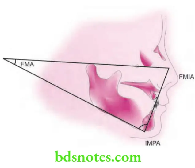
Tweed’s Analysis
It is a type of cephalometric analysis.
- It is given by Charles Tweed.
- Cephalometric points used are:
- Porion: Superiomost point of orbitale.
- Orbital: Inferiomost point of lower border of orbit.
- Tweed’s analysis makes use of three planes.
- The planes used are:
- Frankfort horizontal plane (FH plane): Join porion and orbitale.
- Mandibular plane: A tangent is drawn to lower border of mandible.
- Long axis of lower incisor: A line is drawn along the long axis of incisors.
Objectives of Analysis
- Determination of the position of lower incisor.
- Evaluation of prognosis.
Angle’s Formed
- Angle’s formed by these three planes are:
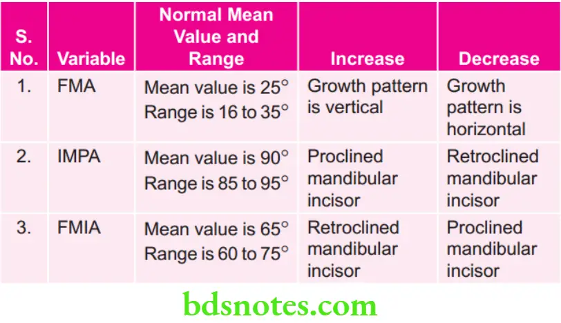
- Frankfort mandibular plane angle (FMA)—It is formed by intersection of the FH plane with the mandibular plane.
- The mean value is 25° in well balanced cases.
- Incisors mandibular plane angle (IMPA)—It is the angle formed by the intersection of the long axis of the lower incisor with the mandibular plane.
- It indicates the inclination of the lower incisor.
- The mean value is 90° in well balanced cases.
- Frankfort mandibular incisor angle (FMIA)—It is the angle formed by the intersection of the long axis of the lower incisor with the FH plane.
- The mean value is 65° in well balanced cases.
- Frankfort mandibular plane angle (FMA)—It is formed by intersection of the FH plane with the mandibular plane.
Interpretation
- FMA>28°: This indicates that patient has high angle and growth of mandible is clockwise.
- FMA<23°: This indicates that patient has low angle and growth of mandible is counterclockwise.
- IMPA>110°: Lower incisors are proclined.
- IMPA<85°: Lower incisors are retroclined.
Summary of Tweed’s Triangle
Clinical Importance
- It is used in orthodontics for classification, diagnosis, treatment planning as well as prognosis.
- Tweed had also mentioned about extraction of teeth for correcting the alveolodental prognathism and also the positioning of mandibular incisors upright over the basal bone.
- If FMA is 20° to 30° the prognosis of orthodontic treatment along with extraction is excellent to good.
- If FMA is 30° to 35° the prognosis of orthodontic treatment along with extraction is excellent lies in the range of good to fair.
- If FMA is 35° to 40° the prognosis of orthodontic treatment along with extraction is unfavorable.
Question 4. List the essential diagnostic aids. Describe in detail the Steiner’s analysis.
Or
Describe Steiner’s cephalometric analysis in detail.
Or
Write short note on Steiner’s analysis.
Answer.
Essential Diagnostic Aids
They are clinical aids that are considered very important for all cases.
- They are simple and do not require expensive equipments.
- Following are the essential diagnostic aids:
- Case history
- Clinical examination
- Study models
- Certain radiographs (lOPA, Bitewing, Panoramic)
- Facial photographs.
Steiner Analysis
It is a cephalometric analysis.
Cecil C Steiner in the year 1930 developed this analysis.
The Steiner analysis is divided into three parts:
- Skeletal analysis.
- Dental analysis.
- Soft tissue analysis.
Skeletal Analysis
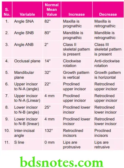
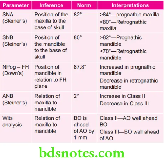
- SNA angle: The angle formed by the intersection of SN plane and a line joining nasion and point A. Indicates position of maxilla in relation to cranium.The mean value is 82°.Value increased in prognathic maxilla (Class 2). Value decreased in retrognathic maxilla (Class 3).
- SNB angle: Angle between SN plane and line joining nasion to point B.
- This angle indicates the position of mandible to cranial base.
- Average value is 80°.
- Value increase in prognathic mandible (Class 3).
- Value decreased in retrognathic mandible (Class 2).
- ANB angle: Angle between line joining point A to nasion and a line joining point B to nasion.
- It indicates position of maxilla and mandible to each other.
- Average value is 2 degree.
- Increased value indicates class II skeletal malocclusion.
- Decreased value indicates class III skeletal malocclusion.
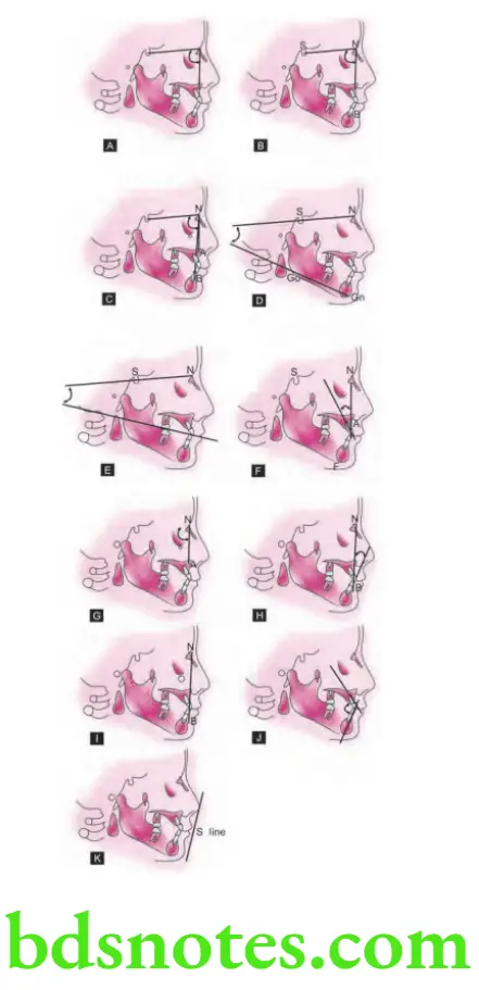
- Mandibular plane angle: It is the angle between SN plane and mandibular plane.
- Average value is 30°.
- Indicates growth pattrn of individual.
- Lower angle indicates horizontal growing pattrn of individual.
- Increased angle indicates vertical growing pattrn of individual.
- Occlusal plane angle: The angle between the SN plane and occlusal plane.
- Occlusal plane is line passing through the overlapping cusps of the fist premolar and fist molar.
- It has a mean value of 14.5°.
- It indicates the relation of occlusal plane to the cranium and face.
- Also indicates growth pattrn in individual.
Dental Analysis
Maxillary Incisor Position
- Upper incisor is related to N-A line for determination of its position.
- Upper incisor to N-A (Angle):It is the angle formed by the intersection of long axis of the upper central incisor and the line joining nasion to point A. Mean value is 22°.
- An increase in angle indicates proclined upper incisors (class II malocclusion) and decrease in the angle is suggestive of retroclination.
- Upper incisors to N-A (Linear): It is a linear measurement between the labial surface of upper central incisor and the line joining nasion to point A. Mean value is 4 mm.
- It increases with proclined upper central incisor and decreases with retroclination.
Mandibular Incisor Position
- Lower incisor is related to N-A line for determination of its position.
- Lower Incisor to N-B (angle): The angle between the N-B plane and the long axis of the lower incisor. Mean value is 25°.
- Increased value indicates proclination of lower incisor while the decreased value indicates upright or retroclined lower incisor.
- Lower Incisor to NB (linear): Linear distance between the labial surface of lower central incisor and the line joining nasion to point B. Mean value is 4 mm.
- Increased value indicate proclined lower central incisor while decreased value indicates of retroclination.
Interincisal Angle
- Angle formed between the long axis of the upper and lower central incisor.
- Mean value is 132°
- When upper and lower incisors are proclined angle is acute.
- When upper and lower incisors are retroclined angle is obtuse.
- These angulations help in detecting incisors with defective angulations.
Soft Tissue Analysis
- In well balanced faces, lip lie along the S line.
- Lips which are located anterior to S line are protrusive. Orthodontic treatment should be carried in order to reduce protrusion.
Summary of Steiner’s Analysis
Question 5. Describe different horizontal and vertical line and planes used in cephalometrics.
Or
Write short note in cephalometric planes.
Answer.
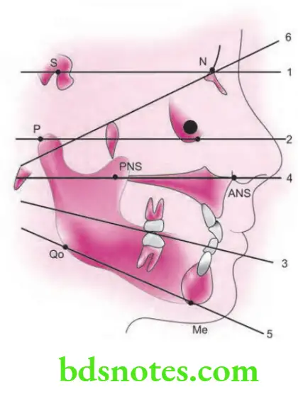
- Cephalometrics uses certain lines or planes
- The planes/lines are obtained by connecting two landmarks.
- The planes can be horizontal/vertical.
Horizontal Planes
- SN plane: It is the cranial line between the sella and the nasion.
- It represent anterior cranial base.
- FH plane (Frankfort Horizontal plane): This plane connects the orbitale to porion (superior point of external auditory meatus).
- Occlusal plane: It is a denture plane bisecting the posterior occlusion of the permanent molar and premolars and extends anteriorly.
- Palatal plane: It is a line between ANS to PNS.
- Mandibular plane: Mandibular plane joining the gonion to gnathion.
- Basion-nasion plane: It represent cranial base, connect nasion to basion.
Vertical Planes
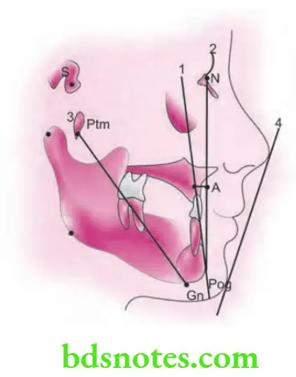
- A- Pog line: A line from point A to pogonion
- Facial plane: A plane from nasion to the pogonion.
- Facial axis: A line from pterygomaxillary fisure to gnathion
- Esthetic plane (E-plane): A line between the most anterior point of the soft tissue nose and soft tissue chin.
Question 6. Write short note on Y-axis.
Answer. The angle is obtained by joining the FH plane with sellagnathion line.
Y-axis is of the two types, i.e. Down’s Y-axis and Rakosi’s Y-axis.
Down’s Y-axis
- Down’s Y-axis is also known as growth axis.
- Y-axis forms an integral part of assessment of skeletal pattern in Down’s analysis.
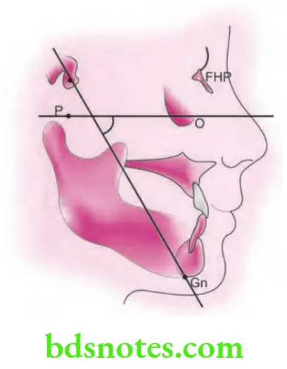
- Y-axis indicates growth pattern of individual.
- Cephalometric plane used in Y-axis is Frankfort horizontal plane.
- Cephalometric landmarks used are sella, gnathion, porion and orbitale.
- Mean value is 59.4° and it ranges from 53° to 66°.
Interpretation
- Increment in Y-axis is indicative of vertical growth pattern.
- Decrease in Y-axis is indicative of horizontal growth pattern.
- It indicates the position of chin in anteroposterior and vertical plane.
- Y-axis indicates the downward, rearward and forward position of chin.
Rakosi’s Y-axis
- It is the measured angle between nasion, sella and gnathion.
- It determines the position of mandible in relation to cranial base.
- Its mean value is 66°
- If angle is greater than 66° it implies retrognathic mandible with vertical growth pattern.
- If angle is lesser than 66° it implies prognathic mandible with horizontal growth pattern.
Question 7. Write briefly on use ofcephalometrics in orthodontics.
Answer. Cephalometry is extensively used for diagnosis and treatment planning purposes.
Role of cephalometry for diagnosis and treatment purpose is divided into four parts:
- Anteroposterior relationships
- Vertical relationships
- Dentoalveolar relationships
- Soft tissue relationships
Analysis of Anteroposterior Relationships
Analysis of vertical Relationship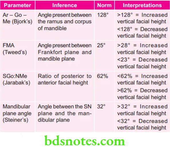

Assessment of Dentoalveolar Relationships
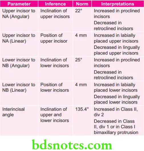
Soft Tissue Relationships
Steiner’s soft tissue analysis
- Steiner’s S line is drawn from middle of S shaped curve formed by lower border of nose to soft tissue contour of chin.
- Lips is well balanced face’s lie along this line
- Lips which are located anterior to this line are protrusive. Orthodontic treatment can be done to reduce protrusion.
Question 8. Write short note on skeletal parameters of Down’s analysis.
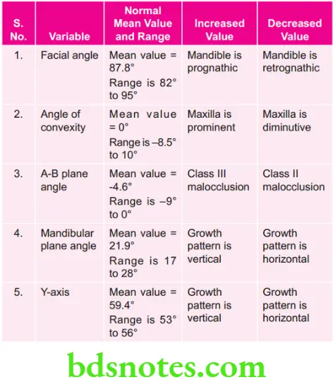
Answer. Following are the skeletal parameters of Down’s analysis:
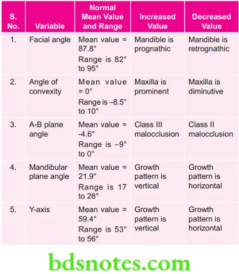
Facial Angle
- It is the inferior inside angle which is formed by intersection of facial line, i.e. nasion-pogonion to the FrankfortHorizontal plane.
- Mean reading for this angle is 87.8°.
- It measures the degree of retrusion or protrusion of lower jaw.
- It denotes the degree of recession or protrusion of mandible in relation to upper face.
- Prominent chin increases this angle while smaller than average angular reading suggests retrusive or retropositioned chin.
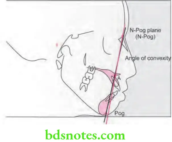
Angle of Convexity
- It is formed by intersection of line N-point A to point A- Pogonion.
- It measures the placement of maxillary basal arch at anterior limit, i.e. Point A relative to total facial profile—Nasion-pogonion.
- This angle should be read in plus or minus degrees starting from zero.
- Mean value is 0°.
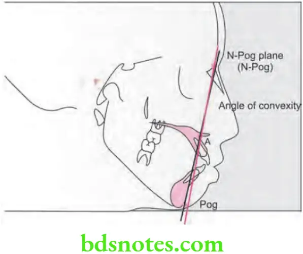
- Positive angle suggests prominence of maxillary denture base relative to mandible while the negative angle is associated with prognathic profie or Class III profile.
AB Plane Angle
- Points A and B are joined by a line which when extended forms an angle with the line Nasion-Pogonion, this is called the A-B plane angle.
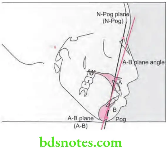
- The A-B plane is a measure of the relation of the anterior limit of the apical bases to each jaw relative to the facial line. Generally point B is positioned behind point A thus this angle is usually negative in value, except in Class 3 malocclusions or Class I occlusions with prominence of the mandible.
- A large negative value suggests a Class II facial pattrn, which can be due to the retropositioned chin or mandible or underdeveloped chin point or a prominent maxilla, i.e. point B located behind point A.
- Mean value of AB-plane angle is 4.6°.
Mandibular Plane Angle
- The Down’s mandibular plane is “tangent to the gonial angle and the lowest point of the symphysis”.
- The mandibular plane angle is established by relating the mandibular plane to the Frankfort horizontal (FH) plane.
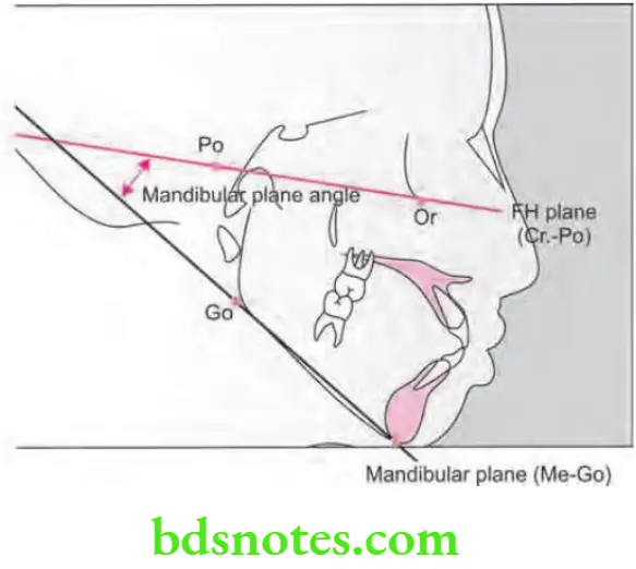
- High mandibular plane angles occur in both retrusive and protrusive faces and are suggestive of unfavorable hyperdivergent facial patterns or ’long face cases’.
- Mean value of mandibular plane angle is 21.9°.
Y(Growth) Axis
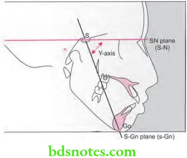
- Growth axis is measured as an acute angle formed by the intersection of a line from sella turcica to Gnathion with the Frankfort horizontal plane.
- This angle is larger in Class II facial pattrns than in those with Class 3 tendencies.
- It indicates the degree of downward, rear ward or forward position of the chin in relation to the upper face.
- A decrease of the Y-axis in serial radiographs may be interpreted as a greater horizontal than vertical growth of the face or a deepening of the bite in orthodontic cases. An increase in the Y-axis is suggestive of vertical growth exceeding horizontal growth of the mandible or an opening of the bite during orthodontic treatment.
- The Y-axis reading also increases with the extrusion of the molars.
- Mean value of Y-axis is 59.4°.
Summary of Skeletal Parameters of Down’s Analysis
Question 9. Write short note on SNA angle.
Answer. The angle formed by the intersection of SN plane and a line joining nasion and point A.
- This angle indicates position of maxilla in relation to cranium.
- The mean value is 82°
- SNA angle assesses the degree of prognathism of maxilla.
- Planes of SNA angle are SN plane which is horizontal plane and NA plane which is vertical plane.
- Value of SNA angle is increased in prognathic maxilla (Class 2).
- Value of SNA angle is decreased in retrognathic maxilla (Class 3).
Question 10. Write briefly on uses of cephalogram.
Answer. Following are the uses of cephalogram:
- Its main use is in the formulation of orthodontic diagnosis by providing the dental, skeletal and soft tissue relationship in craniofacial region.
- It is very useful in predicting the growth of an individual.
- It also formulates the treatment planning, and the response of treatment can be assessed before its beginning.
- Cephalograms helps to assess the functional analysis.
- Growth of skeleton of face is assessed by the cephalograms.
- Cephalogram is useful in estimating the facial type.
- Cephalogram is useful in growth prediction.
- Functional analysis can be carried out by cephalogram.
- Cephalogram predict the growth related changes and changes which are associated with surgical treatment.
- Cephalograms are very useful in planning out the procedures in surgical orthodontics such as skeletal repositioning.
- Cephalogram is a valuable aid in research work which involves craniofacial region.
- Cephalograms are tangible records which are permanent unlike other diagnostic measurements.
- Cephalograms are non-destructive and non-invasive and gives proper information at relatively low cost.
- Cephalograms are very easy to store, transport and reproduce.
Question 11. Write short note on Wits appraisal.
Answer. It is also known as Wits analysis.
- Wits appraisal was introduced by Alexander Jacobson for overcoming the disadvantages of Steiner’s analysis.
- Since the discovery of Wits appraisal was done in University of Witwatersrand. It is known as Wits appraisal.
- In Wits appraisal main aim was to study relationship of maxilla and mandible anteroposteriorly.
Landmarks
- Occlusal plane: Formed by bisecting overlapping cusp of first premolars and fist molars.
- AO point: It is gained when a perpendicular is drawn from point A to occlusal plane.
- BO point: It is gained when a perpendicular is drawn from point B to occlusal plane.
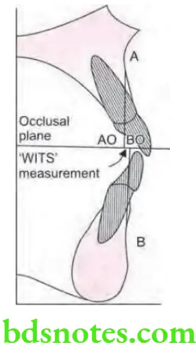
Interpretation
- In normal occlusion:
-
- In men: BO is ahead of AO by 1 mm.
- In women: Both AO and BO coincide in women.
- In Class 2 malocclusion: AO is ahead of BO.
- In Class 3 malocclusion: BO is ahead of AO.
Drawbacks
- In the appraisal readings obtained are solely dependent over occlusal plane inclination.
- When the clockwise rotation of occlusal plane is carried out AO lies behind BO but when counterclockwise rotation of occlusal plane is carried out BO lies behind AO.
- So for making out the anteroposterior relationship the Wits appraisal is combined with other method.
Question 12. Write short note on Down’s analysis.
Answer. For skeletal parametres, refer to Ans 8 of the same chapter.
Dental Analysis
Cant of Occlusal Plane
- It is the measurement of slope of occlusal plane to Frankfort horizontal plane.
- Occlusal plane is formed by drawing a line bisecting overlapping cusps of first molars and overbite of incisors.
- Cant of occlusal plane is gained by measuring angle between occlusal plane and Frankfort horizontal plane.
- Mean value is 9.3°
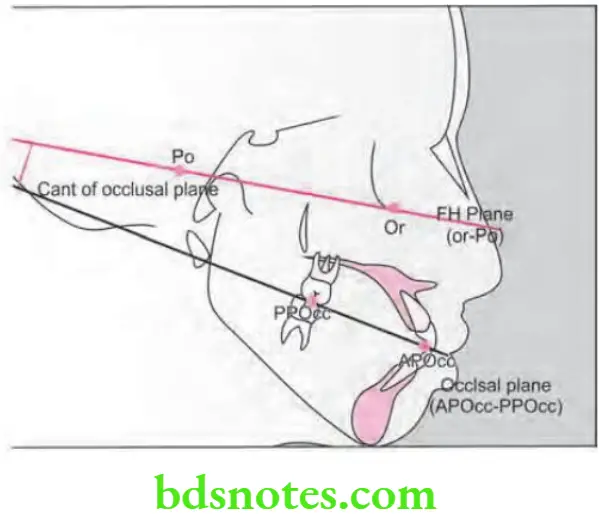
- Decrease in the value occurs in the cases of long ramus while it is increased in cases of short ramus.
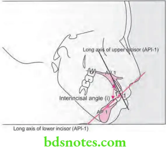
Interincisal Angle
- It is formed by long axis of maxillary and mandibular incisor.
- Mean value is 135.4°.
- Interincisal angle is less in people whose incisors are tipped forward.
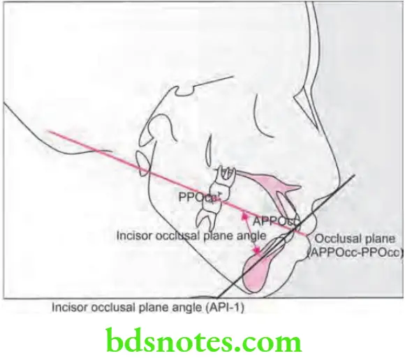
Incisor Occlusal Plane Angle
- It is formed where the occlusal plane and long axis of mandibular incisor meets at an intersection.
- Its mean value is +14.5°.
- At the point of intersection inside the angle can be positive or negative based on deviation from right angle.
- Its value is increased in proclined mandibular incisors and decreased in retroclined mandibular incisors.
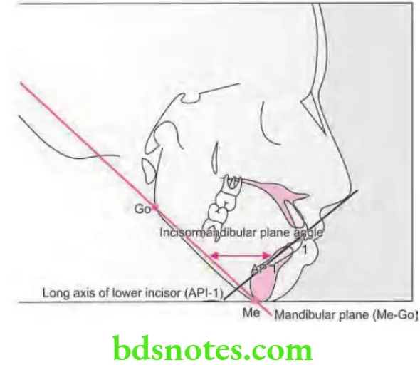
Incisor Mandibular Plane Angle
- It is formed where the mandibular plane and long axis of mandibular incisor meets at an intersection.
- Its mean value is +1.40.
- At the point of intersection inside, the angle can be positive or negative based on deviation from right angle.
- Its value is increased in proclined mandibular incisors and decreased in retroclined mandibular incisors.
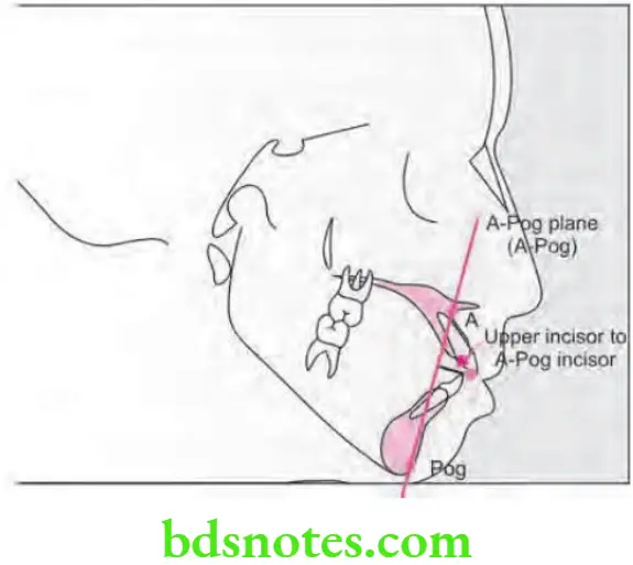
Protrusion of Maxillary Incisors
- It is gained by drawing a line from point A to pogonion and calculating the distance between incisal edge of maxillary central incisor to A-pog line.
- Mean value is + 2.7 mm.
- When incisal edge lies ahead of A-Pog line the value is positive.
- Increase in value is suggestive of protruded maxillary incisors while decrease in value is suggestive on retruded maxillary incisors.
Summary of Dental Parameters of Down’s Analysis
Question 13. What is cephalometrics. Write about Steiner’s analysis in detail.
Or
What is cephalometrics. Describe Steiner’s analysis in detail.
Answer. Cephalo means head and the metric means measurement.
Cephalometries is a radiography. “Cephalometric radiography is a standardized method of producing skull radiographs which are useful in making measurement of cranium and the orofacial complex.”
Question 14. What is Tweed’s analysis. Discuss the clinical considerations of Tweed’s diagnostic setup in planning orthodontic treatment.
Answer. Tweed’s analysis was based on inclination of mandibular incisors to the basal bone and its association with the vertical relation of mandible to cranium.
The mandibular incisors should be placed upright over basal bone for stability and esthetics.
Question 15. Write short note on gonial angle.
Answer. Gonial angle is a skeletal landmark for determination of cephalometric analysis for functional appliances.
- Gonial angle is divided into two parts i.e. upper gonial angle and lower gonial angle when a line is drawn from nasion to gonion.
- Patient having acute or small gonial angle have horizontal growth pattern.
- Acute gonial angle suggests that condition is favorable for anterior positioning of mandible with the functional appliances.
- Patient having obtuse or large gonial angles functional appliance treatment is contraindicated.
- Its mean value is 130° ± 7 degrees.
Question 16. Write short note on 2D and 3D cephalometry.
Answer. Orthodontics is visualized and treatment is planned by using 2D cephalograms, the current paradigm shift in orthodontics and the keen interest in esthetics has resulted in an interest in three-dimensional visualization and diagnosis to plan treatment for what is a three-dimensional structure.
- Except for a few structures of interest which lie in the midsagittl plane it is diffilt to make accurate measurements using 2D cephalograms. Conventional facial photos too lose depth information by projecting images of structures at diffrent heights upon a single plane. Also the one true 3D representation of oral tissues, the dental cast must be integrated into facial images.
- In the late 1970s computerized axial tomography initially referred to as CAT and later CT become available. CT measures X-ray attnuation coeffients as they spatially vary across a section of the anatomy. They are ideal for the visualization of hard osseous structures as these structures attnuate X-rays more than the surrounding sof tissues.
- Upon introduction it was heralded that the CT and the MRI would replace conventional radiography. However, their use in conventional orthodontic treatment has been limited due to the following reasons:
-
- The dose of ionizing radiation has been high.
- Economic costs are prohibitive.
- Slices of relatively thick tissue detail in vertically oriented teeth is quite poor.
- Distortions are produced if CT scans are done with orthodontic appliances in place.
- All 3D imaging systems try to capture the Z-axis and this they achieve by counting the number of slices into which the images are divided.
- Calibration is particularly important when one tries to integrate 3D images and the 2D cephalogram.
- The problem with 3D imaging of face is that the face inherently contains litte detail and it is diffilt to obtain a set of discrete points which can then be used to superimpose and to construct a useful map.
- 3D cephalometric planning is ideal for orthognathic cases where the outcome can be visualized or simulated and executed on patient approval, it has yet to be implemented for routine use in orthodontics particularly when we have limitation like radiation dose.
- The CBCT is ideal tool for 3D diagnosis and treatment planning. We have to train ourselves towards concepts of 3D.
- But still these days practitioners may be talking about 3D and still tend to put 2D landmarks on skull as for years they are trained in 2D landmarks for teeth and jaws.
- 3D cephalometric analysis may be routinely used for 3D assessment of all orthodontic cases in near future and become essential part of treatment planning.
Question 17. Classify diagnostic aids in orthodontics. Describe in detail the uses of cephalometrics in orthodontic diagnosis. Add a note on Tweed’s triangle.
Answer.
Classification of Diagnostic Aids in Orthodontics
Essential Diagnostic Aids
- Case selection
- Clinical examination
- Study models
- Certain radiographs:
- Periapical radiograph
- Bitewing
- Panoramic.
- Facial photographs
Nonessential or Supplemental Diagnostic Aids
- Specialized radiographs:
- Cephalometric radiographs
- Occlusal intraoral films
- Selected lateral jaw view
- Cone shift technique.
- Electromyographic examination of muscle activity.
- Hand and wrist radiographs to assess bone age.
- Endocrine tests.
- Estimation of basal metabolic rate.
- Diagnostic set-up.
- Occlusograms
- Sensitivity (Vitality) test
- Biopsy.

Leave a Reply