Cardiovascular System
Question 1. Define cardiovascular system and mention its functions.
Answer.
The cardiovascular system consists of heart and blood vessels.
Cardiovascular System Functions
- Distribution of nutrients and oxygen to all body cells
- Distribution of hormones to target tissues
- Thermoregulation
- Removal of metabolic waste products and CO2 from all body cells
Classify blood vessels.
The blood vessels are classified into the following types:
- Arteries
- Arterioles
- Capillaries and sinusoids
- Venules
- Veins
Question 2. What are the differences between the arteries and veins?
Answer.
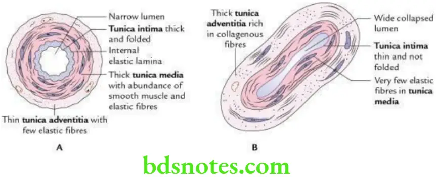
The differences between the arteries and veins.
Differences Between Arteries and Veins


Question 3. What are types of circulation? Describe each type in brief.
Answer.
There are two types of circulation:
- Pulmonary
- Systemic
Pulmonary circulation
In pulmonary circulation, the blood is pumped by the heart into the lungs through pulmonary trunk for oxygenation and then the oxygenated blood is returned to the heart via pulmonary veins.
Systemic circulation
In systemic circulation, the oxygenated blood is pumped by the heart to the entire body through arteries and then the deoxygenated blood is returned to the heart via veins.
Question 4. Discuss the portal circulation in brief.
Answer.
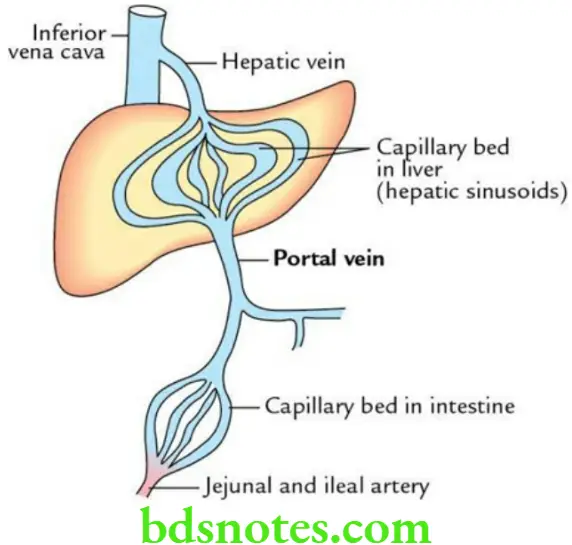
The portal circulation begins and ends with capillaries, i.e. blood passes through two sets of capillaries before it is drained into a systemic vein. The vessels connecting two sets of capillaries are called portal vessels.
The portal circulation is seen in the liver, hypophysis cerebri, kidney and suprarenal gland, where it is termed hepatic portal circulation, hypothalamo-hypophyseal portal circulation, renal portal circulation and suprarenal portal circulation, respectively.
Read And Learn More: Selective Anatomy Notes And Question And Answers
Question 5. Write a short note on hepatic portal circulation.
Answer.
In hepatic portal circulation, the venous blood from capillary bed of GIT (also from spleen and pancreas) passes to the hepatic sinusoids (capillary bed) through the portal vein before it is drained into a systemic vein – the inferior vena cava.

Question 6. What are the various types of arteries?
Answer.
The arteries are of three types:
- Elastic/conducting arteries, e.g. aorta, pulmonary trunk, common carotids, brachiocephalic trunk and subclavian arteries.
- Muscular/distributing arteries (most common), e.g. branches of carotids, axillary, brachial, femoral and popliteal arteries.
- Arterioles (smallest type of muscular arteries with diameter <0.1 mm).
Question 7.Write a short note on arteriole.
Answer.
- These are the smallest muscular arteries.
- The cross-sectional diameter of arteriole is about 100 microns.
- The arteriole at the arterial end possesses all the three coats in its wall.
- The arterioles progressively divide and finally terminate by forming terminal and meta-arterioles.
- Terminal arterioles are covered by continuous coat of smooth muscle cells.
- In meta-arterioles, the smooth muscle coat is replaced by discontinuous contractile cells called pericytes (Rouget cells).
- The passage of blood into capillaries is regulated by precapillary sphincters.
Functions of Arterioles
- Regulate the flow of blood into capillaries by constriction or dilatation of their muscular walls.
- Regulate systolic blood pressure by offering peripheral resistance.
Arterioles Applied anatomy
The persistence in the tone of arterial wall leads to hypertension.
Question 8. Write a note on arterial supply of an artery.
Answer.
- The tunica adventitia and outer two-third of tunica media is supplied by vasa vasorum.
- The tunica intima and inner one-third of tunica media is supplied by luminal blood.
Question 9. Mention the nerve supply of an artery.
Answer.
The arteries are supplied by sympathetic nerves via nervi vasorum. These are vasoconstrictor.
Question 10. What is the difference between capillaries and sinusoids.
Answer.
The differences between capillaries and sinusoids.
Differences Between Capillaries and Sinusoids

Question 11. Enumerate the organs where fenestrated capillaries are present.
Answer.
- Pancreas
- Intestine
- Renal glomeruli
- Endocrine glands
- Skin
Question 12. Enumerate the structures where continuous capillaries are present.
Answer.
- Lungs
- Muscles (skeletal and smooth)
- Fascia
- Brain
- Testis
Question 13. Name the organs where sinusoids are present.
Answer.
- Liver
- Spleen
- Adrenal medulla
- Carotid body
- Bone marrow
Question 14. Describe the arterial anastomosis under the following headings: (a) definition, (b) types and (c) functions.
Answer.
The arterial anastomosis is the communication between the branches of two or more arteries supplying the same organ or region.
The arteries do not always end in capillaries; they unite with one another through their branches forming anastomosis.
Arterial Anastomosis Types
There are two types of anastomoses:
Actual anastomosis: In this, the two arteries connect to each other end-to-end, e.g. anastomosis between the labial branches of the facial arteries in the lips; and right and left gastric arteries along the lesser curvature of the stomach. When the anastomotic channel is cut, the blood spurts with equal force from the both cut ends of the anastomotic vessel (i.e. blood spurts from both directions).
Potential anastomosis: In this, the anastomosis occurs between terminal arterioles of arteries, if given sufficient time. The anastomotic channels formed by the arterioles can dilate and may provide sufficient blood to area supplied (i.e. efficient collateral circulation), e.g. coronary arteries, cortical arteries of cerebral hemispheres and anastomosis around joints of the limbs. When the anastomotic channel is cut, the blood spurts from only the one cut end of the anastomotic vessel.
Arterial Anastomosis Functions
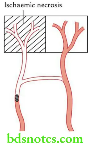
- Provides alternate route (collateral circulation) for blood to reach the tissue/organ when one of the vessels is blocked/compressed.
- Equalizes blood pressure over territories which they supply.
Question 15. Describe the end arteries in brief and mention their clinical significance.
Answer.
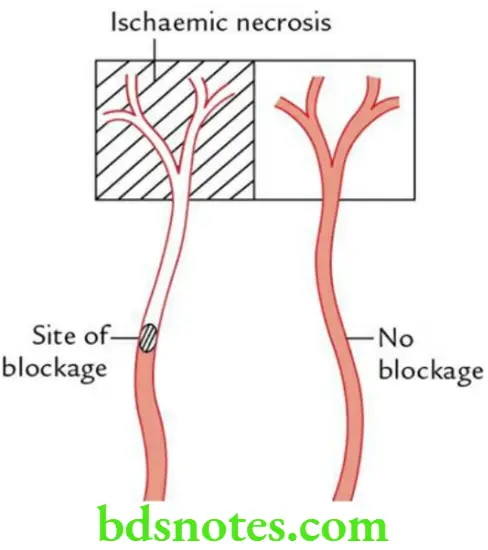
These are the arteries that do not anastomose with neighbouring arteries, e.g. central artery of retina, arteries of spleen, liver, kidneys, metaphyses of long bones, central branches of the cerebral arteries.
Arteries Clinical significance
If an end artery is occluded/blocked, the tissue in area supplied by this vessel is deprived of blood and suffers from ischaemia and may undergo ischaemic cell necrosis called infarction.

Question 16. Write a short note on functional end arteries.
Answer.
These are the arteries which supply the same organ and also have anastomosis between them but if one of them is blocked suddenly, the other is unable to supply the sufficient blood to the organ and maintain its viability, e.g. coronary arteries of the heart.

Question 17. Briefly describe arteriovenous shunts.
Answer.
The arteriovenous shunts are communicating channels between the arteries and veins, and when they open, the blood bypasses the capillaries, e.g. in skin of nose, lips, external ear, mucous membrane of alimentary canal, thyroid gland, palmar skin.
Functions of Arteriovenous Shunts
- Regulation of the regional blood flow
- Regulation of blood pressure
- Regulation of the temperature
Question 18. Enumerate the veins devoid of valves.
Answer.
- Superior vena cava (SVC)
- Inferior vena cava (IVC)
- Hepatic veins
- Portal vein
- Renal veins
- Uterine veins
- Gonadal veins
- Facial veins
- Emissary veins
Question 19. Enumerate the veins devoid of muscular tissue in their walls.
Answer.
- Dural venous sinuses
- Veins of pia mater
- Retinal veins
- Veins of placenta (maternal part)
- Veins of spongy substance of bones
Question 20. Enumerate the factors that facilitate venous return.
Answer.
- Negative intrathoracic pressure (for SVC and IVC)
- Muscular contractions
- Arterial pulsations
- Gravity (for veins of head and neck)

Leave a Reply