Cerebrum And Functional Areas
Question 1: Define the cerebrum and discuss the surfaces of the cerebral hemisphere.
Answer. The cerebrum is the largest part of the brain. It is divided into two equal halves by a median longitudinal cerebral fissure. Each half is called the cerebral hemisphere. The cerebrum is highly evolved in human beings.
Surfaces Of Cerebral Hemisphere Each cerebral hemisphere has three surfaces.
Read And Learn More: Selective Anatomy Notes And Question And Answers
Superolateral Surface: It is convex in conformity with the shape of the skull cap. It is the most convex and most extensive of the three surfaces.
Medial Surface: It is flat and vertical. It is separated from its fellow on the opposite side by the falx cerebri lying in the median longitudinal fissure, but below the falx cerebri the two hemispheres are joined together by a large bundle of white fibres – the commissure called the corpus callosum. In a separated cerebral hemisphere, the corpus callosum is seen as a C-shaped mass of white fibres.
Inferior Surface: It is uneven to adopt the floors of the anterior and middle cranial fossae. It is divided by a deep horizontal fissure (the stem of the lateral sulcus) into an anterior orbital part (related to the floor of the anterior cranial fossa) and a posterior tentorial part (related to the floor of the middle cranial fossa and to the upper surface of the tentorium cerebelli).
Cerebellum Function
Question 2: Name the sulci which help in demarcating the superolateral surface of the cerebral hemisphere into four lobes. Discuss the central sulcus in detail.
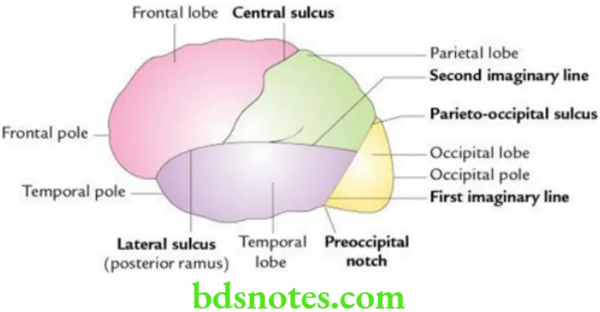
Answer. The sulci help in demarcating the superolateral surface of the cerebral hemisphere into four lobes:
- Central sulcus
- Lateral sulcus (posterior ramus)
- Parieto occipital sulcus (terminal portion)
Central Sulcus (of rolando)
Features Of Central Sulcus
- Presents on the superolateral surface of the cerebral hemisphere
- Begins at the superomedial border of the cerebral hemisphere about 1 cm behind its midpoint
- Runs vertically downwards and slightly forward and ends just above the lateral sulcus
Significance Of Central Sulcus
- Gyri lying in front and behind it are called precentral gyrus and postcentral gyrus, respectively.
- The precentral gyrus is motor in function and most of the fibres of the pyramidal tract arise in this gyrus to supply the opposite half of the body (i.e. contralateral half of the body).
- The postcentral gyrus is sensory in function and receives sensory impulses from the opposite half of the body (i.e. contralateral half of the body).
- The frontal branch of the middle meningeal artery ascends parallel and in front of the central sulcus, just deep into the pterion. The bone is thin here, and fracture at this site causes haemorrhage from the artery and presses upon the precentral gyrus leading to pressure symptoms like hemiplegia.
Cerebellum Function
Question 3: Discuss the demarcation of lobes of the cerebral hemisphere on its superolateral surface.
Answer. The superolateral surface of the cerebral hemisphere is demarcated into four lobes as follows:
- Frontal lobe: It lies anterior to the central sulcus and above the lateral sulcus. It is so named because it is related to the frontal bone of the skull.
- Parietal lobe: It lies posterior to the central sulcus, above the lateral sulcus, and in front of an imaginary vertical line connecting the parieto-occipital sulcus with the occipital notch. It is so named because it is related to the parietal bone of the skull.
- Temporal lobe: It lies below the lateral sulcus and in front of an imaginary vertical line extending from the parieto-occipital sulcus to the occipital notch. It is so-called because it is related to the temporal bone of the skull.
- Occipital lobe: It lies behind the imaginary vertical line connecting the parietal occipital sulcus with the occipital notch. It is so named because it is related to the occipital bone of the skull.

Question 4: Write a short note on the insula.
Answer. The insula is a submerged portion of the cerebral cortex on the floor of the lateral sulcus. It is hidden from surface view by overgrown adjacent areas of frontal, parietal and temporal lobes called opercula/lids.
- It is triangular in shape and surrounded by a circular sulcus.
- It is divided by the central sulcus into the anterior region having small gyri (gyri brevia) and a posterior region bearing long gyri (gyri longa).
- It is believed to be involved in consciousness and linked to emotion.
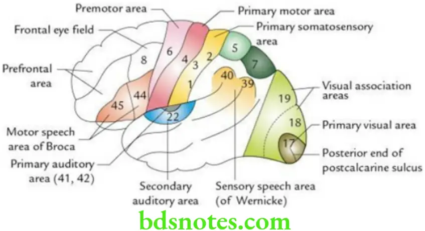
Question 5: Draw a labelled diagram to show the functional areas on the superolateral surface of the cerebral hemisphere.
Answer. Functional areas on the superolateral surface of the cerebral hemisphere.

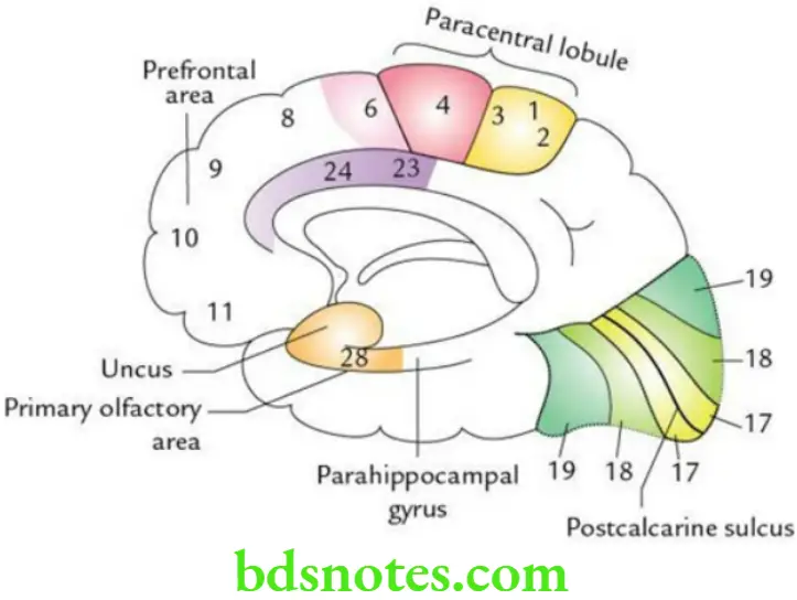
Question 6:Draw a labelled diagram to show the functional areas on the anteromedial surface of the cerebral hemisphere.
Answer:
Cerebellum Function Functional areas on the anteromedial surface of the cerebral hemisphere.

Question 7: Write a short note on the Primary Motor Area.
Answer.
Primary Motor Area Location The primary motor area (Brodmann area 4) is located:
- Precentral gyrus
- Anterior wall of central sulcus
- Anterior part of paracentral lobule
Primary Motor Area Representation of Body
- The body is represented upside down (inverted homunculus) in the primary motor area.
- The sequence of representation of body parts from above to downward is leg, thigh, trunk, upper limb, face, larynx, lips, jaws, tongue and pharynx.
- The area of the cortex representing a part of the body is not proportional to the size of that part but to the skill of movements performed by that part. Thus, movements of the hands, lips, tongue and larynx are represented by relatively large areas of the cortex.
Primary Motor Area Functions
- It controls the voluntary movements of the opposite side of the body.
- It also controls the acts of micturition and defecation.
Primary Motor Area Applied Anatomy A lesion in this area gives rise to upper motor neuron (UMN) type of paralysis on the opposite side of the body.
Question 8: Define the paracentral lobule and mention its functions.
Answer. The paracentral lobule is the area on the medial surface of the cerebral hemisphere around the central sulcus. It is bounded above by the superomedial border of the cerebral hemisphere, below by the cingulate sulcus, posteriorly by the upturned posterior end of the cingulate sulcus and anteriorly by the upturned ramus of the cingulate sulcus.
Paracentral Lobule Functions
- It acts as a cortical centre for micturition and defecation.
- It is responsible for movements of the contralateral foot.
Question 9. Write a short note on the Motor Speech Area.
Answer:
Motor Speech Area Location The motor speech area (Brodmann areas 45 and 44) is located in the pars triangularis (area 45) and pars posterior (area 44) in the inferior frontal gyrus of the frontal lobe in the dominant hemisphere.
Motor Speech Area Function The motor speech area is essential for the production of expressive speech.
Motor Speech Area Applied Anatomy If the motor speech area is damaged, the individual will suffer from motor aphasia. In this condition, there is the inability to articulate properly, though there is no paralysis of the muscles of lips, tongue, palate and vocal cords.
The speech of a person becomes nonfluent, dysarthric, telegraphic and incomprehensive.
Question 10. Write a short note on the Primary Sensory Area.
Answer.
Primary Sensory Area Location The primary sensory area (Brodmann areas 3, 1 and 2) is located in the postcentral gyrus and posterior wall of the central sulcus.
Primary Sensory Area Representation of Body The body is represented upside down in the primary sensory area similar to that in the primary motor area.
Primary Sensory Area Functions The primary sensory area is concerned with the perception of exteroceptive (pain, light touch and temperature) and proprioceptive (muscle and joint sense) sensations from the opposite half of the body.
Primary Sensory Area Applied Anatomy The lesions of the primary sensory area lead to a loss of appreciation of exteroceptive and proprioceptive sensations in the opposite half of the body.
Question 11. Write a short note on the sensory speech area (Wernicke’s area).
Answer. Location Of Sensory Speech Area
The sensory speech area (Brodmann areas 22, 39 and 40) is located in:
- Posterior part of the superior temporal gyrus (Brodmann area 22) of the dominant cerebral hemisphere.
- Parts of the inferior parietal lobule, including the supramarginal and angular gyri correspond to Brodmann areas 40 and 39, respectively.
Functions Of Sensory Speech Area
- Understanding written and spoken languages i.e. is concerned with the understanding and interpretation of language through visual and auditory input.
- Essential for constant availability of learned word patterns.
- Essential for the process of learning such as reading, writing and computing.
Applied Anatomy of Sensory Speech Area If the sensory speech area is damaged, the affected individual will suffer from receptive or sensory aphasia. In this condition, the affected individual cannot understand spoken words though his hearing is normal; consequently, he/she is unaware of the meaning of the words he/she uses.
As a result, he/she uses incorrect words or even nonexistent words. To others, his/her speech sounds like an incomprehensive foreign language.
Other defects seen in sensory aphasia are as follows:
- Alexia: Disability in reading.
- Agraphia: Disability in writing.
- Acalculia: Disability in computing.
- Anomia: Inability in recognition of names of objects.
Question 12. Write a short note on the visual cortex.
Answer.
Visual Cortex. The visual cortex is present over the occipital lobe.
- It is located along the lips and floor of the posterior part of the calcarine sulcus (also called post calcarine sulcus).
- Anteriorly, it extends up to the parieto-occipital sulcus and posteriorly it extends on the outer surface of the occipital pole and is limited by the lunate sulcus.
- It includes the cuneus and lingual gyrus.
Question 13. Write a short note on visual areas.
Answer. Location Of Visual Areas
- The primary visual area (Brodmann area 17) is located in the walls and floor of post calcarine sulcus.
- The secondary visual areas (Brodmann areas 18/peristriate area and 19/parastriate area) surround the primary visual area and occupy most of the remaining visual cortex.
Functions Of Visual Areas
- The primary visual area is concerned with the reception and perception of isolated visual impressions such as colour, size and form.
- The secondary visual areas relate the isolated visual impressions received by the primary visual area with past experience, thus enabling the individual to recognize and interpret what he/she is seeing.
Applied Anatomy Of Visual Areas
- Lesions of the primary visual area result in loss of vision in the opposite visual field (crossed homonymous hemianopia).
- Lesions of the secondary visual areas result in loss of ability to recognize the objects (visual agnosia).
Question 14. Describe cerebral dominance in brief.
Answer. The term cerebral dominance refers to the cerebral hemisphere, which is concerned with the perception and production of language/speech. In 90% of people, the left cerebral hemisphere subserves these functions; hence, it is termed the dominant hemisphere.
There is an important relationship between cerebral dominance and handedness. If the left cerebral hemisphere is dominant, the individual will be right-handed and if the right cerebral hemisphere is dominant, the individual will be left-handed.
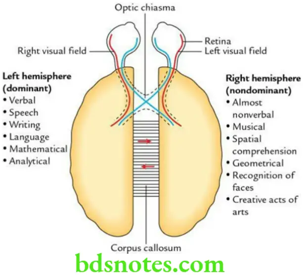
Question 15. Enumerate the main functions of the left and right cerebral hemispheres.
Answer. The main functions of the left and right cerebral hemispheres.


Leave a Reply