Overview Of Brain
Question 1. Define the brain and enumerate its major parts.
Answer. The brain is that part of the CNS, which lies within the cranial cavity (hence it is also called cranial cargo). The brain weighs about 1400 g in males and 1200 g in females. The brain is the highest centre for various sensory and motor functions of the body. It is also the seat of intelligence, cognizance, memory, emotions, etc.
Read And Learn More: Selective Anatomy Notes And Question And Answers
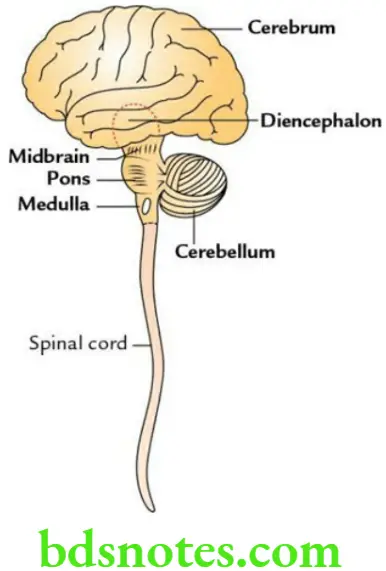
Major Parts Of The Brain The brain consists of six major parts:
- The cerebrum consists of two cerebral hemispheres.
- The diencephalon consists of the thalamus, hypothalamus, metathalamus, subthalamus and epithalamus
- Midbrain
- Pons
- Medulla oblongata
- Cerebellum
Brainstem
Question 2. What is the brainstem? Discuss its functions.
Answer. The brainstem is the stem-like lower part of the brain consisting of three parts – midbrain, pons and medulla oblongata.
Functions Of Brainstem
- Provides passages to various ascending and descending tracts.
- Contains vital (autonomic) centres – cardiac centre, vasomotor centre and respiratory centre.
- Contains reticular formation responsible for consciousness and sleep.
- Provides attachments to the last 10 cranial nerves and contains their nuclei.
Medulla Oblongata
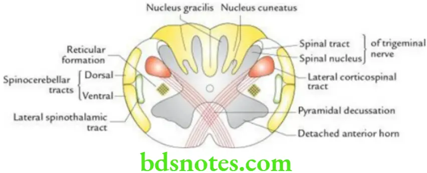
Question 3. Enumerate the main structures seen in the transverse section of the medulla oblongata at the level of pyramidal decussation.
Answer. The main structures seen in the transverse section of the medulla oblongata at the level of pyramidal decussation are as follows:
- Decussation of pyramidal tracts
- Appearance of nucleus gracilis and nucleus cuneatus
- Formation of the nucleus of the spinal tract of the trigeminal nerve
- Appearance of reticular formation
- Detachment of anterior horns due to decussation of pyramidal tracts
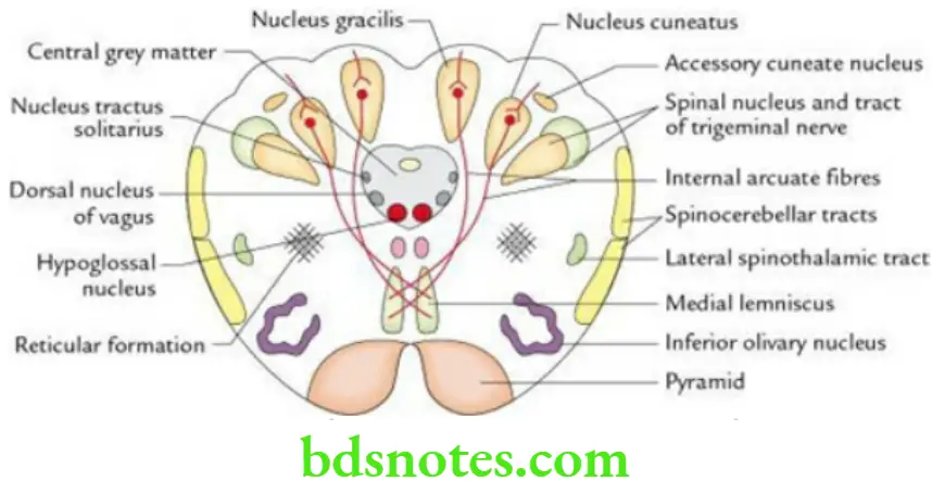
Question 4. Enumerate the main structures seen in the transverse section of the medulla oblongata at the level of sensory decussation.
Answer. The main structures seen in the transverse section of the medulla oblongata at the level of sensory decussation are as follows:
- Sensory decussation (decussation of arcuate fibres)
- Formation of medial lemnisci
- Formation of nucleus gracilis and nucleus cuneatus
- Formation of the nucleus of the spinal tract of the trigeminal nerve
- The appearance of accessory cuneate nucleus
- Formation of central grey matter and nuclei within it (e.g. hypoglossal nucleus, dorsal nucleus of vagus and nucleus tractus solitarius)
- Appearance of inferior olivary nucleus
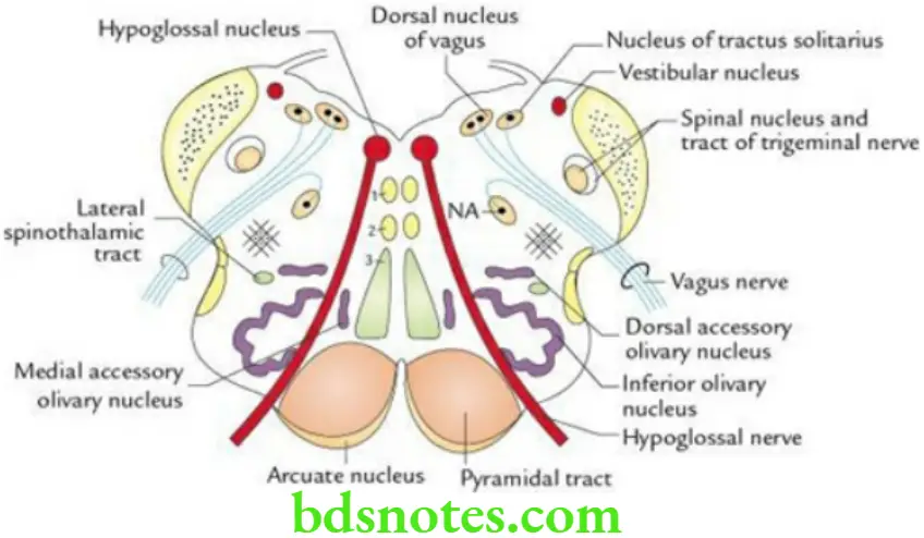
Question 5. Enumerate the main structures seen in the transverse section of the medulla oblongata at the level of the upper part of olives.
Answer. The main structures seen in the transverse section of the medulla oblongata at the level of the upper part of olives are as follows:
- Fully developed inferior olivary nucleus.
- Appearance of medial and dorsal accessory olivary nuclei.
- The appearance of the hypoglossal nucleus, dorsal nucleus of the vagus and vestibular nuclei underneath the floor of the 4th ventricle.
- Appearance of nucleus ambiguus.
- Appearance of nucleus tractus solitarius.
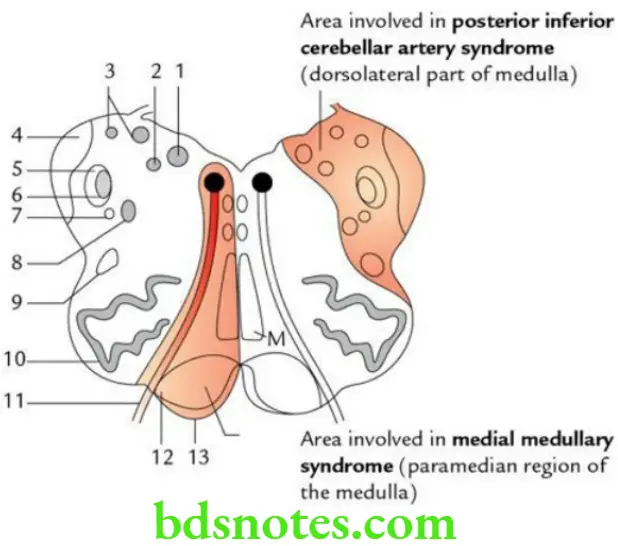
Question 6. Write a short note on lateral medullary syndrome (posterior inferior cerebellar artery syndrome of Wallenberg).
Answer. Causes Of Lateral Medullary Syndrome
Ischaemia of the posterolateral part of the medulla due to occlusion of the posterior inferior cerebellar artery.
Clinical features Of Lateral Medullary Syndrome
- Contralateral loss of pain and temperature in the body due to involvement of the spinothalamic tract.
- Ipsilateral loss of pain and temperature on the face due to involvement of spinal nucleus and tract of CN 5.
- Ipsilateral paralysis of palatal, pharyngeal and laryngeal muscles due to involvement of nucleus ambiguus.
- Ipsilateral ataxia due to involvement of inferior cerebellar peduncle.
- Nausea and vertigo due to involvement of vestibular nuclei.
Question 7. Write a short note on medial medullary syndrome.
Answer. Causes Of Medial Medullary Syndrome
Ischaemia of the medial region of the medulla due to thrombosis of the anterior spinal artery.
Clinical Features Of Medial Medullary Syndrome
- Contralateral hemiplegia (UMN type of paralysis) due to involvement of pyramid (pyramidal tract)
- Ipsilateral paralysis of the tongue due to the involvement of the hypoglossal nerve (LMN type of paralysis)
- Contralateral loss of proprioceptive sensations due to involvement of medial meniscus
Pons
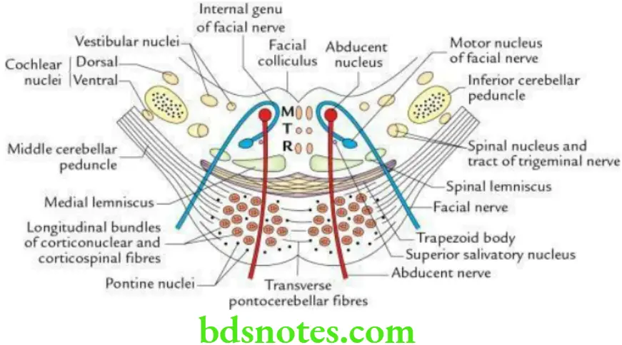
Question 8. Enumerate the main structures seen in the transverse section of pons at the level of facial colliculi.
The main structures seen in the transverse section of pons at the level of facial colliculi are as follows:
- Abducent nucleus
- Motor nucleus of the facial nerve
- Internal genu of the facial nerve
- Dorsal and ventral cochlear nuclei
- Trapezoid body
Question 9. Enumerate the main structures seen in the transverse section through the upper part of the pons.
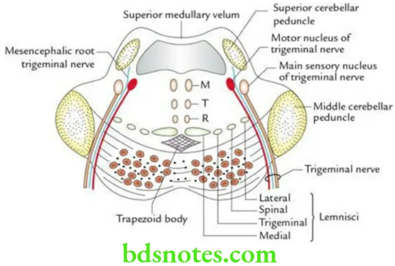
Answer. The main structures seen in the transverse section through the upper part of the pons are as follows:
- The chief (main) sensory nucleus of the trigeminal nerve
- Motor nucleus of trigeminal nerve
- The mesencephalic root of the trigeminal nerve
- Trigeminal lemniscus
- Lateral lemniscus
Question 10. Write a short note on pontocerebellar angle syndrome.
Answer. Causes Of Pontocerebellar Angle Syndrome
Involvement of lateral region of caudal part of pons near pontocerebellar angle by Schwannoma of CN 8.
Clinical features Of Pontocerebellar Angle Syndrome
- Ipsilateral deafness due to involvement of cochlear nerve.
- Ipsilateral imbalance and asynergia due to involvement of vestibular nerve.
- Ipsilateral lower motor neuron type of facial palsy due to involvement of facial nerve.
- Ipsilateral loss of pain and temperature on the face due to the involvement of the spinal nucleus and tract of CN 5.
Midbrain
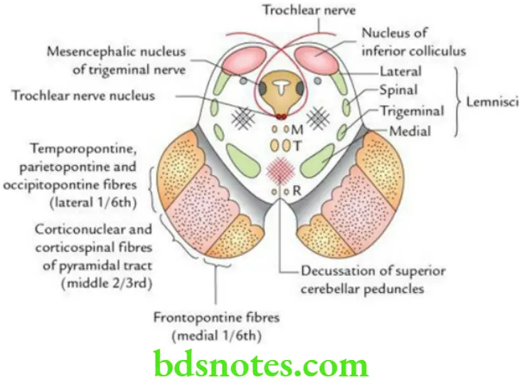
Question 11. Enumerate the main structures seen in the transverse section of the midbrain at the level of the inferior colliculus.
Answer. The main structures seen in the transverse section of the midbrain at the level of the inferior colliculus are as follows:
- Decussation of superior cerebellar peduncles
- Trochlear nerve nucleus
- The mesencephalic nucleus of the trigeminal nerve
- Termination of lateral lemniscus
- Nucleus of inferior colliculus
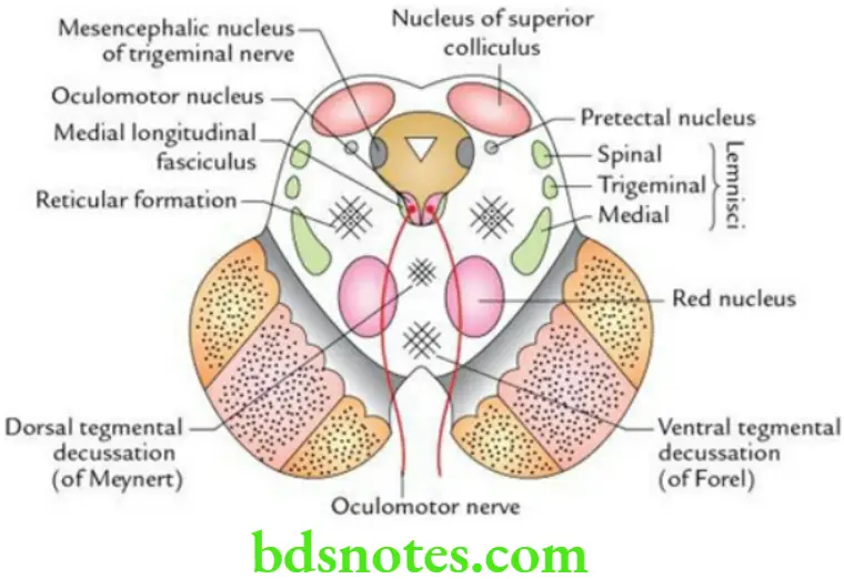
Question 12. Enumerate the main structures seen in the transverse section of the midbrain at the level of the superior colliculus.
Answer. The main structures seen in the transverse section of the midbrain at the level of the superior colliculus are as follows:
- Oculomotor nucleus
- Red nucleus
- Nucleus of superior colliculus
- Dorsal tegmental decussation (of Meynert)
- Ventral tegmental decussation (of Forel)
Question 13. Write a short note on the medial longitudinal fasciculus/medial longitudinal bundle.
Answer. The medial longitudinal fasciculus (MLF) also called medial longitudinal bundle (MLB) is an important association tract on either side of the median plane in the brainstem. It extends cranially to the interstitial nucleus of Cajal (accessory oculomotor nucleus) and caudally it becomes continuous with the intersegmental fasciculus of the spinal cord.
- It connects the nuclei of the cranial nerves that move the eyeballs and in this way, it coordinates the movements of both eyeballs.
- It connects the nuclei of the cranial nerves responsible for articulation, and in this way, it associates together the movements of the organs responsible for articulation.
- It connects both the vestibular and cochlear nuclei with the nuclei of the nerves of the eyeball and with the anterior horn cells of the spinal nerves; in this way, it associates movements of eyes and those of the body in response to movements of the head or in response to the sound.
Functions Of Medial Longitudinal Fasciculus
- Coordination of the conjugate movements of the eyeball.
- Coordination of the movements of head and neck in response to audiovisual reflexes.

Leave a Reply