Biology Of Tooth Movement
Question 1. Describe histology of tooth movement or tissue changes incident to orthodontic treatment.
Or
Describe in detail various biological reactions in the periodontium in response to optimal orthodontic forces.
Or
Briefly describe tissue changes during orthodontic tooth movements.
Or
Write about tissue changes taking place incidental to orthodontic tooth movement.
Answer. The histological or tissue changes incidental to orthodontic treatment are explained under the following headings:
- Tissue changes at pressure zone.
- Tissue changes at tension zone.
- Tissue changes in other areas, i.e. pulp, dentin, cementum, gingiva and TMJ.
Read And Learn More: Orthodontics Question And Answers
Tissue Changes at Pressure Zone
- If light forces are applied frontal resorption will occur.
- If heavy forces are applied hyalinization and undermining resorption will occur.
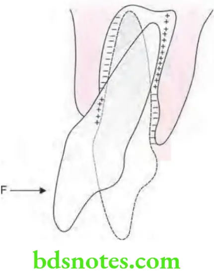
Frontal Resorption
- As orthodontic tooth movement starts osteoclasts get activated.
- Osteoclasts are derived from local population or from blood supply.
- Activated osteoclasts starts resorption by resorbing adjacent lamina dura. This process of bone resorption is also known as frontal resorption.
- Resorption starts from PDL side of alveolar bone.
- Frontal resorption takes place after two days of orthodontic force application.
Frontal Resorption In Orthodontics
Hyalinization
- If the orthodontic force increases more than the capillary pulse pressure, i.e. 20–26 gm/cm2 blood vessels get compressed or occluded.
- As PDL get compressed the blood supply of compressed area get cut off
- Now the cells which become activated osteoclasts remain inactive.
- Sterile necrotic area is visible in compressed PDL.
- This is when seen in light microscope appear as area devoid of cells and this is known as hyalinized area and process is known as hyalinization.
- Hyalinization is a reversible process.
- The effect of hyalinization is that it does not allow tooth to move.
- In hyalinization tooth can be moved in the condition in which bone beneath the hyalinized area undergo resorption.
- Hyalinization lasts for one to two weeks after which resorption occur by undermining resorption.
Undermining Resorption
- It is also known as indirect resorption.
- As there is occurrence of hyalinization, chances of frontal resorption are diminished.
- After some days hyalinized zone is invaded by the cells from adjacent normal areas of PDL.
- With this osteoclasts also appear in adjacent marrow spaces.
- These osteoclasts resorbs bone adjacent to hyalinized PDL zone from underside. This is known as undermining resorption as bone resorption occurs from underside of lamina dura.
- Resorption of bone occurs from endosteal part.
- Hyalinization and undermining resorption lead to delayed tooth movement while tooth movement is efficient with frontal resorption.
- Bone deposition occurs at the rate of 15µm/day
Tissue Changes at Tension Zone
- In areas of tension cellular activity becomes delayed as compared to pressure zone.
- In tension zone 30 hours are required for increased cellular activity.
- Macrophages are abundant in tension zone.
- Inflammation and remodeling of firous elements over evident in tension zone.
- New unmineralized matrix is laid down in the fiers which are close to alveolar wall.
- Later on osteoid which is synthesized by osteoblasts is laid down on complete alveolar wall over tension zone.
- Bone deposition occurs at the rate of 30µm/day.
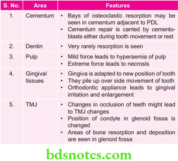
Tissue Changes in Other Areas
Question 2. Describe in detail about tissue changes in response to mild and heavy orthodontic force.
Or
Describe the histological changes occurring in PDL and alveolar bone when a mild orthodontic force is applied to a tooth, what would happen if a force applied turn in heavy force.
Answer. When force is applied on a tooth during orthodontic treatment it leads to formation of areas of pressure and tension around teeth
- Areas of pressure are formed in the direction of tooth movement.
- Areas of tension form in the opposite direction of tooth movement.
- Bone surface subjected to tension shows bone deposition.
- Bone surface subjected to pressure shows bone resorption.
- The histological changes associated with tooth movements depend on amount and duration of force.
- The histological changes associated with tooth movements are studied under following headings.
- Changes following application of mild force.
- Changes following application of extreme force.
Frontal Resorption In Orthodontics
Changes Following Application of Mild/Light Force
Changes on Pressure Side
- Periodontal ligament in direction of tooth movement is compressed 1/3rd of its original thickness.
- Vascularity of PDL is increased due to increased capillary blood supply.
- Increased blood supply leads to mobilization of firoblasts and osteoclasts.
- Osteoclasts line up along the socket wall on the pressure side.
- Osteoclasts lie in Howship’s lacunae.
- Orientation of bony trabeculae changes (They becomeThe osteoclasts that lies within Howship’s lacunae start resorbing bone.
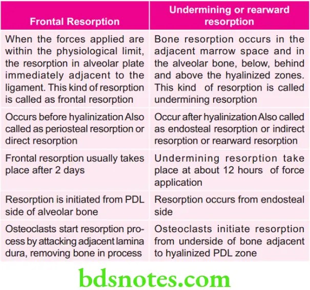
When the forces applied are within the physiological limit, the resorption in alveolar plate immediately adjacent to the ligament. This kind of resorption is called—frontal resorption.
Changes on the Tension Side
- Periodontal membrane get stretched.
- Periodontal fiers get stretched.
- Vascularity increased leads to mobilizations of firoblasts and osteoblasts.
- In response to traction, osteoid is laid down by osteoblasts in the periodontal ligament and it become slightly calcified and is known as woven bone.
Secondary Remodeling Changes
Bony changes elsewhere (other than the area of tension and pressure) to maintain the width of alveolar bone.
Changes Following Application of Extreme/Heavy Force
On Pressure Side
- Root closely approximate the lamina dura.
- Compresses the periodontal ligament.
- Occlusion of blood vessels, leads to less nutritional supply leading to regressive changes called hyalinization.
- Bone resorption occur in the adjacent marrow space and in the alveolar bone, below, behind and above the hyalinized zones this kind of resorption is called undermining or rearward resorption.
On Tension Side
- PDL is overstretched.
- Tearing of blood vessels and ischemia occur.
- Osteoblastic activation is increased.
- Tooth becomes loose in socket.
- Pain and hyperemia of gingiva occur.
Frontal Resorption In Orthodontics
Question 3. Write short note on undermining resorption.
Answer.
- It is also known as indirect resorption or rearward resorption.
- It is named by Sandstedt.
- It occurs due to application of extreme forces for movement of tooth.
- As there is occurrence of hyalinization, chances of frontal resorption are diminished.
- After some days hyalinized zone is invaded by the cells from adjacent normal areas of PDL.
- With this osteoclasts also appear in adjacent marrow spaces.
- These osteoclasts resorb bone adjacent to hyalinized PDL zone from underside. This is known as undermining resorption as bone resorption occurs from underside of lamina dura.
- Resorption of bone occurs from endosteal part.
- Undermining resorption occur in all the cases where root is not moved parallel to bony surface with too much compression.
Question 4. Write short note on frontal resorption.
Or
Describe briefly frontal resorption.
Or
Write short answer on frontal resorption.
Answer. Frontal resorption is also known as periosteal resorption or direct resorption or forward resorption.
Frontal resorption is the type of tissue change at the pressure zone in orthodontic tooth movement which follows the application of light force.
When the forces applied are within the physiological limit, the resorption in alveolar plate immediately adjacent to the ligament. This kind of resorption is called frontal resorption.
Changes Occurring on Pressure Side
- Periodontal ligament in direction of tooth movement is compressed 1/3rd of its original thickness.
- Vascularity of PDL is increased due to increased capillary blood supply.
- Increased blood supply leads to mobilization of firoblasts and osteoclasts.
- Osteoclasts line up along the socket wall on the pressure side.
- Osteoclasts lie in Howship’s lacunae.
- Orientation of bony trabeculae changes (They become parallel to orthodontic force).
- The osteoclasts that lies within Howship’s lacunae start resorbing bone.
In frontal resorption, resorption starts from PDL side of alveolar bone.
Frontal resorption takes place after two days of orthodontic force application.
Question 5. Write short note on piezoelectronic theories of tooth movement.
Answer.
- Farrar (l876) fist noted deformation or bending of the interseptal alveolar walls.
- According to Farrar bone bending may be a possible mechanism for bringing about tooth movement.
- Piezoelectricity is a phenomenon observed in many crystalline materials in which a deformation of crystal structure produces a flw of electric current because of displacement of electrons from one part of the crystal lattce to the other. A small electric current is generated when bone gets mechanically deformed.Possible sources of electric current are:
- Collagen: Inside the bone, collagen occurs in crystalline state and so can be the source of piezoelectricity whenget deformed.
- Hydroxyapatite: It is crystalline in form and produce electricity when deformed.
- Collagen hydroxyapatite interface: It is the junction which lies between the collagen and hydroxyapatit crystals and when it is bent, it can be a source of piezoelectricity.
- Mucopolysaccharide: Fraction of ground substance is not crystalline but it may also possess the ability to generate electric current when get deformed.
- When crystal structure is deformed, the electrons migrate from one location to another which results in electric charge.
- When force is released, the crystals return to their original shape and a reverse flow of electrons is observed.
- Two unusual characteristics which piezoelectric signals possess are:
- Quick decay rate: As force is applied, a piezoelectric signal is produced. This electric charge quickly dies away to zero even though the force is maintained.
- When the force is released, electron flow in the opposite direction is seen.
- On application of force on a tooth, the adjacent alveolar bone bends.
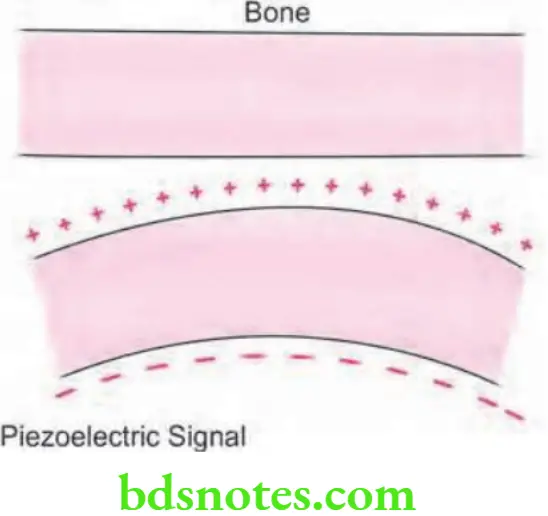
- Areas of concavity in bone associated with negative charge, these evoke bone deposition.
- Areas of convexity associated with positive charge evoke bone resorption.
- On application of force on the tooth both alveolar and medullary cortical plates of bone move together closely and the bone becomes less concave and an electrical signal associated with the resorption get established.
- Bone which is deformed by stress get electrically charged. Concave surfaces attain negative polarity and convex surfaces a positive polarity.
- As a result of these electrical signals, a remodelling response is evoked; bone is added to concave surfaces and resorbed from convex surfaces.
Question 6. Define optimal orthodontic force. Describe in detail the biologic changes in response to light and heavy orthodontic forces.
Answer.
Definition of Optimal Orthodontic Force
“Optimal orthodontic force is one which moves teeth most rapidly in the desired direction, with the least possible damage to tissue and with minimum patient discomfort.” For light forces refer to heading “mild forces” and for heavy forces refer to heading “extreme forces”.
Question 7. Write short note on hyalinization.
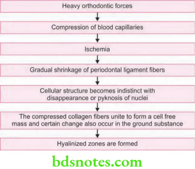
Answer. If the orthodontic force increases more than the capillary pulse pressure, i.e. 20–26 gm/cm2 blood vessels get compressed or occluded.
- As PDL get compressed the blood supply of compressed area get cut off
- Now the cells which become activated osteoclasts remain inactive.
- A sterile necrotic area is visible in compressed PDL.
- This is when seen in light microscope appear as area devoid of cells and this is known as hyalinized area and process is known as hyalinization.
- Hyalinization is a reversible process.
- The effect of hyalinization is that it does not allow tooth to move.
- In hyalinization tooth can be moved in the condition in which bone beneath the hyalinized area undergo resorption.
- Hyalinization last for one to two weeks after which resorption occur by undermining resorption.
Frontal Resorption In Orthodontics
Summary of Hyalinization
Question 8. Briefly differentiate Frontal and undermining resorption.
Or
Differentiate between frontal vs Rear resorption.
Or
Briefly differentiate rear and frontal resorption.
Or
Write short note on frontal vs undermining resorption.
Or
Briefly differentiate between frontal and undermining resorption.
Answer.
Question 9. Write short note on pressuretension theory.|
Answer. It is one of the theories of tooth movement
- Pressure-Tension theory is given by Schwarz in 1932.
- Although Sandstedt (1904) and Oppenheim (1911) investigated the phenomenon of tooth movement by histological examination of the supporting structure, Schwartz is credited with Pressure-Tension theory which has been widely accepted.
- Whenever a tooth is subjected to the orthodontic force, it leads to the areas of pressure and tension.
- Areas of the periodontium in direction of tooth movement are under the pressure while area of periodontium opposite to tooth movement is under tension.
- According to Schwarz, areas of pressure show bone resorption while areas of tension show bone deposition.
Question 10. What are the types of orthodontic tooth movements? Discuss the various theories related to tissue changes incident to orthodontic tooth movement.
Answer.
Orthodontic Tooth Movements:
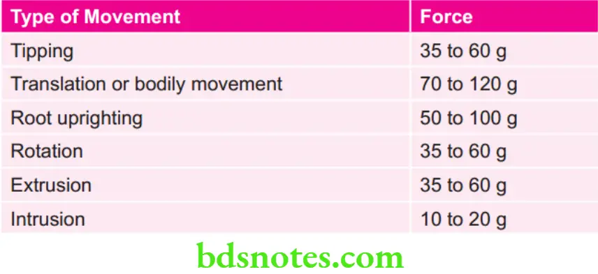
- Tipping: It is a simple type of a tooth movement where a single force is applied to the crown which results in movement of crown in direction of the force and the root in opposite direction. It is the simplest among the tooth movements. It is of two types
- Controlled tipping: Controlled tipping of the tooth occurs when a tooth tips about a center of rotation at its apex. Here there is a lingual movement of the crown with minimal involvement of the root in labial direction.
- Uncontrolled tipping: Uncontrolled tipping of the tooth describes the movement of a tooth that occurs about center of rotation apical to and very close to the center of resistance. It is characterized by the crown moving in one direction while the root moves in opposite direction.
- Bodily movement: If the line of action of an applied force passes through the center of resistance of a tooth, all the points on the tooth will move an equal distance in the same direction signifying a bodily displacement. This is called translation.
- Intrusion: It is a bodily displacement of a tooth along its long axis in an apical direction.
- Extrusion: It is a bodily displacement of a tooth along its long axis in an occlusal direction.
- Rotation: Rotations are labial or lingual movements of a tooth around its long axis.
- Torquing: It can be considered as a reverse tipping characterized by the lingual movement of the root.
- Uprighting: During orthodontic treatment crowns of the certain teeth will be tipped in a mesiodistal direction with the roots tipped in opposite way. Tipping these roots back to get a parallel orientation is termed uprighting.
Theories Related to Tissue Changes Incident to Orthodontic Tooth Movement
Theories are:
- Pressure tension theory: Refer to Ans 9 of same chapter.
- Piezoelectricity: Refer to Ans 5 of same chapter.
Fluid Dynamic Theory
- It is also known as blood flow theory and is proposed by Bein.
- As per this theory, tooth movement occurs due to alteration in fluid dynamics inside the periodontal ligament.
- Periodontal ligament occupies the periodontal space between the tooth and the alveolar socket.
- Periodontal space has a fluid system which is made of interstitial fluid, cellular elements, blood vessels and the ground substance. In addition to the periodontal fibers.
- When force of the greater magnitude and direction is applied during orthodontic tooth movement, interstitial fluid inside the periodontal ligament squeezes out and move to the apex as well as cervical margins and leads to decrease in tooth movement which is known as squeeze film effect by Bien.
- As orthodontic force is applied it leads to compression of periodontal ligament.
- Blood vessels of the periodontal ligament get trapped between the principal fiers which lead to stenosis. Vessel above the stenosis, then balloons which leads to the formation of aneurysm.
- Bien had suggested that there is alteration in the chemical environment at the site of vascular stenosis because of decrease in oxygen level in compressed area as compared to the tension side.
- Formation of such aneurysms and vascular stenosis causes blood gases to escape in the interstitial fluid which create a favorable local environment for the resorption.
Frontal Resorption In Orthodontics
Question 11. What are the characteristic features of orthodontic force. Describe the deleterious effects of orthodontic force on root.
Answer.
Characteristic Features of Orthodontic Force
Clinically
- Orthodontic force should produce rapid tooth movement.
- Orthodontic force should cause minimum patient discomfort.
- Orthodontic force should be such that the lag phase of tooth movement should be shorter.
- As orthodontic force is applied and the tooth is moved it should be loosened in its socket.
Histologically
- After application of an orthodontic force vitality of tooth and PDL is maintained.
- Orthodontic force should initiate maximum cellular response.
- Orthodontic force should produce frontal resorption.
- Orthodontic force is applied such that the integrity of periodontal attachment is maintained by reorganization of fibers.
Deleterious Effects of Orthodontic Force on Root
- Tissues lying adjacent to the root start undergoing remodeling as tooth movement occur.
- Cementum or root remodeling also starts.
- Resorption of cementum occurs next to hyalinized areas so this means that root start resorbing at the place when the heavy forces are implicated.
- Root remodeling restore proper length of root at the time of orthodontic tooth movement.
- As islands of cementum are formed the repair of root does not take place and there is permanent loss of root structure at apex.
- There are two types of root resorption, i.e. localized and generalized
Localized Root Resorption
- Application of excessive orthodontic force as well as excess long duration of treatment leads to the enhancement in chances of resorption.
- Susceptibility of upper incisors is more for resorption to occur.
- As the roots are pressed against cortical plate root resorption occur.
Generalized Root Resorption
- It is divided into two types, i.e. moderate and severe.
- Moderate generalized root resorption: In most of the patients generalized shortening of root will occur due to orthodontic treatment. Resorption of maxillary incisors is highest.
- Severe generalized root resorption: It occurs in the condition like hypothyroidism, in such cases orthodontic treatment is contraindicated.
Question 12. What are the various types of tooth movements. What do you understand by optimum force in orthodontics. Describe briefl mechanism of tooth movement.
Or
Write short answer on optimum orthodontic force.
Or
What is optimum orthodontic forces. Give detail about theories of tooth movement.
Answer.
Optimal Orthodontic Force in Orthodontics
- Oppenheim and Schwarz had discovered optimum orthodontic force.
- Optimal orthodontic force is one which moves teeth most rapidly in the desired direction, with the least possible damage to tissue and with minimum patient discomfort.”
- It is also known as ideal orthodontic force.
- Optimal orthodontic force should be less than capillary blood pressure.
- Range of optimal orthodontic force is 20–26 gm/cm2 of root surface.
- According to Schwarz value of optimum orthodontic force for tooth movement is 15 to 20 mm Hg of capillary pressure.
- Clinically, optimal orthodontic force produces rapid tooth movement with minimum patient discomfort and minimum lag phase and there is no marked mobility of teeth being moved.
- Histologically, vitality of tooth and supporting periodontal ligament is maintained and this produces direct frontal resorption.
Optimum Forces for Orthodontic Tooth Movement
Advantages of Optimum Orthodontic Force
- Tooth movement is efficient.
- Resorption is of frontal type.
- There is elimination of lag phase and hyalinized zone.
- There is less pain and no damage to supporting structures.
- Chances for the root resorption are very less.
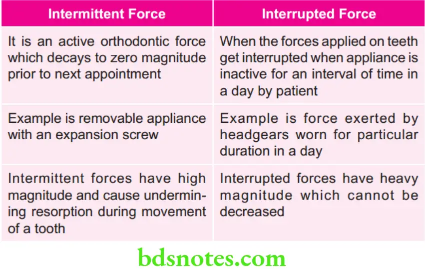
Question 13. Distinguish between intermittent force vs interrupted force.
Or
Briefly differentiate between intermittent and interrupted force.
Answer.
Question 14. Define continuous, interrupted and intermittent orthodontic force. Describe pressure tension theory of tooth movement in detail.
Answer.
Continuous Orthodontic Force
It is defined as the force which is maintained at some appreciable fraction of original force between two successive visits of the patient.
Interrupted Orthodontic Force
It is defined as the force in which the level reduces to zero between two successive visits.
Intermittent Orthodontic Force
It is defied as sudden drop of force to zero level when the orthodontic appliance is removed by the patient.
Pressure Tension Theory
- Whenever a tooth is subjected to the orthodontic force, it leads to the areas of pressure and tension.
- Areas of the periodontium in direction of tooth movement are under the pressure while area of periodontium opposite to tooth movement is under tension.
- According to Schwarz, areas of pressure show bone resorption while areas of tension show bone deposition.
Biochemical Principles Involved in Orthodontic Tooth Movement
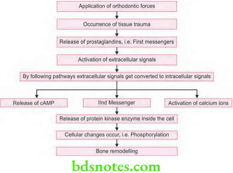
- As orthodontic force is applied on a tooth, it leads to number of biophysical events i.e. compression of periodontal ligament, bone deformation and tissue injury.
- Decrease in vascularity and overstretching of periodontal ligament leads to the chemical changes and inflmmatory response is elicited.
- Biophysical events cause various biochemical reactions at cellular level which causes release of some extracellular signaling molecules known as fist messengers.
- These consists of hormones such as parathormone, local chemical mediators i.e. prostaglandins, neurotransmitters such as substance P and vasoactive intestinal polypeptide.
Frontal Resorption In Orthodontics
First Messenger
- Prostaglandin is the fist messenger, mainly prostaglandin E leads to cellular diffrentiation.
- Other fist messengers are parathormone, substance P and vasoactive intestinal polypeptide.
- They all bind to the cell surface receptors and activate the extracellular signals.
Release of Second Messenger
- Now the extracellular signal is converted to intracellular signal.
- Calcium ions and cAMP are the second messengers.
- Conversion occurs via two pathways, i.e. conversion of ATP into cyclic AMP and other is opening of calcium ion gated channel and activation of calcium ions.
- When continuous pressure is applied for 4 hours the second messenger get activated.
- So it is mandatory to wear any orthodontic appliance for 4 hours to produce effct.
Release of Third Messenger
- Now cAMP and calcium ions act over protein kinase enzyme inside the cell.
- The protein kinase enzyme is the third messenger.
- Protein kinases cause cellular changes in form of phosphorylation.
- Phosphorylation leads to diffrentiation as well as activation of osteoblasts and osteoclasts which causes bony remodeling.
- As remodeling start tooth movements begin.
Question 15. Describe theories of tooth movement. Discuss histology of tooth movement.
Answer. Following are the accepted theories of tooth movement:
- Pressure tension theory by Schwarz (1932)
- Fluid dynamic theory or Blood flow theory by Bein
- Bone bending piezoelectric theory by Farrar (1876)
Question 16. Write short answer on orthodontic and orthopedic force.
Answer.
Orthodontic Force
- It is one which is applied to teeth for bringing about the tooth movement.
- Magnitude of orthodontic force is lesser than orthopedic force.
- Orthodontic forces bring about tooth movement by remodeling and adaptive changes in paradental tissues.
- It is brought about by the use of small amount of force i.e. 20 to 150 gm/tooth.
Orthopedic Force
- Orthopedic force is a force of higher magnitude when delivered via teeth for 12 to 16 hours a day.
- It produces the skeletal effct on maxillofacial complex.
- Orthodontic forces deliver high magnitude of mechanical forces which are more than 300 gms.
- The heavy forces help to modify form of craniofacial bones.
- Part time wear of orthopaedic appliances is recommended to produce more skeletal effcts

Leave a Reply